Descargar la presentación
La descarga está en progreso. Por favor, espere

1
Tratamiento de la Angina Crónica Estable
Dr Eduardo R Perna Subjefe Unidad de Cuidados Intensivos Coronarios Consultorio de Insuficiencia Cardíaca Instituto de Cardiología “J. F. Cabral”. Corrientes Comité de IC de FAC

2
Temario Manifestaciones de la Enfermedad aterosclerótica
Enfermedad coronaria Clasificación Fisiopatología Angina Crónica Estable Epidemiología Evaluación clínica Estudios complementarios Tratamiento Coronary heart disease Restriction of blood flow to the myocardium may be caused by an atherosclerotic plaque narrowing the lumen of the coronary arteries. If the diameter of the coronary artery is reduced by more than 50%, ischaemia will develop and the patient will experience tightness or crushing pain in the chest (angina pectoris). However, pain does not always accompany myocardial ischaemia: this is called silent ischaemia. Coronary plaque rupture and erosion have been shown to result in thrombus formation within coronary arteries. If blood flow is completely obstructed, either due to a thrombus or by a large atherosclerotic plaque, death to part of the myocardium may ensue, resulting in an MI. If damage to the myocardium is severe the pumping action of the heart will be impaired resulting in congestive heart failure or, if very severe, sudden cardiac death.1 Cerebrovascular disease Narrowing of the carotid, vertebral and cerebral arteries supplying blood to the brain can cause a brief interruption in the blood supply to the brain resulting in a transient ischaemic attack. This may cause temporary impairment of vision, speech, sensation or movement and may be followed by a stroke. A stroke may also be caused by formation of a thrombus or embolus, arterial rupture or haemorrhage of the cerebral arteries stopping the oxygen supply to parts of the brain. Sudden loss of consciousness often occurs with subsequent paralysis of parts of the body. It can lead to permanent damage and disability and sudden death.1 Peripheral vascular disease When the lumen of arteries such as the femoral and iliac arteries supplying blood to the legs has been significantly narrowed, by 60% or more, the symptoms of intermittent claudication become evident. These include an aching or cramping pain, most often in the legs when walking, which occurs when insufficient oxygen is reaching the muscles in the legs. In advanced cases of peripheral vascular disease, blood supply to the legs may become completely blocked, possibly by thrombus formation, and painful leg and foot ulcers may develop. If left untreated gangrene may eventually ensue, requiring amputation of the affected limb.1 Reference 1. In: Statins - The HMG-CoA Reductase Inhibitors in Perspective. Eds Gaw A, Packard CJ, Shepherd J. Martin Dunitz 2000,
. However, pain does not always accompany myocardial ischaemia: this is called silent ischaemia. Coronary plaque rupture and erosion have been shown to result in thrombus formation within coronary arteries. If blood flow is completely obstructed, either due to a thrombus or by a large atherosclerotic plaque, death to part of the myocardium may ensue, resulting in an MI. If damage to the myocardium is severe the pumping action of the heart will be impaired resulting in congestive heart failure or, if very severe, sudden cardiac death.1. Cerebrovascular disease Narrowing of the carotid, vertebral and cerebral arteries supplying blood to the brain can cause a brief interruption in the blood supply to the brain resulting in a transient ischaemic attack. This may cause temporary impairment of vision, speech, sensation or movement and may be followed by a stroke. A stroke may also be caused by formation of a thrombus or embolus, arterial rupture or haemorrhage of the cerebral arteries stopping the oxygen supply to parts of the brain. Sudden loss of consciousness often occurs with subsequent paralysis of parts of the body. It can lead to permanent damage and disability and sudden death.1. Peripheral vascular disease When the lumen of arteries such as the femoral and iliac arteries supplying blood to the legs has been significantly narrowed, by 60% or more, the symptoms of intermittent claudication become evident. These include an aching or cramping pain, most often in the legs when walking, which occurs when insufficient oxygen is reaching the muscles in the legs. In advanced cases of peripheral vascular disease, blood supply to the legs may become completely blocked, possibly by thrombus formation, and painful leg and foot ulcers may develop. If left untreated gangrene may eventually ensue, requiring amputation of the affected limb.1. Reference. 1. In: Statins - The HMG-CoA Reductase Inhibitors in Perspective. Eds Gaw A, Packard CJ, Shepherd J. Martin Dunitz 2000,")
3
Manifestaciones de la enfermedad aterosclerótica

4
Manifestaciones clínicas de la aterosclerosis
Enfermedad coronaria Angina pectoris, infarto de miocardio, muerte súbita Enfermedad cerebrovascular Ataque isquémico transitorio, stroke Enfermedad vascular periférica Claudicación intermitente, gangrena Coronary heart disease Restriction of blood flow to the myocardium may be caused by an atherosclerotic plaque narrowing the lumen of the coronary arteries. If the diameter of the coronary artery is reduced by more than 50%, ischaemia will develop and the patient will experience tightness or crushing pain in the chest (angina pectoris). However, pain does not always accompany myocardial ischaemia: this is called silent ischaemia. Coronary plaque rupture and erosion have been shown to result in thrombus formation within coronary arteries. If blood flow is completely obstructed, either due to a thrombus or by a large atherosclerotic plaque, death to part of the myocardium may ensue, resulting in an MI. If damage to the myocardium is severe the pumping action of the heart will be impaired resulting in congestive heart failure or, if very severe, sudden cardiac death.1 Cerebrovascular disease Narrowing of the carotid, vertebral and cerebral arteries supplying blood to the brain can cause a brief interruption in the blood supply to the brain resulting in a transient ischaemic attack. This may cause temporary impairment of vision, speech, sensation or movement and may be followed by a stroke. A stroke may also be caused by formation of a thrombus or embolus, arterial rupture or haemorrhage of the cerebral arteries stopping the oxygen supply to parts of the brain. Sudden loss of consciousness often occurs with subsequent paralysis of parts of the body. It can lead to permanent damage and disability and sudden death.1 Peripheral vascular disease When the lumen of arteries such as the femoral and iliac arteries supplying blood to the legs has been significantly narrowed, by 60% or more, the symptoms of intermittent claudication become evident. These include an aching or cramping pain, most often in the legs when walking, which occurs when insufficient oxygen is reaching the muscles in the legs. In advanced cases of peripheral vascular disease, blood supply to the legs may become completely blocked, possibly by thrombus formation, and painful leg and foot ulcers may develop. If left untreated gangrene may eventually ensue, requiring amputation of the affected limb.1 Reference 1. In: Statins - The HMG-CoA Reductase Inhibitors in Perspective. Eds Gaw A, Packard CJ, Shepherd J. Martin Dunitz 2000,
. However, pain does not always accompany myocardial ischaemia: this is called silent ischaemia. Coronary plaque rupture and erosion have been shown to result in thrombus formation within coronary arteries. If blood flow is completely obstructed, either due to a thrombus or by a large atherosclerotic plaque, death to part of the myocardium may ensue, resulting in an MI. If damage to the myocardium is severe the pumping action of the heart will be impaired resulting in congestive heart failure or, if very severe, sudden cardiac death.1. Cerebrovascular disease Narrowing of the carotid, vertebral and cerebral arteries supplying blood to the brain can cause a brief interruption in the blood supply to the brain resulting in a transient ischaemic attack. This may cause temporary impairment of vision, speech, sensation or movement and may be followed by a stroke. A stroke may also be caused by formation of a thrombus or embolus, arterial rupture or haemorrhage of the cerebral arteries stopping the oxygen supply to parts of the brain. Sudden loss of consciousness often occurs with subsequent paralysis of parts of the body. It can lead to permanent damage and disability and sudden death.1. Peripheral vascular disease When the lumen of arteries such as the femoral and iliac arteries supplying blood to the legs has been significantly narrowed, by 60% or more, the symptoms of intermittent claudication become evident. These include an aching or cramping pain, most often in the legs when walking, which occurs when insufficient oxygen is reaching the muscles in the legs. In advanced cases of peripheral vascular disease, blood supply to the legs may become completely blocked, possibly by thrombus formation, and painful leg and foot ulcers may develop. If left untreated gangrene may eventually ensue, requiring amputation of the affected limb.1. Reference. 1. In: Statins - The HMG-CoA Reductase Inhibitors in Perspective. Eds Gaw A, Packard CJ, Shepherd J. Martin Dunitz 2000,")
5
Factores de riesgo No modificables Historia Familiar Edad Sexo
Hipercolesterolemia HTA Tabaquismo Hipertrofia del VI Sedentarismo Diabetes Sobrepeso/Obesidad Alcoholismo Inciertos Hipertrigliceridemia Lp (a) lipoproteina Acido úrico Microalbuminuria Fibrinógeno Renina Hiperhomocisteinemia Proteina C reactiva Grech ED. BMJ 2003;326;
 lipoproteina. Acido úrico. Microalbuminuria. Fibrinógeno. Renina. Hiperhomocisteinemia. Proteina C reactiva. Grech ED. BMJ 2003;326;")
6
Factores de riesgo Categoría 1: La intervención ha probado el riesgo CV Tabaquismo Colesterol LDL Dieta rica en grasas/colesterol Hipertensión Hipertrofia del VI Factores trombogénicos (afectados por AAS) Categoría 2: La intervención es probable que el riesgo CV Diabetes mellitus Sedentarismo Colesterol HDL Trigliceridos LDL pequeña y densa Obesidad Mujeres Postmenopáusica Categoría 3: FdeR asociados q si son modificados pueden bajar el riesgo Factores psicosociales Lipoprotein(a) Homocisteina Estrés Oxidativo No consumo de alcohol Categoría 4: No modificables Edad Sexo masculino Bajo nivel socioeconómico Historia familiar temprana Fuster V, Pearson TA, Co-Chairs. J Am Coll Cardiol 1996;27(5):957–1047.
 Categoría 2: La intervención es probable que el riesgo CV. Diabetes mellitus. Sedentarismo. Colesterol HDL. Trigliceridos. LDL pequeña y densa. Obesidad. Mujeres Postmenopáusica. Categoría 3: FdeR asociados q si son modificados pueden bajar el riesgo. Factores psicosociales. Lipoprotein(a) Homocisteina. Estrés Oxidativo. No consumo de alcohol. Categoría 4: No modificables. Edad. Sexo masculino. Bajo nivel socioeconómico. Historia familiar temprana. Fuster V, Pearson TA, Co-Chairs. J Am Coll Cardiol 1996;27(5):957–1047.")
7
Wilson PWF, et al. Circulation. 1998;97:1837-1847.
Factores de riesgo Wilson PWF, et al. Circulation. 1998;97:

8
Enfermedad coronaria Clasificación

9
Enfermedad coronaria Es un proceso crónico
Evoluciona típicamente en forma cíclica, a través de los siguientes cuadros clínicos Asintomático Isquemia silente Angina estable Síndrome coronario agudo Sin supradesnivel del ST: Angina Inestable Infarto de miocardio sin onda Q Con supradesnivel del ST Infarto de miocardio con onda Q

10
Enfermedad coronaria Síndrome Definición clínica Fisiopatología
Angina crónica estable Dolor típico que ↑ con el ejercicio/ estrés psicológico, ↓ con reposo/nitro ↑ demanda miocárdica de oxígeno con limitación del flujo epicárdico Angina aislada Similar a ACE sin IM o revasc. previa Similar a ACE Angina resistente a revascularización Similar a ACE con revascularización exitosa Disfunción endotelial + ang. microvascular + síndrome de corazón sensible Angina refractaria Similar a ACE pero con revascularización previa no exitosa ↑ demanda miocárdica de oxígeno con limitación marcada del flujo epicárdico Angina persistente Similar a ACE con recurrencia bajo tratamiento médico Similar a ACE con resistencia farmacológica y angina microvascular Angina microvascular (Síndrome X) Síntomas pueden o no ser similares a ACE con evidencias de isquemia y angiogramas “normales” Alteración en la nocicepción, disfunción endotelial, ↓ en la dilatación microvascular + disfunción del músculo liso vascular Angina variante (Prinzmetal) Similar a ACE, usualmente en reposo y a la mañana temprano, vasoespasmo coronario con ↑ ST Hipersensibilidad vasoconstrictora del músculo liso vascular + ↓ factores de relajación derivado del endotelio. Isquemia silente Episodios asintomáticos en pacientes con EC obstructiva con o sin ACE Defectos en la nocicepción debida a narcóticos, neuropatía o edad avanzada Síndrome del corazón sensible Falta de evidencias de isquemia con dolor cardíaco ↓ umbral nociceptivo + disbalance simpáticovagal Jolicoeur EM et al. Am Heart J 2008;155:418-34
 Síntomas pueden o no ser similares a ACE con evidencias de isquemia y angiogramas normales Alteración en la nocicepción, disfunción endotelial, ↓ en la dilatación microvascular + disfunción del músculo liso vascular. Angina variante (Prinzmetal) Similar a ACE, usualmente en reposo y a la mañana temprano, vasoespasmo coronario con ↑ ST. Hipersensibilidad vasoconstrictora del músculo liso vascular + ↓ factores de relajación derivado del endotelio. Isquemia silente. Episodios asintomáticos en pacientes con EC obstructiva con o sin ACE. Defectos en la nocicepción debida a narcóticos, neuropatía o edad avanzada. Síndrome del corazón sensible. Falta de evidencias de isquemia con dolor cardíaco. ↓ umbral nociceptivo + disbalance simpáticovagal. Jolicoeur EM et al. Am Heart J 2008;155:")
11
Enfermedad coronaria Fisiopatología

12
Progresión de la placa aterosclerótica Pared de una arteria coronaria
Fisiopatología Progresión de la placa aterosclerótica Pared de una arteria coronaria Grech ED. BMJ 2003;326;

13
Fisiopatología Abrams J. N Engl J Med 2005;352:

14
Fisiopatología

15
Angina Crónica Estable
Epidemiología

16
Angina Dolor o molestia en el tórax o región adyacente causado por una flujo sanguíneo inadecuado al músculo cardíaco.

17
Bhatt AB et al. Curr Opin Cardiol 21:492–502.
Epidemiología Prevalencia: 3.5% (6.8 millones) Hombres: 2.7% Mujeres: 4.3% Incidencia: x año Sólo 20% de ataques cardíacos están precedidos por ACE Mortalidad: 1.6 a 3.2% anual Heart disease and stroke statistics Update – American Heart Association Bhatt AB et al. Curr Opin Cardiol 21:492–502.
 Hombres: 2.7% Mujeres: 4.3% Incidencia: x año. Sólo 20% de ataques cardíacos están precedidos por ACE. Mortalidad: 1.6 a 3.2% anual. Heart disease and stroke statistics Update – American Heart Association. Bhatt AB et al. Curr Opin Cardiol 21:492–502.")
18
Epidemiología Heart disease and stroke statistics Update – American Heart Association

19
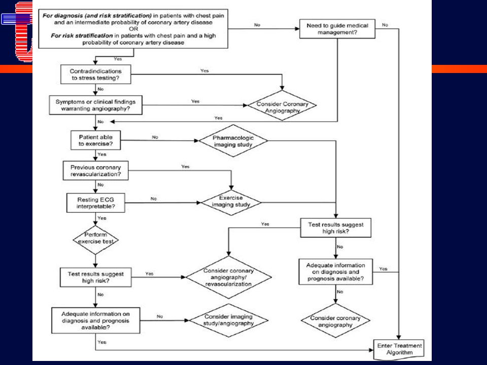
Guías de Manejo AHA/ACC ESC J Am Coll Cardiol 1999; 33, 7:2092-2197
Updates: J Am Coll Cardiol 2003;41:159–68 J Am Coll Cardiol 2007;50:2264 –74. ESC Eur Heart J 2006; 27: 1341–1381

20
1-Historia y examen físico
Evaluación clínica 1-Historia y examen físico

21
Evaluación y Diagnóstico
En pacientes con dolor de pecho Historia detallada de los síntomas Examen físico Evaluación de los factores de riesgo Estimar la probabilidad de enfermedad coronaria importante (baja, intermedia, alta)
")
22
Dolor precordial Calidad - “opresivo," “constricitvo," “pesadez," “ardor" o “malestar“. No se modifica con la posición o respiración. Duración – Generalmente dura minutos. Un dolor muy prolongado es raramente anginoso Localización - Subesternal, irradiación al cuello, mandíbula, epigastrio, miembros. Fuera de estos lugares, es muy raro. Provocación – Ejercicio o estrés emocional, aliviado por el reposo o por NTG sublingual (30” a minutos).
.")
23
Clasificación clínica del dolor precordial
Angina típica (definida) Dolor substernal Provocado por ejercicio o estrés emocional Aliviada por reposo o NTG Angina atípica (probable) Tiene dos de las características Dolor de pecho no cardíaco Reúne 1 de estas características J Am Coll Cardiol. 1983;1:574, Letter
 Dolor substernal. Provocado por ejercicio o estrés emocional. Aliviada por reposo o NTG. Angina atípica (probable) Tiene dos de las características. Dolor de pecho no cardíaco. Reúne 1 de estas características. J Am Coll Cardiol. 1983;1:574, Letter.")
24
Clasificación Canadiense
I “Actividad ordinaria no causa angina” Ej: Caminar o subir escaleras. Angina con ejercicio extenuante, rápido o prolongado en el trabajo o durante actividad recreativa. II “Limitación leve con la actividad ordinaria” Ej: Caminar o subir escaleras rápidamente, después de comer, con el frío, viento. Angina con caminar más de dos cuadras o subir más de un piso. III. “Limitación importante en la actividad ordinaria” Ej: Caminar una o dos cuadras o subir un piso de escalera en condiciones habituales. IV “Incapacidad de realizar cualquier actividad física sin angina – Puede aparecer angina en reposo” Circulation 1976; 54:

25
Dx alternativos CV no isquémico Pulmonar Pared torácica
Disección aórtica Pericarditis Pulmonar TEP Neumotórax Neumonía Pleuritis Pared torácica Costocondritis Fibrositis Fractura de costilla Artritis esternoclavicular Herpes zoster Gastrointestinal Esófago Esofagitis Espasmo Reflujo Biliar Cólico Cholecistitis Coledocolitiasis Colangitis Ulcera péptica Pancreatitis Psiquiátricos Desórdenes de ansiedad Hiperventilación Crisis de pánico Ansiedad primaria Desórdenes afectivos Depresión Desórdenes somatiformes

26
Factores desencadenantes
Incremento demanda O2 No Cardíacos Hipertermia Hipertiroidismo Toxicidad simpaticomimética (cocaine) Hipertensión Ansiedad Fístula arteriovenosa Cardíacas Miocardiopatía hipertrófica Estenosis aórtica Miocardiopatía dilatada Miocardiopatía chagásica Taquiarritmias Disminución aporte O2 No Cardíacos Anemia Hipoxemia neumonía, asma, EPOC, HTP, fibrosis pulmonar, apnea del sueño Hiperviscosidad policitemia, leucemia, trombocitosis, hipergamaglobulinemia Cardíacas Estenosis aórtica Miocardiopatía hipertrófica
 Hipertensión. Ansiedad. Fístula arteriovenosa. Cardíacas. Miocardiopatía hipertrófica. Estenosis aórtica. Miocardiopatía dilatada. Miocardiopatía chagásica. Taquiarritmias. Disminución aporte O2. No Cardíacos. Anemia. Hipoxemia. neumonía, asma, EPOC, HTP, fibrosis pulmonar, apnea del sueño. Hiperviscosidad. policitemia, leucemia, trombocitosis, hipergamaglobulinemia. Cardíacas. Estenosis aórtica. Miocardiopatía hipertrófica.")
27
Probabilidad de EC Análisis bayesiano
Baja (5%) – El valor predictivo positivo de un test anormal es sólo 21%. Intermedia (50%) – Un test positivo incrementa la probabilidad de enfermedad a 83% y uno negativo disminuye a 36%. Alta (90%) – Un test positivo incrementa la probabilidad a 98% y un resultado negativo la disminuye a 83%.
 – El valor predictivo positivo de un test anormal es sólo 21%. Intermedia (50%) – Un test positivo incrementa la probabilidad de enfermedad a 83% y uno negativo disminuye a 36%. Alta (90%) – Un test positivo incrementa la probabilidad a 98% y un resultado negativo la disminuye a 83%.")
28
“The Diamond and Forrester approach “
Probabilidad de EC “The Diamond and Forrester approach “ La evaluación clínica simple del tipo de dolor, edad y génro fueron potentes predictores de probabilidad de enfermedad coronaria Un hombre de 64 años con angina típica tiene una probabilidad de enfermedad coronaria significativa de 94% Una mujer de 32 años con dolor precordial no anginoso tiene una probabilidad de EC de 1% N Engl J Med 1979;300:1350-8

29
*Cada valor representa el porcentaje de EC en la CCG
Probabilidad de EC Probabilidad Pretest de EC en pacientes sintomáticos según edad y sexo (Datos combinados de Diamond/Forrester y CASS) Dolor Edad no anginoso Angina atípica Angina típica años Hombre Mujer Hombre Mujer Hombre Mujer *Cada valor representa el porcentaje de EC en la CCG
 Dolor Edad no anginoso Angina atípica Angina típica años Hombre Mujer Hombre Mujer Hombre Mujer *Cada valor representa el porcentaje de EC en la CCG.")
30
Modelos de Duke y Stanford
Probabilidad de EC Modelos de Duke y Stanford Los predictores más importantes fueron: edad, sexo y tipo de dolor Otros predictores fueron Tabaquismo Ondas Q o cambios del ST Hiperlipidemia Diabetes Am J Med 1983;75: ; Am J Med 1990;89: Ann Intern Med 1993;118:81-90

31
Probabilidad de EC

32
Probabilidad de EC Nomograma de probablidad de enfermedad coronaria severa basada en un score de 5 puntos y la edad. Los 5 factores son: Sexo masculino Angina típica Evidencias de infarto (ECG e historia) Diabetes Uso de insulina
 Diabetes. Uso de insulina.")
33
Modelos de Duke y Stanford
Probabilidad de EC Modelos de Duke y Stanford La probabilidad de EC de una mujer <55 años con angina atípica y sin factores de riesgo es <10%; pero si es diabética, tabaquista y dislipidemia, aumenta a 40%. Am J Med 1983;75: ; Am J Med 1990;89: Ann Intern Med 1993;118:81-90

34
Estratificación de riesgo
Historia Demografía: Edad y sexo Factores de riesgo coronario: HTA, DBT, hipercolesterolemia, tabaquismo, enfermedad vascular periférica e infarto previo Examen físico Vasculopatía (disminución de pulsos, soplos, fondo de ojo) HTA crónica (presión arterial, fondo de ojo) Estenosis aórtica o miocardiopatía hipertrófica subaórtica (soplo sistólico, pulsos anormales) Fallo cardíaco izquierdo y derecho
 HTA crónica (presión arterial, fondo de ojo) Estenosis aórtica o miocardiopatía hipertrófica subaórtica (soplo sistólico, pulsos anormales) Fallo cardíaco izquierdo y derecho.")
35
2-Laboratorio, ECG y Pruebas de provocación de isquemia
Evaluación clínica 2-Laboratorio, ECG y Pruebas de provocación de isquemia

36
Recomendaciones Clase I Class IIa Class IIb Hemoglobina
Glucemia en ayunas Lipidograma (Colesterol total, HDL, TG, LDL) ECG ECG durante la angina Rx Tórax: IC, valvulopatía, enfermedad pericárdica, o aneurisma/disección aórtica Class IIa Rx tórax en pacientes con enfermedad pulmonar Class IIb Rx >Tórax en el resto de los pacientes 1/01 medslides.com
 ECG. ECG durante la angina. Rx Tórax: IC, valvulopatía, enfermedad pericárdica, o aneurisma/disección aórtica. Class IIa. Rx tórax en pacientes con enfermedad pulmonar. Class IIb. Rx >Tórax en el resto de los pacientes. 1/01. medslides.com.")
37
ECG en reposo Debe ser realizado en todos los pacientes con síntomas sugestivos Normal en 50% Un ECG normal no excluye EC severa; pero implica función del VI normal, con pronóstico favorable

38
ECG en reposo anormal Signos de infarto previo (onda Q o R en V1)
Inversión persistente del ST-T, especialmente en V1 a V3, está asociado con eventos coronarios agudos futuros y pobre pronóstico La HVI se asocia con mayor morbi-mortalidad Otros signos asociados con mal pronóstico son: BCRI Bloqueo bifascicular Bloqueo AV FA Arritmias ventriculares Am J Cardiol 1982;49:

39
Rx de Tórax Generalmente normal Poco útil como test rutinario
Hallazgos asociados con peor pronóstico Cardiomegalia Aneurisma de VI Congestión venosa pulmonar Agrandamiento de AI Calcificación de arterias coronarias

40
Test de estrés Ergometría X Eco estrés X SPECT X CCG X

41
Test de estrés Abrams J. N Engl J Med 2005;352:

42
Test de estrés Abrams J. N Engl J Med 2005;352:

43
Test de ejercicio Indicaciones Contraindicaciones
Confirmación de sospecha de angina Evaluación de la extensión de la isquemia y el pronóstico Estratificación de riesgo post-infarto Detección de síntomas inducidos por el ejercicio (arritmias, síncope) Evaluación de resultados de la revascularización Evaluación de trasplante cardíaco Rehabilitación y motivación del paciente Insuficiencia cardíaca Enfermedad febril Miocardiopatía hipertrófica u obstrucción al tracto de salida del VI Estenosis mitral o aórtica severas HTA no controlada Hipetensión pulmonar Taquiarritmias severas Disección de aorta Lesión de tronco Bloqueo AV completo (enadultos Grech ED. BMJ 2003;326;
 Evaluación de resultados de la revascularización. Evaluación de trasplante cardíaco. Rehabilitación y motivación del paciente. Insuficiencia cardíaca. Enfermedad febril. Miocardiopatía hipertrófica u obstrucción al tracto de salida del VI. Estenosis mitral o aórtica severas. HTA no controlada. Hipetensión pulmonar. Taquiarritmias severas. Disección de aorta. Lesión de tronco. Bloqueo AV completo (enadultos. Grech ED. BMJ 2003;326;")
44
Test de ejercicio Principales puntos finales en un test de Ejercicio
Características indicativas de test fuertemente (+) FC > 85% de la predicha ST > 1mm (plano o descendente > valor que ascendente) Recuperación del ST más lenta que lo normal (> 5 min) TAS > 20 mm Hg TAD > 15 mm Hg o progresivo del ST ST > 3 mm sin dolor Arritmias (FE, TV) Angina limitante < 6 min del protocolo de Bruce Falta de de TAS > 10 mm Hg, o con evidencia de isquemia ST > 3 mm extenso Tiempo de recuperación del ST > 6 min Desarrollo de TV ST en ausencia de infarto Grech ED. BMJ 2003;326;
 FC > 85% de la predicha. ST > 1mm (plano o descendente > valor que ascendente) Recuperación del ST más lenta que lo normal (> 5 min) TAS > 20 mm Hg. TAD > 15 mm Hg. o progresivo del ST. ST > 3 mm sin dolor. Arritmias (FE, TV) Angina limitante < 6 min del protocolo de Bruce. Falta de de TAS > 10 mm Hg, o con evidencia de isquemia. ST > 3 mm extenso. Tiempo de recuperación del ST > 6 min. Desarrollo de TV. ST en ausencia de infarto. Grech ED. BMJ 2003;326;")
45
Test de ejercicio Trazado ECG en reposo basal
Trazado luego de 2.24 min del protocolo de Bruce, acompañado de angina de pecho Grech ED. BMJ 2003;326;

46
Máxima capacidad de ejercicio
PEG: Marcadores de riesgo Máxima capacidad de ejercicio Es el marcador pronóstico más fuerte y consistente Se mide con: Duración máxima de ejercicio, máximo MET alcanzado, carga máxima, frecuencia cardíaca y doble producto. Es afectada por función del VI, edad, condición física previa, comorbilidades y estado sicológico (depresión) La utilización de unidades de carga de trabajo (METs) facilita el uso de una medidad estándar independiente del tipo de prueba y protocolo.
 La utilización de unidades de carga de trabajo (METs) facilita el uso de una medidad estándar independiente del tipo de prueba y protocolo.")
47
Isquemia inducida por el ejercicio
PEG: Marcadores de riesgo Isquemia inducida por el ejercicio Depresión o elevación del segmento ST (en derivaciones sin ondas Q patológicas): brinda la mejor información relacionada a isquemia Variables de menor valor: Angina Número de derivaciones con depresión del ST Tipo de depresión del ST (descendente, horizontal o ascendente) Duración en la fase de recuperación Ann Intern Med 1987;106:
: brinda la mejor información relacionada a isquemia. Variables de menor valor: Angina. Número de derivaciones con depresión del ST. Tipo de depresión del ST (descendente, horizontal o ascendente) Duración en la fase de recuperación. Ann Intern Med 1987;106:")
48
PEG: Marcadores de riesgo
Score de Duke Duke Treadmill Score = Tiempo de ejercicio en minutos en Protocolo de Bruce Menos 5x el desvío del segmento ST (durante o después del ejercicio, en milímetros) 4x el índice de angina (“0” no angina, “1” si hay angina, y "2" si la angina es la causa de detención). N Engl J Med 1991;325:849-53
 4x el índice de angina ( 0 no angina, 1 si hay angina, y 2 si la angina es la causa de detención). N Engl J Med 1991;325:")
49
PEG: Marcadores de riesgo
Score de Duke Grupo Riesgo Score Total Sobrevida Mortalidad 4 años anual Bajo +5 62% % % Moderado (-10 to +4) 34% % % Alto (< -10) % % % N Engl J Med 1991;325:849-53
 34% 95% 1.25% Alto (< -10) 4% 79% 5.00% N Engl J Med 1991;325:")
50
ECO vs Cámara gamma Ventajas del ECO estrés Ventajas de la perfusión
1. Mayor especificidad 2. Versatilidad: evalúa anatomía y función 3. Mayor conveniencia / eficacia / disponibilidad 4. Menor costo Ventajas de la perfusión 1. Mayor tasa de éxito técnico 2. Mayor sensibilidad 3. Mejor exactitud para evaluar isquemia cuando están presentes múltiples anormalidades de la motilidad

51
Isquemia reversible de cara anterolateral
Cámara gamma Isquemia reversible de cara anterolateral

52
Estrategia escalonada
Ergometría Simple, bajo costo, más familiar Es el test inicial en pacientes sin digoxina, ECG normal en reposo y con posibilidades de realizarla Imágenes Para pacientes con alteraciones difusas del ST (>1 mm), BCRI, marcapasos o pre-excitación
, BCRI, marcapasos o pre-excitación.")
53
Perfusión: Marcadores de riesgo
Talio normal Pronóstico benigno, incluso en pacientes con EC conocida Tasa de muerte o infarto de 0.9% por año En un estudio de 5,183 pacientes consecutivos, con un seguimiento de 642 ± 226 días, un talio normal se asoció con un riesgo de muerte cardíaca e infarto <0.5% por año Excepción: PEG de alto riesgo y Talio normal Circulation 1998;97:533-43

54
Perfusión: Marcadores de riesgo
Alto riesgo Número, tamaño y localización de las anormalidades de perfusión Captación pulmonar de Talio 201 luego del estrés Dilatación isquémica del VI

55
Estratificación de riesgo
El riesgo de un paciente es función de 4 características: Función del VI: Fracción de eyección Extensión y severidad de la anatomía coronaria Evidencia de un accidente de placa reciente: alto riesgo a corto plazo Estado general y comorbilidades

56
Función del VI La mayoría de los pacientes con ACE no necesitan ECO
En pacientes con infarto previo Selección del tratamiento médico o de revascularización, nivel de actividad física recomendado, rehabilitación y actividad laboral En pacientes con IC Mecanismo fisiopatológico para guiar el tratamiento (sistólica vs diastólica) Una FE <35% se asocia con una mortalidad anual > 3%
 Una FE <35% se asocia con una mortalidad anual > 3%")
57
Alto (Mortalidad anual >3%)
Evaluación no invasiva del riesgo Alto (Mortalidad anual >3%) 1. FE del VI en reposo < 35% 2. Score de treadmill -11 3. FE del VI en esfuerzo < 35% 4. Defecto importante de perfusión 5. Múltiples defectos de perfusión moderados 6. Defectos grandes fijos con dilatación del VI o aumento de la captación pulmonar 7. Defectos moderados inducidos por el ejercicio con dilatación del VI o aumento de la captación pulmonar 8. ECO estrés con > 2 segmentos con anormalidades de la motilidad a bajas dosis de dobutamina ( 10 mg/kg/min) o baja FC (< 120 beats/min) 9. Isquemia extensa en el ECO estrés
 1. FE del VI en reposo < 35% 2. Score de treadmill FE del VI en esfuerzo < 35% 4. Defecto importante de perfusión. 5. Múltiples defectos de perfusión moderados. 6. Defectos grandes fijos con dilatación del VI o aumento de la captación pulmonar. 7. Defectos moderados inducidos por el ejercicio con dilatación del VI o aumento de la captación pulmonar. 8. ECO estrés con > 2 segmentos con anormalidades de la motilidad a bajas dosis de dobutamina ( 10 mg/kg/min) o baja FC (< 120 beats/min) 9. Isquemia extensa en el ECO estrés.")
58
Intermedio (Mortalidad anual<3%) Bajo (Mortalidad annual < 1%)
Evaluación no invasiva del riesgo Intermedio (Mortalidad anual<3%) 1. FE del VI de 35% a 49% 2. Score en treadmill –11 a 5 3. Defectos de perfusión moderados sin dilatación del VI o captación pulmonar 4. Isquemia con ECO pequeña o con altas dosis de dobutamina involucrando 2 segmentos Bajo (Mortalidad annual < 1%) 1. Score 5 2. Talio normal o defectos pequeños 3. Eco normal
 1. FE del VI de 35% a 49% 2. Score en treadmill –11 a 5. 3. Defectos de perfusión moderados sin dilatación del VI o captación pulmonar. 4. Isquemia con ECO pequeña o con altas dosis de dobutamina involucrando 2 segmentos. Bajo (Mortalidad annual < 1%) 1. Score Talio normal o defectos pequeños. 3. Eco normal.")
60
Angiografía coronaria

61
Angiografía coronaria
CCG diagnóstica Cuando los test no invasivos están contraindicados o son inadecuados por otras enfermedades o incapacidades. Ej: EPOC Test no invasivo anormal, pero no claramente diagnóstico Actividad del paciente con riesgo personal o para otros Alta probabilidad de EC severa Diabéticos oligosintomáticos por neuropatía sensorial o disautonomía

62
Estratificación de riesgo
Angiografía coronaria Estratificación de riesgo Los predictores más poderosos son: extensión y severidad de la EC, así como la función del VI Estenosis proximales Lesión severa de tronco de coronaria izquierda Sobrevida a 12 años en el CASS con tratamiento médico Arterias coronarias Fracción de eyección Normales % 50% % Un vaso % % % Dos vasos % <35% % Tres vasos 40% Circulation 1994;90:

63
Angiografía coronaria

64
Indicaciones de CCG

65
Angiografía coronaria
By pass previo La progresión de la EC nativa no es común El desarrollo de lesiones en los puentes venosos son propensas a progresión rápida y oclusiones trombóticas Se recomiendo bajo umbral para una CCG en pacientes que desarrollan ACE >5 años luego de la cirugía, especialmente en presencia de isquemia en la zona del puente, puente venoso a DA, o múltiples puentes venosos La evolución puede ser mejorada con reintervenciones (cirugía o procedimientos endovasculares)
")
66
Test de Ejercicio post ATC
PEG > 6 meses de revascularización Post cirugía El dolor es habitualmente atípico El ECG es habitualmente anormal Se prefieren los test de imágenes 30% tienen una PEG anormal tempranamente luego de la cirugía Test de Ejercicio post ATC La PEG es poco sensible (40% a 55%) para predecir re-estenosis; se prefieren test de imágenes Escasos datos para justificar alguna frecuencia de estudio Se deberían evaluar pacientes con cambios significativos en la angina Puede ser atractivo para pacientes de alto riesgo: disfunción del VI, múltiples vasos, DA proximal, muerte súbita previa, DBT, ocupación de riesgo, resultados subóptimos
 para predecir re-estenosis; se prefieren test de imágenes. Escasos datos para justificar alguna frecuencia de estudio. Se deberían evaluar pacientes con cambios significativos en la angina. Puede ser atractivo para pacientes de alto riesgo: disfunción del VI, múltiples vasos, DA proximal, muerte súbita previa, DBT, ocupación de riesgo, resultados subóptimos.")
67
Manejo de la isquemia miocárdica

68
Síndrome coronario agudo
Espectro de la enfermedad coronaria Síndrome coronario agudo Infarto de miocardio Angina inestable ATC Cirugía Estabilización post SCA Asintomático Isquemia miocárdica Remodelamiento Angina crónica Dilatación ventricular Enfermedad coronaria Insuficiencia cardíaca Aterosclerosis HVI Angina refractaria no revascularizable Factores de riesgo

69
Síndrome coronario agudo
Pacientes con cardiopatía isquémica crónica Síndrome coronario agudo Infarto de miocardio Angina inestable ATC Cirugía Estabilización post SCA Asintomático Isquemia miocárdica Remodelamiento Angina crónica Dilatación ventricular Enfermedad coronaria Insuficiencia cardíaca Aterosclerosis HVI Angina refractaria no revascularizable Factores de riesgo

70
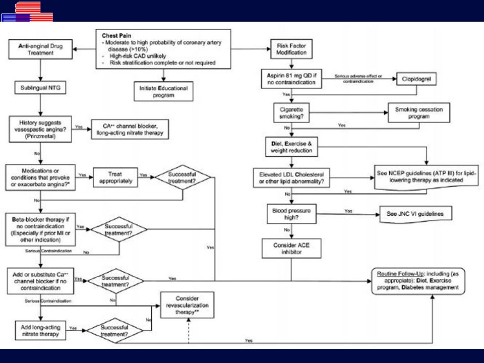
Objetivos del tratamiento
Reducir la isquemia y aliviar los síntomas Prevenir el desarrollo de infarto y reducir la mortalidad Control de factores de riesgo Retrasar la progresión e inducir la regresión Controlar la angina refractaria

71
Prevención de infarto y reducción de mortalidad
Tratamiento Prevención de infarto y reducción de mortalidad

72
Agregación plaquetaria
GP IIb-IIIa inhibitors displace fibrinogen in existing thrombi to disaggregate thrombus and prevent further platelet cross-linking and thrombosis GP IIb-IIIa inhibitors prevent platelet activation by blocking GP IIb-IIa (outside-in signaling) High-dose heparin stimulates PAF which activates platelets Platelets can be activated by many platelet agonists, such as thrombin, adenosine diphosphate, collagen, serotonin, epinephrine, CD40 ligand, and as many as 70 other mediators. Each of these agonists activates a separate signal transduction pathway within platelets. All of these pathways ultimately converge on the platelet receptor GP IIb-IIIa, converting it from an inactive into an active form. The platelet receptor GP IIb-IIIa is the final common pathway to platelet aggregation, which involves binding of a single molecule of fibrinogen to two GP IIb-IIIa molecules on the surface of adjacent platelets. _________________________________________________ 1. Phillips DR, Scarborough RM. Am J Cardiol 1997;80(4A):11B-20B. White HD. Am J Cardiol 1997; 80:2B-10B. Schafer A. J Clin Invest 1986; 78:73-79. DeJong MJ, et al. Critical Care Nursing Clin of N Am 1999; 11: Moser M, et al. J Cardiovasc Pharmacol 2003;41: Phillips DR, Scarborough RM. Am J Cardiol 1997;80(4A):11B-20B.
 High-dose heparin stimulates PAF which activates platelets. Platelets can be activated by many platelet agonists, such as thrombin, adenosine diphosphate, collagen, serotonin, epinephrine, CD40 ligand, and as many as 70 other mediators. Each of these agonists activates a separate signal transduction pathway within platelets. All of these pathways ultimately converge on the platelet receptor GP IIb-IIIa, converting it from an inactive into an active form. The platelet receptor GP IIb-IIIa is the final common pathway to platelet aggregation, which involves binding of a single molecule of fibrinogen to two GP IIb-IIIa molecules on the surface of adjacent platelets. _________________________________________________. 1. Phillips DR, Scarborough RM. Am J Cardiol 1997;80(4A):11B-20B. White HD. Am J Cardiol 1997; 80:2B-10B. Schafer A. J Clin Invest 1986; 78: DeJong MJ, et al. Critical Care Nursing Clin of N Am 1999; 11: Moser M, et al. J Cardiovasc Pharmacol 2003;41: Phillips DR, Scarborough RM. Am J Cardiol 1997;80(4A):11B-20B.")
73
Agentes antiplaquetarios
Clase I Aspirina (75 a 162 mg) Uso rutinario en ausencia de contraindicaciones Inhibe ciclo-oxigenasa y síntesis de thromboxano A2 En >3.000 pacientes con angina estable, redujo un 33% el riesgo de eventos cardiovasculares En angina inestable o IAM no Q, 51-72% de reducción del riesgo de muerte o IAM En prevención primaria se asoció con reducción en la incidencia de infarto (Physicians' Health Study) Aspirina (75 a 325 mg) debería usarse rutinariamente daily should be used routinely in all patients with acute and chronic ischemic heart disease with or without manifest symptoms in the absence of contraindications aspirin exerts an antithrombotic effect by inhibiting cyclo-oxygenase and synthesis of platelet thromboxane A2 in >3,000 patients with stable angina, aspirin reduced the risk of adverse cardiovascular events by 33% in patients with unstable angina, aspirin decreases the short and long-term risk of fatal and nonfatal MI in the Physicians' Health Study, aspirin (325 mg), given on alternate days to asymptomatic persons, was associated with a decreased incidence of MI BMJ 1995;308:81-106
 Uso rutinario en ausencia de contraindicaciones. Inhibe ciclo-oxigenasa y síntesis de thromboxano A2. En >3.000 pacientes con angina estable, redujo un 33% el riesgo de eventos cardiovasculares. En angina inestable o IAM no Q, 51-72% de reducción del riesgo de muerte o IAM. En prevención primaria se asoció con reducción en la incidencia de infarto (Physicians Health Study) Aspirina (75 a 325 mg) debería usarse rutinariamente daily should be used routinely in all patients with acute and chronic ischemic heart disease with or without manifest symptoms in the absence of contraindications. aspirin exerts an antithrombotic effect by inhibiting cyclo-oxygenase and synthesis of platelet thromboxane A2. in >3,000 patients with stable angina, aspirin reduced the risk of adverse cardiovascular events by 33% in patients with unstable angina, aspirin decreases the short and long-term risk of fatal and nonfatal MI. in the Physicians Health Study, aspirin (325 mg), given on alternate days to asymptomatic persons, was associated with a decreased incidence of MI. BMJ 1995;308:")
74
Agentes antiplaquetarios
Clase IIa Las tienopiridinas inhiben irreversiblemente la unión del ADP al receptor plaquetario Ticlopidina: no ha demostrado reducción de eventos cardiovasculares (puede inducir neutropenia y púrpura trombocitopénica trombótica) Clopidogrel: Estudio CAPRIE Pacientes con IM previo, stroke y enfermedad vascular periférica Clopidogrel fue levemente más efectivo que aspirina en reducir el punto final combinado de IM, muerte vascular o stroke isquémico Lancet 1996;348:
 Clopidogrel: Estudio CAPRIE. Pacientes con IM previo, stroke y enfermedad vascular periférica. Clopidogrel fue levemente más efectivo que aspirina en reducir el punto final combinado de IM, muerte vascular o stroke isquémico. Lancet 1996;348:")
75
Clopidogrel: Estudio CAPRIE
384 Centros: 16 países Criterios de exclusión: < 21 años, stroke incapacitante, CI generales Criterios de inclusión Historia de Stroke, infato o enf. Vascular periférica The HOPE (Heart Outcomes Prevention Evaluation) study was a double-blind, randomized multinational clinical trial. Patients, 55 years or older, at high risk of cardiovascular events (history of either coronary artery disease, stroke, or peripheral vascular disease, or of diabetes and at least one additional cardiovascular disease risk factor) were recruited from 267 centers in 19 countries. Exclusion criteria included heart failure, known low ejection fraction (<0.40), uncontrolled hypertension or overt nephropathy, myocardial infarction or stroke within 4 weeks of study entry, and current use of an angiotensin-converting enzyme inhibitor or vitamin E. Of the 10,576 patients entering the run-in phase, 9,541 were eligible for randomization to treatment. A small subset (244 patients) were randomized to treatment with ALTACE 2.5 mg, given once daily. The remaining 9,297 patients were randomized to treatment with once daily ALTACE (4,645) or placebo (4,652). All patients randomized to the main treatment group (ALTACE) or placebo were included in the main study analyses. The Heart Outcomes Prevention Evaluation Study Investigators. Effects of an angiotensin-converting enzyme inhibitor, ramipril, on cardiovascular events in high-risk patients. N Engl J Med. 2000; 342: Pacientes Randomizados N=19158 Clopidogrel Aspirina CAPRIE Trial Lancet 1996; 348: 11
 study was a double-blind, randomized multinational clinical trial. Patients, 55 years or older, at high risk of cardiovascular events (history of either coronary artery disease, stroke, or peripheral vascular disease, or of diabetes and at least one additional cardiovascular disease risk factor) were recruited from 267 centers in 19 countries. Exclusion criteria included heart failure, known low ejection fraction (<0.40), uncontrolled hypertension or overt nephropathy, myocardial infarction or stroke within 4 weeks of study entry, and current use of an angiotensin-converting enzyme inhibitor or vitamin E. Of the 10,576 patients entering the run-in phase, 9,541 were eligible for randomization to treatment. A small subset (244 patients) were randomized to treatment with ALTACE 2.5 mg, given once daily. The remaining 9,297 patients were randomized to treatment with once daily ALTACE (4,645) or placebo (4,652). All patients randomized to the main treatment group (ALTACE) or placebo were included in the main study analyses. The Heart Outcomes Prevention Evaluation Study Investigators. Effects of an angiotensin-converting enzyme inhibitor, ramipril, on cardiovascular events in high-risk patients. N Engl J Med. 2000; 342: Pacientes Randomizados N= Clopidogrel. Aspirina. CAPRIE Trial Lancet 1996; 348:")
76
Estudio CAPRIE Reducción de eventos
-35 -30 -25 -20 -15 -10 -5 Muerte CV Muerte CV, IM,Stroke %RR Muerte CV, IM,Stroke, Amp 8.7%* 7.6% 2.2% 7.6% Each of the outcomes in the primary composite outcome was analyzed separately. A number of secondary outcomes, including all-cause mortality, were also analyzed. The relative risks of myocardial infarction (MI), death from cardiovascular (CV) causes, and stroke were significantly reduced (P=0.0001) by 20% (95% CI, ), 26% (95% CI, ), and 32% (95% CI: ), respectively, in the ALTACE group as compared to the placebo group. The relative risk of death from any cause was also significantly reduced (P=0.005) by 16% (95% CI, ) in the ALTACE group as compared to the placebo group. Notably, treatment with ALTACE was beneficial among patients who were already receiving a number of effective CV risk-reduction medications, including aspirin, beta-blockers, and lipid-lowering agents. *P = 0.043 CAPRIE Trial Lancet 1996; 348: 14
, death from cardiovascular (CV) causes, and stroke were significantly reduced (P=0.0001) by 20% (95% CI, ), 26% (95% CI, ), and 32% (95% CI: ), respectively, in the ALTACE group as compared to the placebo group. The relative risk of death from any cause was also significantly reduced (P=0.005) by 16% (95% CI, ) in the ALTACE group as compared to the placebo group. Notably, treatment with ALTACE was beneficial among patients who were already receiving a number of effective CV risk-reduction medications, including aspirin, beta-blockers, and lipid-lowering agents. *P = CAPRIE Trial Lancet 1996; 348:")
77
Agentes antiplaquetarios
Clase III Dipiridamol: Causa indirectamente vasodilatación coronaria por inhibir la captación celular de adenosina Tiene también efecto antitrombótico PRECAUCION: El dipiridamol no debería usarse como un agente antiplaquetario Aún dosis orales pueden aumentar la isquemia inducida por el ejercicio en pacientes con angina crónica Am J Cardiol 1990;66:275-8

78
IECA en enfermedad coronaria: IAM - Trabajos a corto plazo
Deaths (n)/Randomized (n) ACEI better Control better ACEI Control O-E Variance CONSENSUS-II* 220/3044 (7.23%) 192/3046 (6.30%) 14.07 96.05 This meta-analysis included data from all randomized trials with more than 1000 patients in which an ACE inhibitor was initiated during the acute phase of an MI (0–36 hours) and continued for 4 to 6 weeks thereafter.1 At 30 days, there were 3501 deaths (7.1%) and 3740 deaths (7.6%) in the ACE inhibitor and control groups, respectively. The odds reduction was 7% (95% CI, 2%–11%). GISSI-3 570/9682 (5.89%) 650/9712 (6.69%) –39.06 285.83 ISIS-4 2035/29,028 (7.01%) 2171/29,022 (7.48%) –68.22 975.33 CCS-1 676/7460 (9.06%) 727/7489 (9.71%) –24.14 317.85 3501/49,214 (7.11%) 3740/49,269 (7.59%) Odds reduction (± SD) 7 ± 2 –117.35 Total 0.5 0.75 1.0 1.25 1.5 Odds ratio (95% CI) Test for Heterogeneity: 2 5.8 (2p = 0.1) df = 3 Treat Eff: 2 (2p = 0.004) *IV infusion followed by oral therapy ACE Inhibitor MI Collaborative Group. Circulation. 1998;97: 1. ACE Inhibitor Myocardial Infarction Collaborative Group. Indications for ACE inhibitors in the early treatment of acute myocardial infarction: Systematic overview of individual data from 100,000 patients in randomized trials. Circulation. 1998;97:
/Randomized (n) ACEI better. Control better. ACEI. Control. O-E Variance. CONSENSUS-II* 220/3044 (7.23%) 192/3046 (6.30%) This meta-analysis included data from all randomized trials with more than 1000 patients in which an ACE inhibitor was initiated during the acute phase of an MI (0–36 hours) and continued for 4 to 6 weeks thereafter.1. At 30 days, there were 3501 deaths (7.1%) and 3740 deaths (7.6%) in the ACE inhibitor and control groups, respectively. The odds reduction was 7% (95% CI, 2%–11%). GISSI /9682 (5.89%) 650/9712 (6.69%) – ISIS /29,028 (7.01%) 2171/29,022 (7.48%) – CCS /7460 (9.06%) 727/7489 (9.71%) – /49,214 (7.11%) 3740/49,269 (7.59%) Odds reduction (± SD) 7 ± 2. – Total Odds ratio (95% CI) Test for Heterogeneity: 2 5.8 (2p = 0.1) df = 3. Treat Eff: 2 (2p = 0.004) *IV infusion followed by oral therapy. ACE Inhibitor MI Collaborative Group. Circulation. 1998;97: ACE Inhibitor Myocardial Infarction Collaborative Group. Indications for ACE inhibitors in the early treatment of acute myocardial infarction: Systematic overview of individual data from 100,000 patients in randomized trials. Circulation. 1998;97:")
79
IECA en enfermedad coronaria: IAM - Trabajos a largo plazo
AIRE 27% 0.002 TRACE 22% 0.001 Duration of follow-up in these trials ranged from 15 to 42 months. Observed risk reductions for MI ranged from 11% to 25%. These findings provide a basis for the design of large-scale trials in high-risk CAD patients with normal LV function.1-5 SOLVD (Treatment) 16% 0.0036 SOLVD (Prevention) 8% 0.30 SAVE 19% 0.019 10 5 15 20 25 30 Risk reduction in total mortality (%) 1. AIRE Study Investigators. Effect of ramipril on mortality and morbidity of survivors of acute myocardial infarction with clinical evidence of heart failure. Lancet. 1993;342: 2. Køber L, Torp-Pedersen C, Carlsen JE, Bagger H, Eliasen P, Lyngborg K, et al. A clinical trial of the angiotensin-converting-enzyme inhibitor trandolapril in patients with left ventricular dysfunction after myocardial infarction. Trandolapril Cardiac Evaluation (TRACE) Study Group. N Engl J Med. 1995;333: 3. The SOLVD Investigators. Effect of enalapril on survival in patients with reduced left ventricular ejection fractions and congestive heart failure. N Engl J Med. 1991;325: 4. The SOLVD Investigators. Effect of enalapril on mortality and the development of heart failure in asymptomatic patients with reduced left ventricular ejection fractions. N Engl J Med. 1992;327: 5. Pfeffer MA, Braunwald E, Moye LA, Basta L, Brown EJ Jr, Cuddy TE, et al. Effect of captopril on mortality and morbidity in patients with left ventricular dysfunction after myocardial infarction. Results of the survival and ventricular enlargement trial. N Engl J Med. 1992;327: AIRE Study Investigators. Lancet. 1993;342:821-8. Køber L et al. N Engl J Med. 1995;333: SOLVD Investigators. N Engl J Med. 1991;325: SOLVD Investigators. N Engl J Med. 1992;327: Pfeffer MA et al. N Engl J Med. 1992;327:
 16% SOLVD. (Prevention) 8% SAVE. 19% Risk reduction in total mortality (%) 1. AIRE Study Investigators. Effect of ramipril on mortality and morbidity of survivors of acute myocardial infarction with clinical evidence of heart failure. Lancet. 1993;342: Køber L, Torp-Pedersen C, Carlsen JE, Bagger H, Eliasen P, Lyngborg K, et al. A clinical trial of the angiotensin-converting-enzyme inhibitor trandolapril in patients with left ventricular dysfunction after myocardial infarction. Trandolapril Cardiac Evaluation (TRACE) Study Group. N Engl J Med. 1995;333: The SOLVD Investigators. Effect of enalapril on survival in patients with reduced left ventricular ejection fractions and congestive heart failure. N Engl J Med. 1991;325: The SOLVD Investigators. Effect of enalapril on mortality and the development of heart failure in asymptomatic patients with reduced left ventricular ejection fractions. N Engl J Med. 1992;327: Pfeffer MA, Braunwald E, Moye LA, Basta L, Brown EJ Jr, Cuddy TE, et al. Effect of captopril on mortality and morbidity in patients with left ventricular dysfunction after myocardial infarction. Results of the survival and ventricular enlargement trial. N Engl J Med. 1992;327: AIRE Study Investigators. Lancet. 1993;342: Køber L et al. N Engl J Med. 1995;333: SOLVD Investigators. N Engl J Med. 1991;325: SOLVD Investigators. N Engl J Med. 1992;327: Pfeffer MA et al. N Engl J Med. 1992;327:")
80
Bloqueo de Aldosterona y receptor AT1 Post-IM/Disfunción VI o IC
RALES EPHESUS 1.00 15% Risk reduction RR 0.85 (0.75–0.96) P = 0.008 30% Risk reduction RR 0.70 (0.60–0.82) P < 0.001 22 Placebo 0.90 18 The Randomized Aldactone Evaluation Study (RALES) randomized patients with NYHA class III or IV heart failure to placebo or spironolactone 25 mg.1 All patients were treated with an ACE inhibitor and loop diuretic; most patients also received digoxin. The trial was terminated early. There was a 30% relative risk reduction in mortality with spironolactone compared with placebo (relative risk [RR], 0.70; 95% CI, 0.60 to 0.82; P < 0.001). The Eplerenone Post-Acute Myocardial Infarction Heart Failure Efficacy and Survival Study (EPHESUS) randomized 6632 patients with post-MI LV dysfunction and heart failure to placebo or the selective aldosterone blocker eplerenone 50 mg.2 Subjects were eligible for randomization 3 to 14 days after the index event. At baseline, 87% of subjects were receiving ACE inhibitors or AT1 receptor blockers, 75% beta-blockers, 60% diuretics, and 88% aspirin. At study end, there was a 15% relative risk reduction in all-cause death associated with eplerenone compared with placebo (RR, 0.85; 95% CI, 0.75 to 0.96; P = 0.008). The Valsartan in Acute Myocardial Infarction (VALIANT) trial randomized 14,703 patients with post-MI LV dysfunction and heart failure to valsartan 160 mg 2 daily, valsartan 80 mg 2 daily plus captopril 50 mg 3 daily, or captopril 50 mg daily.3 Subjects were eligible for randomization the day of or up to 10 days after the index events. At study end, there was no difference among the group with regard to all-cause mortality. 0.75 Cumulative incidence (%) 14 Eplerenone Probability of survival Spironolactone 10 0.60 6 Placebo 0.45 2 0.00 6 12 18 24 30 36 6 12 18 24 30 36 Months Months VALIANT 0.4 0% RR V vs C HR 1.00 (0.90–1.11) P = 0.98 2% RR V/C vs C HR 0.98 (0.89–1.09) P = 0.73) 0.3 Valsartan Probability of event Captopril 0.2 Valsartan/captopril 0.1 Pitt B et al. N Eng J Med. 1999;341: Pitt B et al. N Eng J Med. 2003;348: Pitt B et al. N Eng J Med. 2003;349: 0.0 6 12 18 24 30 36 1. Pitt B, Zannad F, Remme WJ, Cody R, Castaigne A, Perez A, et al, for the Randomized Aldactone Evaluation Study (RALES) Investigators. The effect of spironolactone on morbidity and mortality in patients with severe heart failure. N Engl J Med. 1999;341: Pitt B, Remme WJ, Zannad F, Neaton J, Martinez F, Roniker B, et al, for the Eplerenone Post-Acute Myocardial Infarction Heart Failure Efficacy and Survival Study Investigators. Eplerenone, a selective aldosterone blocker, in patients with left ventricular dysfunction after myocardial infarction. N Engl J Med. 2003;348: Pfeffer MA, McMurray JJV, Velazquez EJ, Rouleau J-L, Køber L, Maggioni AP, et al, for the Valsartan in Acute Myocardial Infarction Trial Investigators. Valsartan, Captopril, or both in myocardial infarction complicated by heart failure, left ventricular dysfunction, or both. N Engl J Med. 2003;349: Months
 P = % Risk reduction. RR 0.70 (0.60–0.82) P < Placebo The Randomized Aldactone Evaluation Study (RALES) randomized 1633 patients with NYHA class III or IV heart failure to placebo or spironolactone 25 mg.1 All patients were treated with an ACE inhibitor and loop diuretic; most patients also received digoxin. The trial was terminated early. There was a 30% relative risk reduction in mortality with spironolactone compared with placebo (relative risk [RR], 0.70; 95% CI, 0.60 to 0.82; P < 0.001). The Eplerenone Post-Acute Myocardial Infarction Heart Failure Efficacy and Survival Study (EPHESUS) randomized 6632 patients with post-MI LV dysfunction and heart failure to placebo or the selective aldosterone blocker eplerenone 50 mg.2 Subjects were eligible for randomization 3 to 14 days after the index event. At baseline, 87% of subjects were receiving ACE inhibitors or AT1 receptor blockers, 75% beta-blockers, 60% diuretics, and 88% aspirin. At study end, there was a 15% relative risk reduction in all-cause death associated with eplerenone compared with placebo (RR, 0.85; 95% CI, 0.75 to 0.96; P = 0.008). The Valsartan in Acute Myocardial Infarction (VALIANT) trial randomized 14,703 patients with post-MI LV dysfunction and heart failure to valsartan 160 mg 2 daily, valsartan 80 mg 2 daily plus captopril 50 mg 3 daily, or captopril 50 mg daily.3 Subjects were eligible for randomization the day of or up to 10 days after the index events. At study end, there was no difference among the group with regard to all-cause mortality Cumulative. incidence. (%) 14. Eplerenone. Probability. of survival. Spironolactone Placebo Months. Months. VALIANT % RR V vs C. HR 1.00 (0.90–1.11) P = % RR V/C vs C. HR 0.98 (0.89–1.09) P = 0.73) 0.3. Valsartan. Probability. of event. Captopril Valsartan/captopril Pitt B et al. N Eng J Med. 1999;341: Pitt B et al. N Eng J Med. 2003;348: Pitt B et al. N Eng J Med. 2003;349: Pitt B, Zannad F, Remme WJ, Cody R, Castaigne A, Perez A, et al, for the Randomized Aldactone Evaluation Study (RALES) Investigators. The effect of spironolactone on morbidity and mortality in patients with severe heart failure. N Engl J Med. 1999;341: Pitt B, Remme WJ, Zannad F, Neaton J, Martinez F, Roniker B, et al, for the Eplerenone Post-Acute Myocardial Infarction Heart Failure Efficacy and Survival Study Investigators. Eplerenone, a selective aldosterone blocker, in patients with left ventricular dysfunction after myocardial infarction. N Engl J Med. 2003;348: Pfeffer MA, McMurray JJV, Velazquez EJ, Rouleau J-L, Køber L, Maggioni AP, et al, for the Valsartan in Acute Myocardial Infarction Trial Investigators. Valsartan, Captopril, or both in myocardial infarction complicated by heart failure, left ventricular dysfunction, or both. N Engl J Med. 2003;349: Months.")
81
IECA en enfermedad coronaria sin IC
ACE inhibitor Key inclusion criteria Primary outcome EUROPA N = 12,218 (4.2 years) Perindopril 8 mg CAD No heart failure Age ≥18 years CV death, MI, cardiac arrest Four major randomized placebo-controlled clinical trials examined the efficacy of different ACE inhibitors in high-risk patients with stable CAD and normal LV function: HOPE, EUROPA, PEACE, and Quinapril Ischemic Event Trial (QUIET). HOPE studied the effects of ramipril 10 mg in 9297 high-risk patients (age ≥55 years) with vascular diseases (80% with CAD) or with diabetes plus ≥1 other CV risk factor but without LV dysfunction or heart failure.1 EUROPA studied the effects of perindopril 8 mg in 12,218 patients (ages ≥18 years) with CAD and without heart failure.2 PEACE was a somewhat smaller trial that studied the effect of trandolapril 4 mg in 8290 patients (ages ≥50 years) with CAD and preserved LV function.3 Originally, the primary outcome of PEACE was CV death or nonfatal MI, but the trial was not powered to determine this outcome. After randomizing 1584 patients, the Steering Committee decided that recruiting the necessary 14,000 patients was not feasible. At this point, the sample size was reduced to 8100 and the primary outcome expanded to include coronary revascularization. QUIET randomized 1750 patients with CAD and LVEF ≥40% to quinapril or placebo. At baseline, all patients had undergone successful coronary angioplasty or atherectomy.4 HOPE N = 9297 (4.5 years) Ramipril 10 mg Vascular disease (80% had CAD) LVEF ≥40%, or No heart failure Age ≥55 years CV death, MI, stroke PEACE N = 8290 (4.8 years) Trandolapril 4 mg CAD LVEF ≥40% Age ≥50 years CV death, MI, coronary revascularization QUIET N = (2.25 years) Quinapril 20 mg PTCA, atherectomy Normal LVEF CV death, MI, coronary revasc, cardiac arrest, hosp for angina EUROPA Investigators. Lancet. 2003;362:782-8. HOPE Study Investigators. N Engl J Med. 2000;342: PEACE Trial Investigators. N Engl J Med. 2004;351: Pitt B et al. Am J Cardiol. 2001;87: 1. HOPE Study Investigators. Effects of an angiotensin-converting enzyme inhibitor, ramipril, on cardiovascular events in high-risk patients. N Engl J Med. 2000;342: EUROPA Investigators. Efficacy of perindopril in reduction of cardiovascular events among patients with stable coronary artery disease: Randomised, double-blind, placebo-controlled, multicentre trial (the EUROPA study). Lancet. 2003;362: PEACE Trial Investigators. Angiotensin-converting-enzyme inhibition in stable coronary artery disease. N Engl J Med. 2004;351: Pitt B, O’Neill B, Feldman R, Ferrari R, Schwartz L, Mudra H, et al, for the QUIET Study Group. The Quinapril Ischemic Event Trial (QUIET): Evaluation of chronic ACE inhibitor therapy in patients with ischemic heart disease and preserved left ventricular function. Am J Cardiol. 2001;87:
 Perindopril 8 mg. CAD. No heart failure. Age ≥18 years. CV death, MI, cardiac arrest. Four major randomized placebo-controlled clinical trials examined the efficacy of different ACE inhibitors in high-risk patients with stable CAD and normal LV function: HOPE, EUROPA, PEACE, and Quinapril Ischemic Event Trial (QUIET). HOPE studied the effects of ramipril 10 mg in 9297 high-risk patients (age ≥55 years) with vascular diseases (80% with CAD) or with diabetes plus ≥1 other CV risk factor but without LV dysfunction or heart failure.1. EUROPA studied the effects of perindopril 8 mg in 12,218 patients (ages ≥18 years) with CAD and without heart failure.2. PEACE was a somewhat smaller trial that studied the effect of trandolapril 4 mg in 8290 patients (ages ≥50 years) with CAD and preserved LV function.3. Originally, the primary outcome of PEACE was CV death or nonfatal MI, but the trial was not powered to determine this outcome. After randomizing 1584 patients, the Steering Committee decided that recruiting the necessary 14,000 patients was not feasible. At this point, the sample size was reduced to 8100 and the primary outcome expanded to include coronary revascularization. QUIET randomized 1750 patients with CAD and LVEF ≥40% to quinapril or placebo. At baseline, all patients had undergone successful coronary angioplasty or atherectomy.4. HOPE. N = (4.5 years) Ramipril 10 mg. Vascular disease (80% had CAD) LVEF ≥40%, or. No heart failure. Age ≥55 years. CV death, MI, stroke. PEACE. N = (4.8 years) Trandolapril 4 mg. CAD. LVEF ≥40% Age ≥50 years. CV death, MI, coronary revascularization. QUIET. N = 1750 (2.25 years) Quinapril 20 mg. PTCA, atherectomy. Normal LVEF. CV death, MI, coronary revasc, cardiac arrest, hosp for angina. EUROPA Investigators. Lancet. 2003;362: HOPE Study Investigators. N Engl J Med. 2000;342: PEACE Trial Investigators. N Engl J Med. 2004;351: Pitt B et al. Am J Cardiol. 2001;87: HOPE Study Investigators. Effects of an angiotensin-converting enzyme inhibitor, ramipril, on cardiovascular events in high-risk patients. N Engl J Med. 2000;342: EUROPA Investigators. Efficacy of perindopril in reduction of cardiovascular events among patients with stable coronary artery disease: Randomised, double-blind, placebo-controlled, multicentre trial (the EUROPA study). Lancet. 2003;362: PEACE Trial Investigators. Angiotensin-converting-enzyme inhibition in stable coronary artery disease. N Engl J Med. 2004;351: Pitt B, O’Neill B, Feldman R, Ferrari R, Schwartz L, Mudra H, et al, for the QUIET Study Group. The Quinapril Ischemic Event Trial (QUIET): Evaluation of chronic ACE inhibitor therapy in patients with ischemic heart disease and preserved left ventricular function. Am J Cardiol. 2001;87:")
82
I-ECA: Estudio HOPE 267 Centros: USA, Europe, Canada, Central America
Criterios de exclusión: ICC, FE < 0.40; IAM, stroke < 4 sem; IECA o vit E actual Criterios de inclusión Edad 55, historia: CAD, stroke, vasc. periférica o diabetes + 1 factor de riesgo CV The HOPE (Heart Outcomes Prevention Evaluation) study was a double-blind, randomized multinational clinical trial. Patients, 55 years or older, at high risk of cardiovascular events (history of either coronary artery disease, stroke, or peripheral vascular disease, or of diabetes and at least one additional cardiovascular disease risk factor) were recruited from 267 centers in 19 countries. Exclusion criteria included heart failure, known low ejection fraction (<0.40), uncontrolled hypertension or overt nephropathy, myocardial infarction or stroke within 4 weeks of study entry, and current use of an angiotensin-converting enzyme inhibitor or vitamin E. Of the 10,576 patients entering the run-in phase, 9,541 were eligible for randomization to treatment. A small subset (244 patients) were randomized to treatment with ALTACE 2.5 mg, given once daily. The remaining 9,297 patients were randomized to treatment with once daily ALTACE (4,645) or placebo (4,652). All patients randomized to the main treatment group (ALTACE) or placebo were included in the main study analyses. The Heart Outcomes Prevention Evaluation Study Investigators. Effects of an angiotensin-converting enzyme inhibitor, ramipril, on cardiovascular events in high-risk patients. N Engl J Med. 2000; 342: Pacientes Randomizados N=9297 Ramipril Placebo The HOPE Study Investigators. N Engl J Med. 2000;342: 11
 study was a double-blind, randomized multinational clinical trial. Patients, 55 years or older, at high risk of cardiovascular events (history of either coronary artery disease, stroke, or peripheral vascular disease, or of diabetes and at least one additional cardiovascular disease risk factor) were recruited from 267 centers in 19 countries. Exclusion criteria included heart failure, known low ejection fraction (<0.40), uncontrolled hypertension or overt nephropathy, myocardial infarction or stroke within 4 weeks of study entry, and current use of an angiotensin-converting enzyme inhibitor or vitamin E. Of the 10,576 patients entering the run-in phase, 9,541 were eligible for randomization to treatment. A small subset (244 patients) were randomized to treatment with ALTACE 2.5 mg, given once daily. The remaining 9,297 patients were randomized to treatment with once daily ALTACE (4,645) or placebo (4,652). All patients randomized to the main treatment group (ALTACE) or placebo were included in the main study analyses. The Heart Outcomes Prevention Evaluation Study Investigators. Effects of an angiotensin-converting enzyme inhibitor, ramipril, on cardiovascular events in high-risk patients. N Engl J Med. 2000; 342: Pacientes Randomizados N=9297. Ramipril. Placebo. The HOPE Study Investigators. N Engl J Med. 2000;342:")
83
HOPE: Punto final primario: IM, Stroke, o muerte CV
0.20 Placebo 22% Reducción en eventos P=.0001* 0.15 Ramipril % de pacientes con eventos 0.10 15% Reducción en eventos a 1 año 0.05 The primary endpoint in the HOPE (Heart Outcomes Prevention Evaluation) study was a composite outcome that included myocardial infarction, stroke, or death from cardiovascular causes. This landmark trial was halted early, after an average treatment duration of 4.5 years, due to the highly significant risk reductions seen with ALTACE for the primary endpoint. Of the 4,645 patients randomized to ALTACE, 651 (14%) reached the primary endpoint; 826 (17.8%) of the 4,652 randomized to placebo reached the primary endpoint. The relative risk of reaching the composite endpoint in the ALTACE group as compared to the placebo group was 0.78 (95% confidence interval, 0.70 to 0.86) (P=0.0001), a 22% reduction. The reduction in risk was evident in the ALTACE group at the end of 1 year: 169 patients and 198 patients in the ALTACE and placebo groups, respectively, reached the endpoint (relative risk: 0.85; 95% confidence interval, 0.70 to 1.05), a 15% reduction. Package Insert, Altace Prescribing Information as of September 2000 500 1000 1500 Días de seguimiento The HOPE Study Investigators. N Engl J Med. 2000;342: 13
 study was a composite outcome that included myocardial infarction, stroke, or death from cardiovascular causes. This landmark trial was halted early, after an average treatment duration of 4.5 years, due to the highly significant risk reductions seen with ALTACE for the primary endpoint. Of the 4,645 patients randomized to ALTACE, 651 (14%) reached the primary endpoint; 826 (17.8%) of the 4,652 randomized to placebo reached the primary endpoint. The relative risk of reaching the composite endpoint in the ALTACE group as compared to the placebo group was 0.78 (95% confidence interval, 0.70 to 0.86) (P=0.0001), a 22% reduction. The reduction in risk was evident in the ALTACE group at the end of 1 year: 169 patients and 198 patients in the ALTACE and placebo groups, respectively, reached the endpoint (relative risk: 0.85; 95% confidence interval, 0.70 to 1.05), a 15% reduction. Package Insert, Altace Prescribing Information as of September Días de seguimiento. The HOPE Study Investigators. N Engl J Med. 2000;342:")
84
HOPE: Reducción de eventos
• Aspirina • Beta-bloqueantes • Hipolipemiantes La terapia basal incluyó • Diuréticos • Antiplaquetarios • Bloqueantes cálcicos Muerte CV IM no fatal Mortalidad total Stroke -5 Each of the outcomes in the primary composite outcome was analyzed separately. A number of secondary outcomes, including all-cause mortality, were also analyzed. The relative risks of myocardial infarction (MI), death from cardiovascular (CV) causes, and stroke were significantly reduced (P=0.0001) by 20% (95% CI, ), 26% (95% CI, ), and 32% (95% CI: ), respectively, in the ALTACE group as compared to the placebo group. The relative risk of death from any cause was also significantly reduced (P=0.005) by 16% (95% CI, ) in the ALTACE group as compared to the placebo group. Notably, treatment with ALTACE was beneficial among patients who were already receiving a number of effective CV risk-reduction medications, including aspirin, beta-blockers, and lipid-lowering agents. -10 %RR -15 16%** -20 20%* -25 *P = 26%* -30 **P = 0.005 32%* -35 The HOPE Study Investigators. N Engl J Med. 2000;342: 14
, death from cardiovascular (CV) causes, and stroke were significantly reduced (P=0.0001) by 20% (95% CI, ), 26% (95% CI, ), and 32% (95% CI: ), respectively, in the ALTACE group as compared to the placebo group. The relative risk of death from any cause was also significantly reduced (P=0.005) by 16% (95% CI, ) in the ALTACE group as compared to the placebo group. Notably, treatment with ALTACE was beneficial among patients who were already receiving a number of effective CV risk-reduction medications, including aspirin, beta-blockers, and lipid-lowering agents %RR %** %* -25. *P = %* -30. **P = %* -35. The HOPE Study Investigators. N Engl J Med. 2000;342:")
85
HOPE, QUIET, EUROPA, PEACE: Resultados primarios
14 Placebo Placebo 20 22% Risk reduction RR 0.78 (0.70–0.86) P = 0.001 12 20% Risk reduction RR 0.80 (0.71–0.91) P = 10 15 Ramipril 10 mg % Patients 8 Perindopril 8 mg Four major trials have been conducted in high-risk CAD patients with normal LV function. HOPE demonstrated the benefit of ramipril 10 mg in high-risk stable CAD patients without LV dysfunction or heart failure. The primary outcome (CV death, MI, and stroke) was reduced 15% after 1 year and 22% at the end of the study.1 EUROPA, conducted in lower-risk patients with stable CAD and no heart failure, demonstrated a 20% reduction with perindopril 8 mg in the primary outcome (CV death, MI, and cardiac arrest).2 Thus, HOPE and EUROPA demonstrated comparable benefits with long-term treatment. In contrast, PEACE demonstrated a neutral effect of trandolapril 4 mg on the primary outcome (CV death, MI, and revascularization) in lower-risk patients with stable CAD and no LV dysfunction.3 QUIET was conducted in 1750 patients who had undergone coronary angioplasty or atherectomy at baseline.4 Subjects were randomized to quinapril 20 mg or placebo. This study also demonstrated a neutral effect for the ACE inhibitor studied. Various reasons have been suggested for the difference in outcome—including the low-risk population, the drug or dose used, and notably, the study was underpowered to provide evidence of a reduction in hard outcomes such as MI and CV death.4,5 These issues as they relate to the optimal use of ACE inhibition in high-risk CAD patients without LV dysfunction are discussed in following slides. 10 6 4 5 2 1 2 3 4 1 2 3 4 5 Time (years) Time (years) PEACE QUIET 30 50 25 4% Risk reduction HR 0.96 (0.88–1.06) P = 0.43 4% Risk increase RR 1.04 (0.89–1.22) P = 0.6 Quinapril 20 mg 40 20 30 % Patients Placebo Trandolapril 4 mg 15 Placebo 20 10 10 5 1 2 3 1 2 3 4 5 6 Time (years) Time (years) HOPE Study Investigators. N Engl J Med. 2000;342: EUROPA Investigators. Lancet. 2003;362:782-8. PEACE Trial Investigators. N Engl J Med. 2004;351: Pitt B et al. Am J Cardiol. 2001;87: 1. HOPE Study Investigators. Effects of an angiotensin-converting-enzyme inhibitor, ramipril, on cardiovascular high-risk patients. N Engl J Med. 2000;342: EUROPA Investigators. Efficacy of perindopril in reduction of cardiovascular events among patients with stable coronary artery disease: Randomised, double-blind, placebo-controlled, multicentre trial (the EUROPA study). Lancet. 2003;362: PEACE Trial Investigators. Angiotensin-converting-enzyme inhibition in stable coronary artery disease. N Engl J Med. 2004;351: Pitt B, O’Neill B, Feldman R, Ferrari R, Schwartz L, Mudra H, et al, for the QUIET Study Group. The Quinapril Ischemic Event Trial (QUIET): Evaluation of chronic ACE inhibitor therapy in patients with ischemic heart disease and preserved left ventricular function. Am J Cardiol. 2001;87: Pitt B. ACE inhibitors for patients with vascular disease without left ventricular dysfunction–May they rest in PEACE? N Engl J Med. 2004;351:
 P = % Risk reduction. RR 0.80 (0.71–0.91) P = Ramipril 10 mg. % Patients. 8. Perindopril 8 mg. Four major trials have been conducted in high-risk CAD patients with normal LV function. HOPE demonstrated the benefit of ramipril 10 mg in high-risk stable CAD patients without LV dysfunction or heart failure. The primary outcome (CV death, MI, and stroke) was reduced 15% after 1 year and 22% at the end of the study.1. EUROPA, conducted in lower-risk patients with stable CAD and no heart failure, demonstrated a 20% reduction with perindopril 8 mg in the primary outcome (CV death, MI, and cardiac arrest).2 Thus, HOPE and EUROPA demonstrated comparable benefits with long-term treatment. In contrast, PEACE demonstrated a neutral effect of trandolapril 4 mg on the primary outcome (CV death, MI, and revascularization) in lower-risk patients with stable CAD and no LV dysfunction.3. QUIET was conducted in 1750 patients who had undergone coronary angioplasty or atherectomy at baseline.4 Subjects were randomized to quinapril 20 mg or placebo. This study also demonstrated a neutral effect for the ACE inhibitor studied. Various reasons have been suggested for the difference in outcome—including the low-risk population, the drug or dose used, and notably, the study was underpowered to provide evidence of a reduction in hard outcomes such as MI and CV death.4,5. These issues as they relate to the optimal use of ACE inhibition in high-risk CAD patients without LV dysfunction are discussed in following slides Time (years) Time (years) PEACE. QUIET % Risk reduction. HR 0.96 (0.88–1.06) P = % Risk increase. RR 1.04 (0.89–1.22) P = 0.6. Quinapril 20 mg % Patients. Placebo. Trandolapril. 4 mg. 15. Placebo Time (years) Time (years) HOPE Study Investigators. N Engl J Med. 2000;342: EUROPA Investigators. Lancet. 2003;362: PEACE Trial Investigators. N Engl J Med. 2004;351: Pitt B et al. Am J Cardiol. 2001;87: HOPE Study Investigators. Effects of an angiotensin-converting-enzyme inhibitor, ramipril, on cardiovascular high-risk patients. N Engl J Med. 2000;342: EUROPA Investigators. Efficacy of perindopril in reduction of cardiovascular events among patients with stable coronary artery disease: Randomised, double-blind, placebo-controlled, multicentre trial (the EUROPA study). Lancet. 2003;362: PEACE Trial Investigators. Angiotensin-converting-enzyme inhibition in stable coronary artery disease. N Engl J Med. 2004;351: Pitt B, O’Neill B, Feldman R, Ferrari R, Schwartz L, Mudra H, et al, for the QUIET Study Group. The Quinapril Ischemic Event Trial (QUIET): Evaluation of chronic ACE inhibitor therapy in patients with ischemic heart disease and preserved left ventricular function. Am J Cardiol. 2001;87: Pitt B. ACE inhibitors for patients with vascular disease without left ventricular dysfunction–May they rest in PEACE N Engl J Med. 2004;351:")
86
IECA en enfermedad coronaria sin IC
Pooled all-cause mortality results No. of deaths/no. of patients (%) ACEI Control P ACEI Control Meta-analysis of the HOPE, EUROPA, and PEACE data showed a significant 14% relative risk reduction in all-cause mortality (odds ratio, 0.86; 95% CI, ; P < 0.001).1 482/4645 (10.4) 569/4652 (12.2) HOPE 0.005 375/6110 (6.1) 420/6108 (6.9) EUROPA 0.098 299/4158 (7.2) 334/4132 (8.1) 0.126 PEACE Total 1156/14,913 (7.8) 1323/14,892 (8.9) < 0.001 0.6 0.8 1.0 3.0 5.0 Odds ratio Yusuf S, Pogue J. N Engl J Med. 2005;352:937-9. 1. Yusuf S, Pogue J. ACE inhibition in stable coronary artery disease. N Engl J Med. 2005;352:
 ACEI. Control. P. ACEI. Control. Meta-analysis of the HOPE, EUROPA, and PEACE data showed a significant 14% relative risk reduction in all-cause mortality (odds ratio, 0.86; 95% CI, ; P < 0.001) /4645 (10.4) 569/4652 (12.2) HOPE /6110 (6.1) 420/6108 (6.9) EUROPA /4158 (7.2) 334/4132 (8.1) PEACE. Total. 1156/14,913 (7.8) 1323/14,892 (8.9) < Odds ratio. Yusuf S, Pogue J. N Engl J Med. 2005;352: Yusuf S, Pogue J. ACE inhibition in stable coronary artery disease. N Engl J Med. 2005;352:")
87
IECA en enfermedad coronaria sin IC
HOPE, EUROPA, PEACE, QUIET Event rate (%) P ACEI Placebo Favors ACEI Favors placebo When the QUIET data were added to the meta-analysis shown on the last slide, the risk reduction for all-cause death was unaffected.1-5 MI and stroke were also significantly reduced. All-cause death 7.5 8.9 0.0004 0.86 MI 6.4 7.7 0.0004 0.86 Stroke 2.1 2.7 0.0004 0.77 Revascularization 15.5 16.3 0.025 0.93 1. EUROPA Investigators. Efficacy of perindopril in reduction of cardiovascular events among patients with stable coronary artery disease: Randomised, double-blind, placebo-controlled, multicentre trial (the EUROPA study). Lancet. 2003;362: HOPE Study Investigators. Effects of an angiotensin-converting enzyme inhibitor, ramipril, on cardiovascular events in high-risk patients. N Engl J Med. 2000;342: PEACE Trial Investigators. Angiotensin-converting-enzyme inhibition in stable coronary artery disease. N Engl J Med. 2004;351: Pitt B, O’Neill B, Feldman R, Ferrari R, Schwartz L, Mudra H, et al, for the QUIET Study Group. The Quinapril Ischemic Event Trial (QUIET): Evaluation of chronic ACE inhibitor therapy in patients with ischemic heart disease and preserved left ventricular function. Am J Cardiol. 2001;87: Pepine CJ, Probstfield JL. A HOPE for PEACE? Update on the role of ACE inhibition in CAD patients. In: Vascular Biology in Clinical Practice: A CME monograph. University of Florida College of Medicine; Gainesville, Fla. 2004;6(3). 0.5 0.75 1 1.25 Odds ratio Pepine CJ, Probstfield JL. Vasc Bio Clin Pract. CME Monograph; UF College of Medicine. 2004;6(3).
 P. ACEI. Placebo. Favors ACEI. Favors placebo. When the QUIET data were added to the meta-analysis shown on the last slide, the risk reduction for all-cause death was unaffected.1-5. MI and stroke were also significantly reduced. All-cause death MI Stroke Revascularization EUROPA Investigators. Efficacy of perindopril in reduction of cardiovascular events among patients with stable coronary artery disease: Randomised, double-blind, placebo-controlled, multicentre trial (the EUROPA study). Lancet. 2003;362: HOPE Study Investigators. Effects of an angiotensin-converting enzyme inhibitor, ramipril, on cardiovascular events in high-risk patients. N Engl J Med. 2000;342: PEACE Trial Investigators. Angiotensin-converting-enzyme inhibition in stable coronary artery disease. N Engl J Med. 2004;351: Pitt B, O’Neill B, Feldman R, Ferrari R, Schwartz L, Mudra H, et al, for the QUIET Study Group. The Quinapril Ischemic Event Trial (QUIET): Evaluation of chronic ACE inhibitor therapy in patients with ischemic heart disease and preserved left ventricular function. Am J Cardiol. 2001;87: Pepine CJ, Probstfield JL. A HOPE for PEACE Update on the role of ACE inhibition in CAD patients. In: Vascular Biology in Clinical Practice: A CME monograph. University of Florida College of Medicine; Gainesville, Fla. 2004;6(3) Odds ratio. Pepine CJ, Probstfield JL. Vasc Bio Clin Pract. CME Monograph; UF College of Medicine. 2004;6(3).")
88
CAMELOT: Reducción de la PA con IECA o AntiCa en EC sin IC
Study design: Randomized, double-blind, multicenter, 24-month trial in patients with angiographically documented CAD, LVEF ≥40%, and no HF (N = 1991) Treatment: Amlodipine (10 mg), enalapril (20 mg), or placebo added to background therapy with -blockers and/or diuretics Primary outcome: Incidence of CV events for amlodipine vs placebo IVUS substudy: Measurement of atherosclerosis progression using IVUS (n = 274) Outcome: Change in percent atheroma volume The Comparison of Amlodipine vs Enalapril to Limit Occurrences of Thrombosis (CAMELOT) study was a randomized, double-blind, multicenter, 24-month comparison of treatment with a calcium channel antagonist (amlodipine 10 mg), an ACE inhibitor (enalapril 20 mg), and placebo in normotensive patients with CAD.1 Entry criteria for CAMELOT included angiographically documented CAD (1 lesions in a native coronary artery with >20% stenosis) plus diastolic BP <100 mm Hg (with or without treatment). Patients with LVEF <40% or moderate to severe HF were excluded. The primary outcome was the incidence of CV events for amlodipine versus placebo. Events included CV death, nonfatal MI, resuscitated cardiac arrest, coronary revascularization, hospitalization for angina or congestive heart failure, fatal/nonfatal stroke or transient ischemic attack, and new diagnosis of peripheral vascular disease. CAMELOT also included a substudy of 274 patients in whom the progression of atherosclerosis was measured by intravascular ultrasound (IVUS). Nissen SE et al. JAMA. 2004;292: 1. Nissen SE, Tuzcu EM, Libby P, Thompson PD, Ghali M, Garza D, et al, for the CAMELOT Investigators. Effect of antihypertensive agents on cardiovascular events in patients with coronary disease and normal blood pressure: The CAMELOT Study: A randomized controlled trial. JAMA. 2004;292:
 Treatment: Amlodipine (10 mg), enalapril (20 mg), or placebo added to background therapy with -blockers and/or diuretics. Primary. outcome: Incidence of CV events for amlodipine vs placebo. IVUS substudy: Measurement of atherosclerosis progression using IVUS (n = 274) Outcome: Change in percent atheroma volume. The Comparison of Amlodipine vs Enalapril to Limit Occurrences of Thrombosis (CAMELOT) study was a randomized, double-blind, multicenter, 24-month comparison of treatment with a calcium channel antagonist (amlodipine 10 mg), an ACE inhibitor (enalapril 20 mg), and placebo in normotensive patients with CAD.1. Entry criteria for CAMELOT included angiographically documented CAD (1 lesions in a native coronary artery with >20% stenosis) plus diastolic BP <100 mm Hg (with or without treatment). Patients with LVEF <40% or moderate to severe HF were excluded. The primary outcome was the incidence of CV events for amlodipine versus placebo. Events included CV death, nonfatal MI, resuscitated cardiac arrest, coronary revascularization, hospitalization for angina or congestive heart failure, fatal/nonfatal stroke or transient ischemic attack, and new diagnosis of peripheral vascular disease. CAMELOT also included a substudy of 274 patients in whom the progression of atherosclerosis was measured by intravascular ultrasound (IVUS). Nissen SE et al. JAMA. 2004;292: Nissen SE, Tuzcu EM, Libby P, Thompson PD, Ghali M, Garza D, et al, for the CAMELOT Investigators. Effect of antihypertensive agents on cardiovascular events in patients with coronary disease and normal blood pressure: The CAMELOT Study: A randomized controlled trial. JAMA. 2004;292:")
89
CAMELOT: Reducción de la PA con IECA o AntiCa en EC sin IC
0.25 HR (95% CI) A vs P: 0.69 (0.54–0.88) E vs P: 0.85 (0.67–1.07) A vs E: 0.81 (0.63–1.04) P = 0.16 0.20 P = 0.003 P = 0.1 The primary outcome occurred in 23.1% of placebo-treated patients, 16.6% of amlodipine-treated patients HR, 0.69; 95% CI, 0.54–0.88; P = vs placebo), and 20.2% of enalapril-treated patients (HR, 0.85; 95% CI, 0.67–1.07; P = 0.16 vs placebo).1 The difference in primary outcome for amlodipine versus enalapril was not significant (HR, 0.81; 95% CI, 0.63–1.04; P = 0.10). 0.15 Cumulative CV events (proportion) 0.10 Placebo Enalapril 0.05 Amlodipine 6 12 18 24 Months No. at risk Placebo 655 588 558 525 488 Enalapril 673 608 572 553 529 Amlodipine 663 623 599 574 535 Primary outcome = incidence of CV events Nissen et al. JAMA. 2004;292: 1. Nissen SE, Tuzcu EM, Libby P, Thompson PD, Ghali M, Garza D, et al, for the CAMELOT Investigators. Effect of antihypertensive agents on cardiovascular events in patients with coronary disease and normal blood pressure: The CAMELOT Study: A randomized controlled trial. JAMA. 2004;292:
 A vs P: 0.69 (0.54–0.88) E vs P: 0.85 (0.67–1.07) A vs E: 0.81 (0.63–1.04) P = P = P = 0.1. The primary outcome occurred in 23.1% of placebo-treated patients, 16.6% of amlodipine-treated patients HR, 0.69; 95% CI, 0.54–0.88; P = vs placebo), and 20.2% of enalapril-treated patients (HR, 0.85; 95% CI, 0.67–1.07; P = 0.16 vs placebo).1 The difference in primary outcome for amlodipine versus enalapril was not significant (HR, 0.81; 95% CI, 0.63–1.04; P = 0.10) Cumulative CV events. (proportion) Placebo. Enalapril Amlodipine Months. No. at risk. Placebo Enalapril Amlodipine Primary outcome = incidence of CV events. Nissen et al. JAMA. 2004;292: Nissen SE, Tuzcu EM, Libby P, Thompson PD, Ghali M, Garza D, et al, for the CAMELOT Investigators. Effect of antihypertensive agents on cardiovascular events in patients with coronary disease and normal blood pressure: The CAMELOT Study: A randomized controlled trial. JAMA. 2004;292:")
90
IECA en enfermedad coronaria sin IC
La evidencia acumulada soporta el uso de IECA en enfermedad coronaria estables con y sin IC No todos los IECAs deben ser vistos con efectos comparables en todas las indicaciones Razones para estas divergencias El beneficio puede depender del nivel de riesgo Dosis inadecuadas Diferencias entre IECAs Diferencias en el riesgo basal Inclusión de revascularización en el punto final primario Falta de poder Pobre adherencia al tratamiento asignado The pooled data from these trials supports the use of ACE inhibition in stable patients with CAD and without clinical signs of heart failure.1 Not all ACE inhibitors can be assumed to have comparable effects on vascular protection. Dose, as well as lipophilicity, tissue specificity, or other features of these agents, may be important.1 Benefits of ACE inhibition may also depend on baseline risk. Patients with well-controlled risk factors may benefit less from ACE inhibition than those whose risk factors are less well-controlled. While the ultimate decision on which agent to use is based on the physician’s judgment, evidence-based medicine and current guidelines should guide treatment decisions. Pitt B et al. Am J Cardiol. 2004;87: Yusuf S, Pogue J. N Engl J Med. 2005;352:937-8. Pitt B. N Engl J Med. 2004;351: Pepine CJ, Probstfield JL. Vasc Bio Clin Pract. CME Monograph; UF College of Medicine. 2004;6(3). 1. Pitt B. ACE inhibitors for patients with vascular disease without left ventricular dysfunction–May they rest in PEACE? N Engl J Med. 2004;351:
. 1. Pitt B. ACE inhibitors for patients with vascular disease without left ventricular dysfunction–May they rest in PEACE N Engl J Med. 2004;351:")
91
cumulative survival (%)
No todos los IECA son iguales en enfermedad coronaria sin IC N = 7512 100 Pilote et al conducted a claims-based nonrandomized study comparing the effectiveness of different ACE inhibitors in reducing mortality in elderly survivors of MI.1 The study included 7512 patients age ≥65 years who filled a prescription for an ACE inhibitor within 30 days of discharge from the hospital after acute MI and who continued to receive the same drug for at least 1 year. Results showed that ramipril and perindopril were associated with larger relative reductions in mortality compared with several other ACE inhibitors. The results suggest that not all drugs within the class of ACE inhibitors should be considered to have the same effect. Given that this was a retrospective, observational study of an administrative database, a large randomized clinical trial or prospective study would be necessary to confirm these results. 90 Unadjusted cumulative survival (%) Ramipril n = 905 Perindopril n = 243 Lisinopril n = 2201 80 Enalapril n = 2577 Quinapril n = 276 Fosinopril n = 889 Captopril n = 421 P < log-rank 70 2 4 6 8 10 12 Months Pilote L et al. Ann Intern Med. 2004;141: 1. Pilote L, Abrahamowicz M, Rodrigues E, Eisenberg MJ, Rahme E. Mortality rates in elderly patients who take different angiotensin-converting enzyme inhibitors after acute myocardial infarction: A class effect? Ann Intern Med. 2004;141:
 Ramipril. n = 905. Perindopril. n = 243. Lisinopril. n = Enalapril. n = Quinapril. n = 276. Fosinopril. n = 889. Captopril. n = 421. P < log-rank Months. Pilote L et al. Ann Intern Med. 2004;141: Pilote L, Abrahamowicz M, Rodrigues E, Eisenberg MJ, Rahme E. Mortality rates in elderly patients who take different angiotensin-converting enzyme inhibitors after acute myocardial infarction: A class effect Ann Intern Med. 2004;141:")
92
IECA en enfermedad coronaria sin IC
IIa IIb III IECA debe ser iniciado y continuado indefinidamente en: FEVI ≤ 40%, HTA, DBT o fallo renal crónico A I IIa IIb III IECA debe ser iniciado y continuado indefinidamente en aquellos no de bajo riesgo B I IIa IIb III IECA es razonable en pacientes de bajo riesgo con deterioro leve de la FVI con FdeR controlados luego de la revascularización B

93
Control de isquemia y alivio de síntomas
Tratamiento Control de isquemia y alivio de síntomas

95
Opciones terapéuticas
Betabloqueantes Antagonistas cálcicos Nitratos, molsidomina Otras drogas Trimetazidina Nicorandil Ivabradina Inhibidores de enzima de conversión Amiodarona

96
Bhatt AB et al. Curr Opin Cardiol 21:492–502.
Opciones terapéuticas Mejora en el tiempo de ejercicio al inicio de angina o descenso del ST con una droga Bhatt AB et al. Curr Opin Cardiol 21:492–502.

97
Betabloqueantes Mecanismo de acción Efectividad Clínica
Reducción del inotropismo y frecuencia sinusal Retraso de la conducción AV Descenso de la demanda de oxígeno miocárdico e incremento del tiempo de perfusión diastólico Efectividad Clínica Mejora la sobrevida en pacientes con infarto reciente Mejora la sobrevida y previene stroke e ICC en hipertensos La dosis debe ajustarse para reducir la frecuencia cardíaca en reposo a lpm El incremento de la FC con el ejercicio no debería exceder el 75% de la FC asociada con el inicio de isquemia

98
Betabloqueantes en ACE
APSIS trial: Angina prognosis study in stockholm reported in Eur Heart J 1996;17:76-81 809 patients, <70 (mean 59 +/- 7, 31% women) followed for 6-75 mos, (mean 3-4yrs) Include: typical angina or atypical w/ mset Exl: MI in last 3 yrs, UA last 1mo TX: metoprolol 200mg qd or verapamil 240mg bid ASA, ACEI, statins, nitrates allowed not required EVENTS: death, CV events (AMI, UA, CVA, PV events) Psych: QOL: psyco-somatic symp, sleep, life satisfaction BASELINE characteristics: 16% MI, 6%CHF, 28% HTN, 7% CABG/PTCA, 9%DM, 22% tob/50% ex meds: 39% ASA (increased to 51%!!), 53% NTG, 54% BB, 16% CB Angina class: II 69%, I 27% 30.8 vs 29.3 combined cv events WD due to SE 11 vs 14% , both w/ improved sleep and psychosomatic ANNUAL CV mort of 1.3%/yr and CV rate of 7% (c/w ASSIST =11%)
 followed for 6-75 mos, (mean 3-4yrs) Include: typical angina or atypical w/ mset. Exl: MI in last 3 yrs, UA last 1mo. TX: metoprolol 200mg qd or verapamil 240mg bid. ASA, ACEI, statins, nitrates allowed not required. EVENTS: death, CV events (AMI, UA, CVA, PV events) Psych: QOL: psyco-somatic symp, sleep, life satisfaction. BASELINE characteristics: 16% MI, 6%CHF, 28% HTN, 7% CABG/PTCA, 9%DM, 22% tob/50% ex. meds: 39% ASA (increased to 51%!!), 53% NTG, 54% BB, 16% CB. Angina class: II 69%, I 27% 30.8 vs 29.3 combined cv events. WD due to SE 11 vs 14% , both w/ improved sleep and psychosomatic. ANNUAL CV mort of 1.3%/yr and CV rate of 7% (c/w ASSIST =11%)")
99
Propiedades de los Beta-Bloqueantes de uso clínico
Droga Selectividad Agonismo parcial Dosis Usual Propranolol No No mg x 2 Metoprolol 1 No mg x 2 Atenolol 1 No mg /d Nadolol No No mg / d Timolol No No 10 mg x 2 Acebutolol 1 Sí mg x 2 Betaxolol 1 No mg / d Bisoprolol 1 No 10 mg / d Esmolol (ev) 1 No ug/kg/min Labetalol No Sí mg x 2 Pindolol No Sí mg x 3
 1 No ug/kg/min. Labetalol No Sí mg x 2. Pindolol No Sí mg x 3.")
100
Betabloqueantes Contraindicaciones Efectos colaterales
Absolutas: bradicardia severa, bloqueo AV de alto grado, enfermedad del nódulo y fallo cardíaco severo e inestable Relativas: asma y enfermedad broncoespástica, depresión severa y enfermedad vascular periférica Uso cauteloso en diabéticos que requieren insulina No deberían usarse en angina de Prinzmetal Efectos colaterales Fatiga, intolerancia al ejercicio, letargia, insomnio, empeoramiento de la claudicación, impotencia (1%), disfunción eréctil (<26%)
, disfunción eréctil (<26%)")
101
Antagonistas cálcicos
Mecanismo de Acción Reduce el transporte de calcio a través de la membrana (canales L, T, o N) Altera la relación aporte demanda de oxígeno al miocardio dilata arterias epicárdicas reduce la contractilidad cardíaca nifedipina >> amlodipina y felodipina disminuye la FC verapamil y diltiazem pueden disminuir al nódulo sinusal y reducir la conducción AV reduce la resistencia vascular sistémica y la presión arterial
 Altera la relación aporte demanda de oxígeno al miocardio. dilata arterias epicárdicas. reduce la contractilidad cardíaca. nifedipina >> amlodipina y felodipina. disminuye la FC. verapamil y diltiazem pueden disminuir al nódulo sinusal y reducir la conducción AV. reduce la resistencia vascular sistémica y la presión arterial.")
102
AntiCa en ACE Estudios retrospectivos en estudios caso-control:
Incremento de IM en pacientes con HTA Acción corta: nifedipine, diltiazem, verapamilo Meta análisis: 16 trabajos con nifedipina de corta acción (13 post-IM) Mortalidad Total RR 1.16 (IC= ), 80mg (IC ) Retrospective case control studies in pts with HTN noted an increase in MI with short acting Nifedipine, diltiazem, verapamil (30,60,60%) Meta analysis of 16 trials with short acting nifedipine in pts with MI and UA, dose related influence on mortality. Furberg, Circ.1995:92:1326
 Mortalidad Total RR 1.16 (IC= ), 80mg (IC ) Retrospective case control studies in pts with HTN noted an increase in MI with short acting Nifedipine, diltiazem, verapamil. (30,60,60%) Meta analysis of 16 trials with short acting nifedipine in pts with MI and UA, dose related influence on mortality. Furberg, Circ.1995:92:1326.")
103
Propiedades de los Bloq. Ca de uso clínico
Droga Dosis Duración acción Efectos colaterales Dihidropiridinas Nifedipina Lib Ráp mg corta hipotensión; mareos; flushing; nausea; constipación; edema Lib. Lenta mg larga Amlodipina 5-10 mg cefalea; edema Felodipina 5-20 mg Isradipina mg x 2 intermedia cefalea; fatiga Nicardipina 20-40 mg x 3 cefalea; flushing; edema Nisoldipina 20-40 mg similar a nifedipina Nitrendipina 20 mg Miscelaneas Diltiazem Lib. Ráp mg x 4 hipotensión; mareos; flushing; bradicardia; edema Lib.lenta mg Verapamil Lib. Ráp mg x 4 hipotensión; depresión miocárdica; fallo cardíaco; edema; bradicardia Lib lenta mg

104
Betabloqueantes y AntiCa en ACE

105
Betabloqueantes y AntiCa en ACE
TIBET 682 pts, for 2 yrs atenolol nifedipine or both mean age 60, 14% women, 7% prior revasc, 33% prior MI no diff in primary endpoints (10%) c/w APSIS and IMAGE (209pts, metoprolol or nifedipine or both clinical effect better with combination due to recruitmento f responders among those not responding to single therapy NOT additive effect.
 c/w APSIS and IMAGE (209pts, metoprolol or nifedipine or both. clinical effect better with combination due to recruitmento f responders among those not responding to single therapy NOT additive effect.")
106
Antagonistas cálcicos
Contraindicaciones Fallo cardíaco (amlodipina / felodipina son bien tolerados en pacientes con baja FE) Bradicardia, disfunción del nódulo sinusal, y bloqueo AV QT largo (contraindicación para el uso de mibefradil y bepridil) Efectos colaterales hipotensión, empeoramiento de ICC Edema periférico y constipación cefalea, “flushing”, síntomas inespecíficos del sistema nervioso central bradicardia, disociación y bloqueo AV, disfunction del NS Bepridil puede inducir TV polimorfa (QT prolongado)
 Bradicardia, disfunción del nódulo sinusal, y bloqueo AV. QT largo (contraindicación para el uso de mibefradil y bepridil) Efectos colaterales. hipotensión, empeoramiento de ICC. Edema periférico y constipación. cefalea, flushing , síntomas inespecíficos del sistema nervioso central. bradicardia, disociación y bloqueo AV, disfunction del NS. Bepridil puede inducir TV polimorfa (QT prolongado)")
107
Activadores de los canales de K+
Propiedades vasodilatadoras (arterial y venosa) Similar a otros agentes – puede tener beneficios adicionales como adjunto (3° o 4° línea) Nicorandil – tiene un componente de nitratos Efectos adversos: Cefaleas Indicaciones: Angina
 Similar a otros agentes – puede tener beneficios adicionales como adjunto (3° o 4° línea) Nicorandil – tiene un componente de nitratos. Efectos adversos: Cefaleas. Indicaciones: Angina.")
108
Nicorandil: Reducción de eventos
Punto final Placebo (n=2561) Nicorandil (n=2565) Hazard ratio IC 95% p 0.72, 0.96 15.5% 13.1% 0.83 0.014 Muerte x CI, IM no fatal, hospitalizaciónpor angina 0.61, 1.02 5.2% 0.068 0.79 4.2% Muerte x CI e IM no fatal Síndrome coronario agudo 6.1% 7.6% 0.79 0.028 Todos eventos CV 17.0% 14.7% 0.85 0.74, 0.98 0.025 IONA Study. Dargie H et al. Lancet 2002;359(9314):
 Nicorandil (n=2565) Hazard ratio. IC 95% p. 0.72, % 13.1% Muerte x CI, IM no fatal, hospitalizaciónpor angina. 0.61, % % Muerte x CI e IM no fatal. Síndrome coronario agudo. 6.1% 7.6% Todos eventos CV. 17.0% 14.7% , IONA Study. Dargie H et al. Lancet 2002;359(9314):")
109
Manejo metabólico de la CI
Alteraciones Bioquímicas a nivel celular Modificaciones mecánicas y electrocardiográficas Incrementa consumo de Glucosa ATP Mantener gradiente iónico homeostasis del Ca Isquemia leve a moderada Disminución de consumo de Glucosa Disminución de ATP Isquemia severa Isquemia y reperfusión del miocardio Producción de Radicales Libres Am J Cardiol 1998;82:50K-53K

110
Acciones de la Trimetazidina
Cardioprotector de isquemia. En CRVM y ATC J Cardiovasc Surg 1992;33: ; Eur Heart J 1992;13: Inhibe la beta-oxidación cuando el sustrato es el palmitato. J Mol Cell Cardiol 1994;26: Aumenta la oxidación de glucosa. Circ Res. 2000;86: Disminuye la elevación del ST durante la oclusión coronaria. J Pharmacol Methods 1991;26:43-51 Su eficacia no depende de: FC, contractilidad o TAS. European Heart Journal 2001, Cito y cardioprotección Son la capacidad de un agente o intervención de aumentar en forma directa la habilidad del miocito de sobrevivir a la injuria de la isquemia y la reperfusión, independientemente de cambios hemodinámicos. Trimetazidina, droga antiisquémica, citoprotectora, clínicamente demostrada Aprobada por FDA Eficacia y Seguridad Una opción más en grupos seleccionados Sola o en combinación No reemplaza a las drogas clásicas Bajo costo Tsk force

111
Eficacia clínica de la trimetazidina
A 2 horas : incremento del trabajo realizado, > duración de Ex y > tiempo para disminuir 1 mm ST Sellier P , Eur J Clin Pharmacol 1987;33: A largo plazo se asoció con mayor esfuerzo realizado, < episodios anginosos y < nitritos Sellier P , Arch Mal Coeur 1986 Tan efectivo como la nifedipina y el propanolol para el control de la isquemia (sintomática o silente). Cardiovasc Drugs Ther 1990;4: Detry JM Br J Clin Pharmacol 1994;37: La adición de TMZ a betabloqueantes o anticálcicos fue superior en el control de la isquemia durante el Ex. Mompere C, Cardiovascular Drugs Ther 1990; 4: Michaelides A, Curr Ther Res 1989;46: Levy S Group of South of France Investigators. Am J Cardiol 1995;76 Suppl. 12B-16B
. Cardiovasc Drugs Ther 1990;4: Detry JM Br J Clin Pharmacol 1994;37: La adición de TMZ a betabloqueantes o anticálcicos fue superior en el control de la isquemia durante el Ex. Mompere C, Cardiovascular Drugs Ther 1990; 4: Michaelides A, Curr Ther Res 1989;46: Levy S Group of South of France Investigators. Am J Cardiol 1995;76 Suppl. 12B-16B.")
112
Inhibidores de los canales If
Nuevo antianginoso - Ivabradine Bloquea los canales If – corriente de ingreso mixto de Na-K activado por hiperpolarización y sistema nervioso autónomo - regula la actividad de marcapasos del nódulo sinusal Reduce la FC – diferente a los BB Efectos adversos: Fenómenos luminosos (los canales Ih de la retina son similares a los If) Indicaciones: Angina
 Indicaciones: Angina.")
113
Sitios de acción de antiisquémicos
Ischemia (Ca2+ overload) ↑ O2 demand Heart rate Blood pressure Preload Contractility ↓ O2 supply Development of ischemia Consequences of ischemia Electrical instability Myocardial dysfunction (↓ systolic function/ ↑ diastolic stiffness) Conventional anti-ischemic medications ß blockers Nitrates Ca++ blockers Ranolazine Compression of nutritive blood vessels
 ↑ O2 demand. Heart rate. Blood pressure. Preload. Contractility. ↓ O2 supply. Development of ischemia. Consequences of ischemia. Electrical instability. Myocardial dysfunction (↓ systolic function/ ↑ diastolic stiffness) Conventional. anti-ischemic. medications. ß blockers. Nitrates. Ca++ blockers. Ranolazine. Compression of nutritive blood vessels.")
114
Consecuencias de la disfunción de la corriente tardía de Na
Diseases (eg, ischemia, heart failure) Pathological milieu (reactive O2 species, ischemic metabolites) Toxins and drugs (eg, ATX-II, etc.) Na+ channel (Gating mechanism malfunction) Ju YK, Saint DA, Gage PW. Hypoxia increases persistent sodium current in rat ventricular myocytes. J Physiol. 1996;497 ( Pt 2): Undrovinas AI, Fleidervish IA, Makielski JC. Inward sodium current at resting potentials in single cardiac myocytes induced by the ischemic metabolite lysophosphatidylcholine. Circ Res. 1992;71: Wu J, Corr PB. Palmitoyl carnitine modifies sodium currents and induces transient inward current in ventricular myocytes. Am J Physiol. 1994;266:H1034-H1046. Ward CA, Giles WR. Ionic mechanism of the effects of hydrogen peroxide in rat ventricular myocytes. J Physiol. 1997;500 ( Pt 3): Huang B, El Sherif T, Gidh-Jain M, Qin D, el Sherif N. Alterations of sodium channel kinetics and gene expression in the postinfarction remodeled myocardium. J Cardiovasc Electrophysiol. 2001;12: Murphy E, Perlman M, London RE, Steenbergen C. Amiloride delays the ischemia-induced rise in cytosolic free calcium. Circ Res. 1991;68: Jansen MA, van Emous JG, Nederhoff MG, van Echteld CJ. Assessment of myocardial viability by intracellular 23Na magnetic resonance imaging. Circulation. 2004;110: Abnormal contraction and relaxation ↑ diastolic tension (↑LV wall stiffness) Mechanical dysfunction Increase ATP consumption Decrease ATP formation Oxygen supply and demand Early after potentials Beat-to-beat ΔAPD Arrhythmias (VT) Electrical instability
 Pathological milieu (reactive O2 species, ischemic metabolites) Toxins and drugs (eg, ATX-II, etc.) Na+ channel. (Gating mechanism malfunction) Ju YK, Saint DA, Gage PW. Hypoxia increases persistent sodium current in rat ventricular myocytes. J Physiol. 1996;497 ( Pt 2): Undrovinas AI, Fleidervish IA, Makielski JC. Inward sodium current at resting potentials in single cardiac myocytes induced by the ischemic metabolite lysophosphatidylcholine. Circ Res. 1992;71: Wu J, Corr PB. Palmitoyl carnitine modifies sodium currents and induces transient inward current in ventricular myocytes. Am J Physiol. 1994;266:H1034-H1046. Ward CA, Giles WR. Ionic mechanism of the effects of hydrogen peroxide in rat ventricular myocytes. J Physiol. 1997;500 ( Pt 3): Huang B, El Sherif T, Gidh-Jain M, Qin D, el Sherif N. Alterations of sodium channel kinetics and gene expression in the postinfarction remodeled myocardium. J Cardiovasc Electrophysiol. 2001;12: Murphy E, Perlman M, London RE, Steenbergen C. Amiloride delays the ischemia-induced rise in cytosolic free calcium. Circ Res. 1991;68: Jansen MA, van Emous JG, Nederhoff MG, van Echteld CJ. Assessment of myocardial viability by intracellular 23Na magnetic resonance imaging. Circulation. 2004;110: Abnormal contraction and relaxation. ↑ diastolic tension (↑LV wall stiffness) Mechanical dysfunction. Increase ATP. consumption. Decrease ATP formation. Oxygen supply and demand. Early after potentials. Beat-to-beat ΔAPD. Arrhythmias (VT) Electrical instability.")
115
Sitios de acción de ranolazina
Ischemia Ranolazine inhibits the late inward Na current ↑ Late INa Na+ overload Ischemia is associated with disruptions in cellular sodium and calcium homeostasis. An enhanced late sodium current is likely to contribute to the sodium overload observed in ischemia. Late phase sodium channels have been shown to remain open longer in ischemic conditions. Sodium overload may result from decreased efflux and increased influx during ischemia, with greater intracellular accumulation of sodium as the duration of ischemia increases. This is followed by an increase in intracellular Calcium through the Na/Ca exchanger on the myocyte wall. Ju YK, Saint DA, Gage PW. Hypoxia increases persistent sodium current in rat ventricular myocytes. J Physiol. 1996;497 ( Pt 2): Murphy E, Perlman M, London RE, Steenbergen C. Amiloride delays the ischemia-induced rise in cytosolic free calcium. Circ Res. 1991;68: Jansen MA, van Emous JG, Nederhoff MG, van Echteld CJ. Assessment of myocardial viability by intracellular 23Na magnetic resonance imaging. Circulation. 2004;110: Ca2+ overload Diastolic relaxation failure (increased diastolic tension) Extravascular compression
: Murphy E, Perlman M, London RE, Steenbergen C. Amiloride delays the ischemia-induced rise in cytosolic free calcium. Circ Res. 1991;68: Jansen MA, van Emous JG, Nederhoff MG, van Echteld CJ. Assessment of myocardial viability by intracellular 23Na magnetic resonance imaging. Circulation. 2004;110: Ca2+ overload. Diastolic relaxation failure. (increased diastolic tension) Extravascular compression.")
116
Ranolazina y efectos sobre el ejercicio. Estudio MARISA
Placebo 500 mg bid 1000 mg bid 1500 mg bid Trough Peak *** *** *** *** ** *** *** *** *** *** *** *** *** *** *** *** *** ** n=175, **p <0.01 vs placebo; ***p <0.001 vs. placebo Chaitman et al JACC 2004;43:1375

117
Ranolazina y efectos sobre el ejercicio. Estudio CARISA
Combination regimen of ranolazine with: ► Atenolol 50 mg qd, or ► Diltiazem 120 mg qd, or ► Amlodipine 5 mg qd Placebo 750 mg bid 1000 mg bid * Trough * Peak ** ** * * *** *** ** * Change from baseline, sec n=791 *p <0.05; **p ≤0.01; ***p ≤0.001 vs placebo. Chaitman et al. JAMA 2004;291:309

118
Ranolazina en angina refractaria. Estudio ERICA
Angina episodes/week NTG consumption/week p=0.18 p=0.48 5.5 p=0.014 p=0.028 6 5.0 4.5 5 4.0 Number of angina episodes/week Number of NTGs consumed/week 3.5 4 3.0 3 2.5 2.0 2 1.5 1.0 1 0.5 0.0 Amlodipine Amlodipine Amlodipine Amlodipine + + + + Placebo Ranolazine Placebo Ranolazine Baseline On placebo On ranolazine Stone et al. Circulation 2005;112:II-748

119
Euro Heart Survey of Stable Angina – 3779 ptes
ACE en el mundo real Euro Heart Survey of Stable Angina – 3779 ptes Daly CA et al. Eur Heart J 2005;26:1011–1022

120
Control de factores de riesgo
Tratamiento Control de factores de riesgo

121
Manejo de los factores de riesgo
Clase I Tratamiento de hipertensión según Joint National Conference VII . (Nivel de Evidencia: A) Terapia para dejar de fumar. (Nivel de Evidencia: B) Control de diabetes. (Nivel de Evidencia: C) Programa de ejercicios. (Nivel de Evidencia: B) Hipolipemiantes en pacientes con CI documentada o sospechada y LDL-C >100 mg/dL, con un objetivo <70 mg/dL. (Nivel de Evidencia: A; IIaA) Reducción de peso en obesos con HTA, dislipidemia o diabetes. (Nivel de Evidencia: C)
 Terapia para dejar de fumar. (Nivel de Evidencia: B) Control de diabetes. (Nivel de Evidencia: C) Programa de ejercicios. (Nivel de Evidencia: B) Hipolipemiantes en pacientes con CI documentada o sospechada y LDL-C >100 mg/dL, con un objetivo <70 mg/dL. (Nivel de Evidencia: A; IIaA) Reducción de peso en obesos con HTA, dislipidemia o diabetes. (Nivel de Evidencia: C)")
122
Pacientes Randomizados N=20536
Estudio HPS 69 Centros 5 años de seg Criterios de inclusión Infarto, CI, DBT, HTA Vascular periférica 40-80 años Col Total > 135 Sin indic. De estatina o Vit E Criterios de exclusión: CI generales, ICC, enf. Vascular severa The HOPE (Heart Outcomes Prevention Evaluation) study was a double-blind, randomized multinational clinical trial. Patients, 55 years or older, at high risk of cardiovascular events (history of either coronary artery disease, stroke, or peripheral vascular disease, or of diabetes and at least one additional cardiovascular disease risk factor) were recruited from 267 centers in 19 countries. Exclusion criteria included heart failure, known low ejection fraction (<0.40), uncontrolled hypertension or overt nephropathy, myocardial infarction or stroke within 4 weeks of study entry, and current use of an angiotensin-converting enzyme inhibitor or vitamin E. Of the 10,576 patients entering the run-in phase, 9,541 were eligible for randomization to treatment. A small subset (244 patients) were randomized to treatment with ALTACE 2.5 mg, given once daily. The remaining 9,297 patients were randomized to treatment with once daily ALTACE (4,645) or placebo (4,652). All patients randomized to the main treatment group (ALTACE) or placebo were included in the main study analyses. The Heart Outcomes Prevention Evaluation Study Investigators. Effects of an angiotensin-converting enzyme inhibitor, ramipril, on cardiovascular events in high-risk patients. N Engl J Med. 2000; 342: Pacientes Randomizados N=20536 Simvastatina Vitamina E,C carotenos Placebo Placebo HPS Trial Lancet 2002; 360:7-32 11
 study was a double-blind, randomized multinational clinical trial. Patients, 55 years or older, at high risk of cardiovascular events (history of either coronary artery disease, stroke, or peripheral vascular disease, or of diabetes and at least one additional cardiovascular disease risk factor) were recruited from 267 centers in 19 countries. Exclusion criteria included heart failure, known low ejection fraction (<0.40), uncontrolled hypertension or overt nephropathy, myocardial infarction or stroke within 4 weeks of study entry, and current use of an angiotensin-converting enzyme inhibitor or vitamin E. Of the 10,576 patients entering the run-in phase, 9,541 were eligible for randomization to treatment. A small subset (244 patients) were randomized to treatment with ALTACE 2.5 mg, given once daily. The remaining 9,297 patients were randomized to treatment with once daily ALTACE (4,645) or placebo (4,652). All patients randomized to the main treatment group (ALTACE) or placebo were included in the main study analyses. The Heart Outcomes Prevention Evaluation Study Investigators. Effects of an angiotensin-converting enzyme inhibitor, ramipril, on cardiovascular events in high-risk patients. N Engl J Med. 2000; 342: Pacientes Randomizados N= Simvastatina Vitamina E,C carotenos. Placebo Placebo. HPS Trial Lancet 2002; 360:")
123
HPS: Reducción de eventos
IM, muerte coronaria Stroke Revasc Muerte -5 -10 -15 %RR -20 Each of the outcomes in the primary composite outcome was analyzed separately. A number of secondary outcomes, including all-cause mortality, were also analyzed. The relative risks of myocardial infarction (MI), death from cardiovascular (CV) causes, and stroke were significantly reduced (P=0.0001) by 20% (95% CI, ), 26% (95% CI, ), and 32% (95% CI: ), respectively, in the ALTACE group as compared to the placebo group. The relative risk of death from any cause was also significantly reduced (P=0.005) by 16% (95% CI, ) in the ALTACE group as compared to the placebo group. Notably, treatment with ALTACE was beneficial among patients who were already receiving a number of effective CV risk-reduction medications, including aspirin, beta-blockers, and lipid-lowering agents. -25 -30 18 %** 27%* 25%* 24%* -35 *P < **P=0.0005 HPS Trial Lancet 2002; 360:7-32 14
, death from cardiovascular (CV) causes, and stroke were significantly reduced (P=0.0001) by 20% (95% CI, ), 26% (95% CI, ), and 32% (95% CI: ), respectively, in the ALTACE group as compared to the placebo group. The relative risk of death from any cause was also significantly reduced (P=0.005) by 16% (95% CI, ) in the ALTACE group as compared to the placebo group. Notably, treatment with ALTACE was beneficial among patients who were already receiving a number of effective CV risk-reduction medications, including aspirin, beta-blockers, and lipid-lowering agents %** 27%* 25%* 24%* -35. *P < **P= HPS Trial Lancet 2002; 360:")
124
HPS: Reducción de eventos

125
Estatinas en enfermedad coronaria

126
Manejo de los factores de riesgo
Clase IIb Terapia de reemplazo hormonal en mujeres post-menopáusicas Reducción de peso en obesos sin HTA, DBT o dislipidemia Folatos en hiperhomocisteinemia Vitamina C y E Control de la depresión Reducción del estrés psicosocial Clase III Quelantes, ajo, acupunctura

128
Tratamiento Revascularización

129
Tto médico vs revascularización

130
Tto médico vs revascularización

131
Estudio COURAGE: Tto médico vs revascularización
Boden WE et al. N Engl J Med 2007;356.

132
Estudio COURAGE: Tto médico vs revascularización
Boden WE et al. N Engl J Med 2007;356.

133
Estudio COURAGE: Tto médico vs revascularización
Boden WE et al. N Engl J Med 2007;356.

134
Estudio COURAGE: Tto médico vs revascularización
Boden WE et al. N Engl J Med 2007;356.

135
Estudio COURAGE: Tto médico vs revascularización
Boden WE et al. N Engl J Med 2007;356.

136
Estudio COURAGE: Tto médico vs revascularización
Boden WE et al. N Engl J Med 2007;356.

137
Cirugía Factores relevantes para elegir el tratamiento
Función ventricular izquierda FE mayor de 40%: 5% de mortalidad FE menor de 20%: 45% de mortalidad Cirugía vs tratamiento médico (sobrevida) FE menor de 50%: 79% vs 61% a 10 años (CASS) FE 26-30%: 66% vs 49% a 6 años (CASS) ns FE menor a 26%: 62% vs 38% a 6 años (CASS)
 FE menor de 50%: 79% vs 61% a 10 años (CASS) FE 26-30%: 66% vs 49% a 6 años (CASS) ns. FE menor a 26%: 62% vs 38% a 6 años (CASS)")
138
Cirugía Factores relevantes para elegir el tratamiento Número de vasos
Mortalidad a 4 años: 1,2 y 3 vasos: 8%,16% y 32% Sobrevida a 15 años: 1,2 y 3 vasos: 58%,35% y 26% Cirugía vs tratamiento médico (sobrevida) Tres vasos: 91% vs 76.7% a 8 años (Europeo) Dos vasos: 85% vs 87.4% a 8 años (Europeo)
 Tres vasos: 91% vs 76.7% a 8 años (Europeo) Dos vasos: 85% vs 87.4% a 8 años (Europeo)")
139
Factores relevantes para elegir el tratamiento
Cirugía Factores relevantes para elegir el tratamiento Localización de la lesión Tronco mayor de 70%: 41% de sobrevida a 3 años Tronco: 69% y 39% de sobrevida a 1 y 5 años Simil tronco: 76% y 57% de sobrevida a 1 y 5 años Tres vasos: 89% y 66% de sobrevida a 1 y 5 años Cirugía vs tratamiento médico (sobrevida) Tronco: 88% vs 65% a 42 meses (VA) Simil tronco: 98.2% vs 75.9% a 5 años
 Tronco: 88% vs 65% a 42 meses (VA) Simil tronco: 98.2% vs 75.9% a 5 años.")
140
Cirugía O’Toole L et al. BMJ 2003;326;

141
Cirugía O’Toole L et al. BMJ 2003;326;

142
Cirugía ACC/AHA Task Force Circulation,1991

143
Cirugía Clase I Lesión de tronco >70%
Enfermedad de 3 vasos (mayor beneficio con FE del VI < 50%). Enfermedad de 2 vasos con DA proximal >70% y FE del VI < 50% O isquemia demostrable
. Enfermedad de 2 vasos con DA proximal >70% y. FE del VI < 50% O isquemia demostrable.")
144
Cirugía o ATC Clase I ATC para lesión de 2 o 3 vasos con DA proximal, con anatomía adecuada, buena función del VI y sin diabetes ATC o cirugía para 1 o 2 vasos con DA proximal con gran área de miocardio viable y alto riesgo pra test no invasivo

145
ATC vs Cirugía en Diabetes

146
Control de angina refractaria
Tratamiento Control de angina refractaria

147
Definición de enfermedad coronaria avanzada
Condición crónica (> 3 meses) caracterizada por: Angina causada por insuficiencia coronaria En presencia de EC No controlable con Tto médico, ATC o cirugía Se debe establecer clínicamente que la isquemia es la causa de los síntomas Enfermedad coronaria de múltiples vasos sintomática, en pacientes que no son candidatos a revascularización percutánea ni qurúrgica Mannheimer C, et al. ESC Joint Study Group on the Treatment of Refractory Angina. Eur Heart J 2002;23: Jolicoeur EM et al. Am Heart J 2008;155:418-34
 caracterizada por: Angina causada por insuficiencia coronaria. En presencia de EC. No controlable con Tto médico, ATC o cirugía. Se debe establecer clínicamente que la isquemia es la causa de los síntomas. Enfermedad coronaria de múltiples vasos sintomática, en pacientes que no son candidatos a revascularización percutánea ni qurúrgica. Mannheimer C, et al. ESC Joint Study Group on the Treatment of Refractory Angina. Eur Heart J 2002;23: Jolicoeur EM et al. Am Heart J 2008;155:")
148
Definición de tratamiento exitoso
Eliminación del dolor anginoso Retorno a actividades normales Capacidad funcional de angina clase I de CCS Cumplimiento adecuado del tratamiento, mínimos efectos colaterales, costo-efectivo Los objetivos deben modificarse de acuerdo a las características clínicas y preferencias de cada paciente

149
Otras opciones terapéuticas
Jolicoeur EM et al. Am Heart J 2008;155:418-34

150
Otras opciones terapéuticas
Jolicoeur EM et al. Am Heart J 2008;155:418-34

151
Nuevas opciones Rehabilitación física
Estimulación eléctrica transcutánea o de médula espinal Anestesia epidural torácica Simpaticectomía torácica endoscópica Bloqueo del ganglio estrellado Contrapulsación externa

152
Revascularización transmiocárdica
Quirúrgica El cirujano usa el laser para realizar entre 20 a 40 canales de 1 mm

153
Revascularización transmiocárdica
Percutánea

154
Revascularización transmiocárdica

155
Contrapulsación externa

156
Seguimiento Pacientes con tratamiento exitoso: evaluación cada meses Durante el primer año: cada 4-6 meses Luego anualmente Glucemia: En ayunas cada 3 años Hemoglobina glicosilada anualmente Colesterol 6-8 semanas luego del inicio (+ función hepática) Primer año: cada 8-12 semanas Luego cada 4-6 años
 Primer año: cada 8-12 semanas. Luego cada 4-6 años.")
157
Seguimiento ECG Según clínica y uso de drogas Test de estrés:
Bajo riesgo: Cada 3 años Riesgo intermedio: Cada 1-3 años Alto riesgo: Anualmente

158
Resumen del manejo de la ACE

159
Conclusiones A= Aspirina y terapia antianginosa
B= Beta-bloqueantes y control de la presión C= Control de cigarrillos y colesterol D= Dieta y Diabetes E= Education y Ejercicio

160
fin

Presentaciones similares