Descargar la presentación
La descarga está en progreso. Por favor, espere

1
El Dolor Torácico en Urgencias
José Ramón González-Juantey Hospital Clínico Universitario. Santiago de Compostela

2
ISCHEMIC SYNDROMES ST elevation MI UA / Non STE MI Stable Angina
Plaque rupture ISCHEMIC SYNDROMES Stable Angina Q wave MI Unstable Angina Non-Q wave MI Antithrombotic Therapy Thrombolysis / PCI ECG: UA / Non STE MI ST elevation MI Cannon CP J T Thrombolysis 1996

3
Immediate clinical evaluation 3- ECG in ≤ 10 minutes
EARLY RISK STRATIFICATION. FAST TRACK SUSPECTED ISCHEMIC CHEST PAIN IN ED 1- Bed rest & Immediate clinical evaluation 3- ECG in ≤ 10 minutes - Correctly read - Ask if in doubt 4- Decisions

4
What is Acute Cardiovascular Care?
Atención pre-hospitalaria URGENCIAS HOSPITAL Cardiología UC UCIC UCIC: Unidad Cuidados Intensivos Cardiacos UC: Unidad Coronaria

5
1- Clínica 2- ECG DIAGNOSTICO
3- Encimas (marcadores séricos de daño miocárdico) 4- Pruebas detección isquemia 5- Coronariografia 6- Otras
 4- Pruebas detección isquemia. 5- Coronariografia. 6- Otras.")
6
Síntomas clave de cardiopatía
Dolor precordial Disnea Síncope Palpitaciones Muerte súbita

7
DOLOR o malestar precordial
1- DOLOR o malestar precordial Donde: Precordial (boca- ombligo) Calidad: opresivo Intensidad: variable Aparición: brusca Irradiado: brazos, mandíbula Desencadenado: esfuerzo, nada Duración: minutos, horas (no dias) Alivio: reposo, NTG Otros síntomas: disnea, mareo, sudor
 Calidad: opresivo. Intensidad: variable. Aparición: brusca. Irradiado: brazos, mandíbula. Desencadenado: esfuerzo, nada. Duración: minutos, horas (no dias) Alivio: reposo, NTG. Otros síntomas: disnea, mareo, sudor.")
8
Differential Diagnosis of STEMI: Other Noncardiac
ED Evaluation of Patients With STEMI Differential Diagnosis of STEMI: Other Noncardiac Gastroesophageal reflux (GERD) and spasm Chest-wall pain Pleurisy Peptic ulcer disease Panic attack Cervical disc or neuropathic pain Biliary or pancreatic pain Somatization and psychogenic pain disorder
 and spasm. Chest-wall pain. Pleurisy. Peptic ulcer disease. Panic attack. Cervical disc or neuropathic pain. Biliary or pancreatic pain. Somatization and psychogenic pain disorder.")
9
CARACTERISTICAS SUGESTIVAS DE DOLOR TORACICO NO ISQUEMICO
- Pinchazos, difuso en todo el torax - ”cuchillo clavado” LOCALIZACION - Area Inframamaria izq. - Hemitorax izquierdo DURACION - Segundos o días PROVOCACION - Agrava con respiración - Reproduce con la presión - Provocado con movimientos del cuerpo ALIVIO - Comida o antiacidos - Cambios de postura

10
ACUTE CORONARY OCLUSION ECG EVOLUTIVE CHANGES
ST Q Q QS minutes hours days - years Bayes de Luna. Clinical Electrocard 1993

11
IAM inferior 1h 24h Evolutionary changes in a patient with acute inferior myocardial infarction. A: 1 hour of evolution of chest pain, ST segment elevation without significant Q waves in leads II, III and F. ST depression in V1-V2 probably indicates posterior wall ischemia and ST elevation in V5.V6 lateral wall involvement. B After 24 hours of evolution ST segment is isoelectric, there is a Q wave in leads II, III and F and T wave inversion is present in II, III, F, V5 - V6. Sometimes this Evolutionary changes in inferior myocardial infarction can be observer just after few hours of evolution.

12
ECG CHANGES and EVOLUTION
Anterior AMI. I V1 I V1 2 febr 4 febr II V2 II V2 III V3 III V3 V4 aVR V4 aVR V5 aVL aVL V5 aVF V6 aVF V6

13
ECG CHANGES and EVOLUTION
Anterior AMI. A B I V1 II V2 III V3 aVR V4 aVL V5 aVF V6

14
NTG s.l. Hombre, 53 años, Dolor torácico Sin dolor torácico I V1 II V2
III NTG s.l. V3 V4 aVR Cambios típicos del ECG durante la crisis de angina: La depresión del segmento ST sólo ocurre mientras existe dolor. Tras la administración sublingual de NTG, el ECG se normaliza. Siempre que sea posible, debe obtenerse un ECG con y sin dolor. Los cambios ECG son particularmente importantes en el diagnóstico de la isquemia aguda cuando el trazado basal es ya patológico, como sucede en los pacientes con infarto de miocardio antiguo. V5 aVL V6 aVF

15
Múltiplos de valor normal
3- Analítica. Marcadores de daño miocárdico 3 1 2 5 10 20 50 3 CK-MB poco específica 2 Troponina, muy específica (de miocardio) 1 Mioglobina, la que se normaliza antes 2 1 Múltiplos de valor normal Límite normal Dias post IAM Wu AH et al. Clin Chem 1999;45:1104.
 1 Mioglobina, la que se normaliza antes Múltiplos de valor normal. Límite normal Dias post IAM. Wu AH et al. Clin Chem 1999;45:1104.")
16
Lab tests (Hb, Crea Clea…) Imaging techniques results (optional)
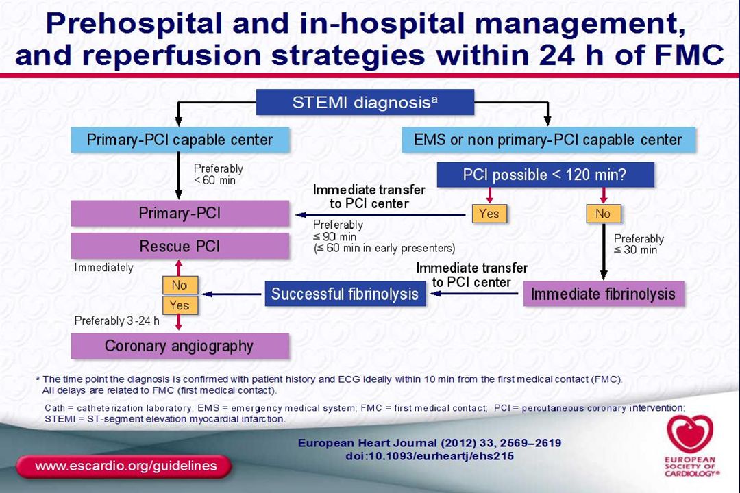
1 Clinical Evaluation 2 Diagnosis / Risk assessment 3 Medical Treatment 4 Invasive Strategy STEMI REPERFUSION Emergent <2 hours Serial ECGs Serial troponin Lab tests (Hb, Crea Clea…) Ischemic risk score (i.e. GRACE) Bleeding risk score (i.e. CRUSADE) Imaging techniques results (optional) Quality of chest pain Probability of CAD Physical examination ECG (↑ST?) Urgent 2-24 hours Anti-ischemic therapy Antiplatelet Anticoagulation NSTE ACS Early 24-72 hours No / Elective ACS unclear (Rule out ACS) Chest Pain Unit No ACS Serial ECGs Serial troponin Lab tests (Hb, Crea Clea…) Ischemic risk score (i.e. GRACE) Bleeding risk score (i.e. CRUSADE) Imaging techniques results (optional) Anti-ischemic therapy Antiplatelet Anticoagulation Emergent <2 hours Urgent 2-24 hours Early 24-72 hours No / Elective
 Ischemic risk score. (i.e. GRACE) Bleeding risk score. (i.e. CRUSADE) Imaging techniques results (optional) Quality of chest pain. Probability of CAD. Physical examination. ECG (↑ST ) Urgent hours. Anti-ischemic. therapy. Antiplatelet. Anticoagulation. NSTE ACS. Early hours. No / Elective. ACS unclear. (Rule out ACS) Chest Pain Unit. No ACS. Serial ECGs. Serial troponin. Lab tests (Hb, Crea Clea…) Ischemic risk score. (i.e. GRACE) Bleeding risk score. (i.e. CRUSADE) Imaging techniques results (optional) Anti-ischemic. therapy. Antiplatelet. Anticoagulation. Emergent. <2 hours. Urgent hours. Early hours. No / Elective.")
18
ST elevation MI PTCA + STENT

20
Oxygen Supplemental oxygen should be administered to patients with arterial oxygen desaturation (SaO2 < 90%). It is reasonable to administer supplemental oxygen to all patients with uncomplicated STEMI during the first 6 hours. I IIa IIb III

21
Nitroglycerin Patients with ongoing ischemic discomfort should receive sublingual NTG (0.4 mg) every 5 minutes for a total of 3 doses, after which an assessment should be made about the need for intravenous NTG. Intravenous NTG is indicated for relief of ongoing ischemic discomfort that responds to nitrate therapy, control of hypertension, or management of pulmonary congestion.
 every 5 minutes for a total of 3 doses, after which an assessment should be made about the need for intravenous NTG. Intravenous NTG is indicated for relief of ongoing ischemic discomfort that responds to nitrate therapy, control of hypertension, or management of pulmonary congestion.")
22
Nitroglycerin Nitrates should not be administered to patients with: Nitrates should not be administered to patients who have received a phosphodiesterase inhibitor for erectile dysfunction within the last 24 hours (48 hours for tadalafil). systolic pressure < 90 mm Hg or ≥ to 30 mm Hg below baseline severe bradycardia (< 50 bpm) tachycardia (> 100 bpm) or suspected RV infarction.
. systolic pressure < 90 mm Hg or ≥ to 30 mm Hg below baseline. severe bradycardia (< 50 bpm) tachycardia (> 100 bpm) or. suspected RV infarction.")
23
Analgesia Morphine sulfate (2 to 4 mg intravenously with increments of 2 to 8 mg intravenously repeated at 5 to 15 minute intervals) is the analgesic of choice for management of pain associated with STEMI.
 is the analgesic of choice for management of pain associated with STEMI.")
24
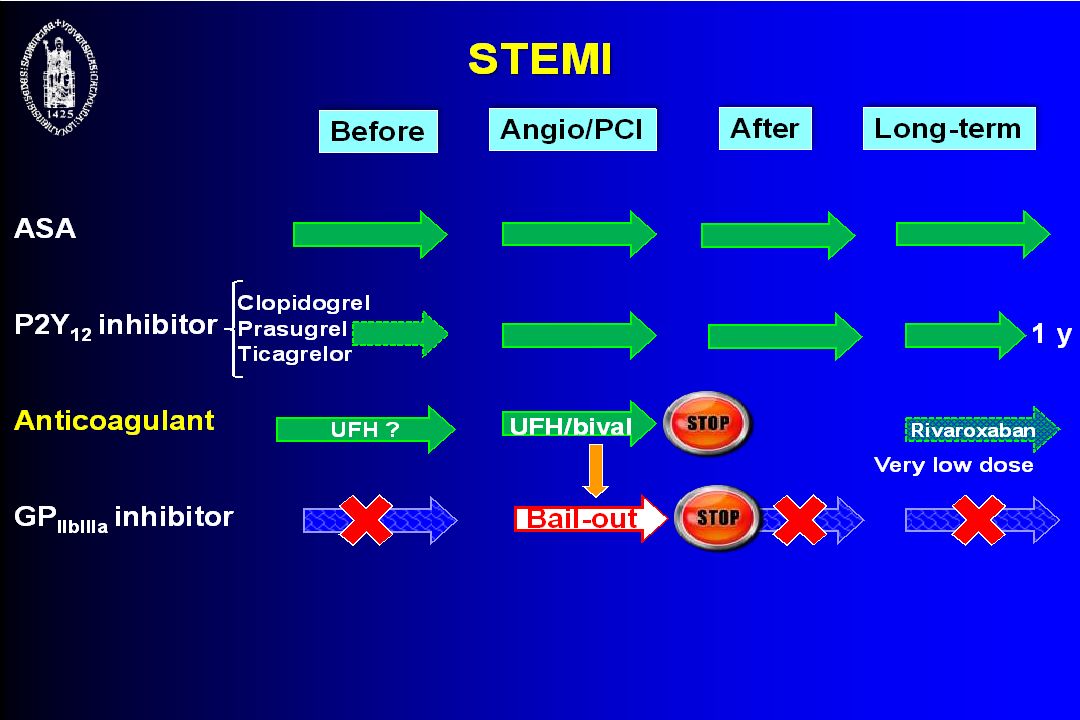
Aspirin/Clopidogrel/Prasugrel/Ticagrelor
Aspirin should be chewed by patients who have not taken aspirin before presentation with STEMI. The initial dose should be 162 mg (Level of Evidence: A) to 325 mg (Level of Evidence: C) Although some trials have used enteric-coated aspirin for initial dosing, more rapid buccal absorption occurs with non–enteric-coated formulations.
 to 325 mg (Level of Evidence: C) Although some trials have used enteric-coated aspirin for initial dosing, more rapid buccal absorption occurs with non–enteric-coated formulations.")
25
Beta-Blockers Oral beta-blocker therapy should be administered promptly to those patients without a contraindication, irrespective of concomitant fibrinolytic therapy or performance of primary PCI. It is reasonable to administer intravenous beta-blockers promptly to STEMI patients without contraindications, especially if a tachyarrhythmia or hypertension is present.

26
Ischemia/Reperfusion Injury
-acute inflammatory response -apoptosis -platelet-neutrofil aggregates (no-reflow)
")
28
Other Pharmacological Measures
Angiotensin converting enzyme (ACE) inhibitors Angiotensin receptor blockers (ARB) Aldosterone blockers Glucose control Magnesium Calcium channel blockers Inhibition of the renin -angiotensin -aldosterone system
 inhibitors. Angiotensin receptor blockers (ARB) Aldosterone blockers. Glucose control. Magnesium. Calcium channel blockers. Inhibition of the renin -angiotensin -aldosterone system.")
Presentaciones similares


>")


