Descargar la presentación
La descarga está en progreso. Por favor, espere

1
BLOQUEO AURICULO-VENTRICULAR
DRA. SILVIA CERDA ADAME TORRE QUALITY JULIO 2015

2
El bloqueo auriculoventricular (AV) es un retardo o interrupción del impulso eléctrico proveniente del nodo sinoauricular a nivel del nodo auriculoventricular.
 es un retardo o interrupción del impulso eléctrico proveniente del nodo sinoauricular a nivel del nodo auriculoventricular.")
3
BLOQUEO A-V CONGENITOS

4
congénito (BAVC) Se presenta en forma aislada o familiar.
uno por cada 20,000 a 25,000 nacidos vivos. la transposición de grandes arterias, síndromes poli-esplénicos, ventrículo único, tumores del miocardio, síndrome de QT-largo Kearn-Soyer (oftalmoplejía externa, retinosis pigmentaria y miopatía mitocondrial) 25%.
 25%.")
5
Son factores de riesgo:
títulos elevados de AC anti-Ro+ (superiores a 1:16) AC anti-Ro+ (SS-A) acompañados de AC anti-La+ (SSB) HLA (HLA DR3) insuficiencia cardiaca
 AC anti-Ro+ (SS-A) acompañados de AC anti-La+ (SSB) HLA (HLA DR3) insuficiencia cardiaca.")
6
Son factores de riesgo:
El síndrome de lupus neonatal incluye: eritemas, leucopenia, anemia y trombocitopenia BAVC, desaparecen de seis a ocho meses de vida extrauterina. Se detecta por Doppler o ecocardiografía fetal entre las 16 y 30 semanas de gestación por bradicardia fetal persistente.

7
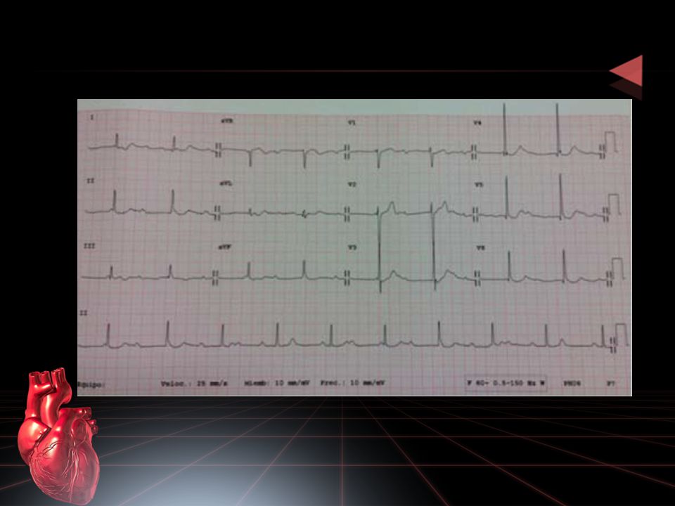
CASO CLINICO Masculino de 33 años de edad, carga genética para diabetes mellitus, hipertensión arterial sistémica y cardiopatías por ambas ramas, producto de gesta III, madre de 28 años, durante la gestación, la cual se le diagnostica artritis reumatoide a los 34 años de edad, diabetes mellitus a los 40 años, carcinoma renal a los 50 años. Ocupación cajero, vida sedentaria, refiere disnea de grandes esfuerzos, edema de miembros inferiores vespertino posicional o con el calor, precordalgia punzante, con duración de tres horas, sin relación con esfuerzos y sin más datos acompañantes, motivo por el cual acudió a consulta y al realizarle electrocardiograma.

10
ANALISIS en caso de bradicardia en paciente joven, generalmente no hacemos diagnóstico diferencial con bloqueo auriculoventricular (AV) completo congénito: monitoreo de Holter, Prueba de esfuerzo ecocardiograma la indicación de marcapasos está relacionado con la clase funcional y la actividad del paciente.
 completo congénito: monitoreo de Holter, Prueba de esfuerzo. ecocardiograma. la indicación de marcapasos está. relacionado con la clase funcional y la. actividad del paciente.")
11
BLOQUEO A-V ADQUIRIDOS

12
ELECTROCARDIOGRAMA Registro de la actividad eléctrica del corazón
Onda P: Despolarización de las aurículas. Intervalo PR: Retardo del impulso en el nodo auriculoventricular. Segmento QRS: Despolarización de los ventrículos. Onda T: Repolarización ventricular. QRS waveform nomenclature The ECG consists of a small deflection called the P wave, arising from the atria, a more complicated deflection called the QRS complex due to ventricular depolarisation and a final T wave resulting from repolarisation of the ventricles. The QRS complex of waves is the largest deflection of the ECG and is always spiky in shape. All sharp deflections resulting from electrical activation of the ventricles are called QRS complexes. However, these waves can vary immensely in size, and arrangement. The QRS complex is very important when diagnosing myocardial infarction. In order to be able to describe these complexes, a nomenclature for the waves is needed. This can be done using combinations of the letters q, r, s, Q, R, S, lower case letters denoting small waves and upper case larger waves. The first positive wave is labelled with r or R Any second positive wave is labelled r´ or R´ A negative wave which follows an R wave or r wave is labelled S or s A negative wave that precedes an R or r wave, is labelled a q or Q wave Any wave that is entirely negative is labelled qs or QS. Using these rules and nomenclature all QRS complexes can be described, enabling more accurate diagnosis.

13
ELECTROCARDIOGRAMA Intervalo PR mide normalmente: 0.12 a 0.20 seg.
Location of infarction and its relation to the ECG: inferior infarction ST elevation in leads II, III and aVF, and often ST depression in I, aVL, and precordial leads are signs of an inferior (lower) infarction. Inferior infarctions may occur due to occlusion of the right circumflex coronary arteries resulting in infarction of the inferior surface of the left ventricle, although damage can be made to the right ventricle and interventricular septum. This type of infarction often results in bradycardia due to damage to the atrioventricular node.
 infarction. Inferior infarctions may occur due to occlusion of the right circumflex coronary arteries resulting in infarction of the inferior surface of the left ventricle, although damage can be made to the right ventricle and interventricular septum. This type of infarction often results in bradycardia due to damage to the atrioventricular node.")
14
ETIOLOGIA DE LOS BLOQUEOS AV
Cardiopatías: Isquémica Valvulopatías Miocardiopatías Enfermedad degenerativa Medicamentos: Metoprolol Digoxina Alteraciones en el K Hiperkalemia Rule 10 In leads I, II, and V2 to V6 the T wave must be upright.

16
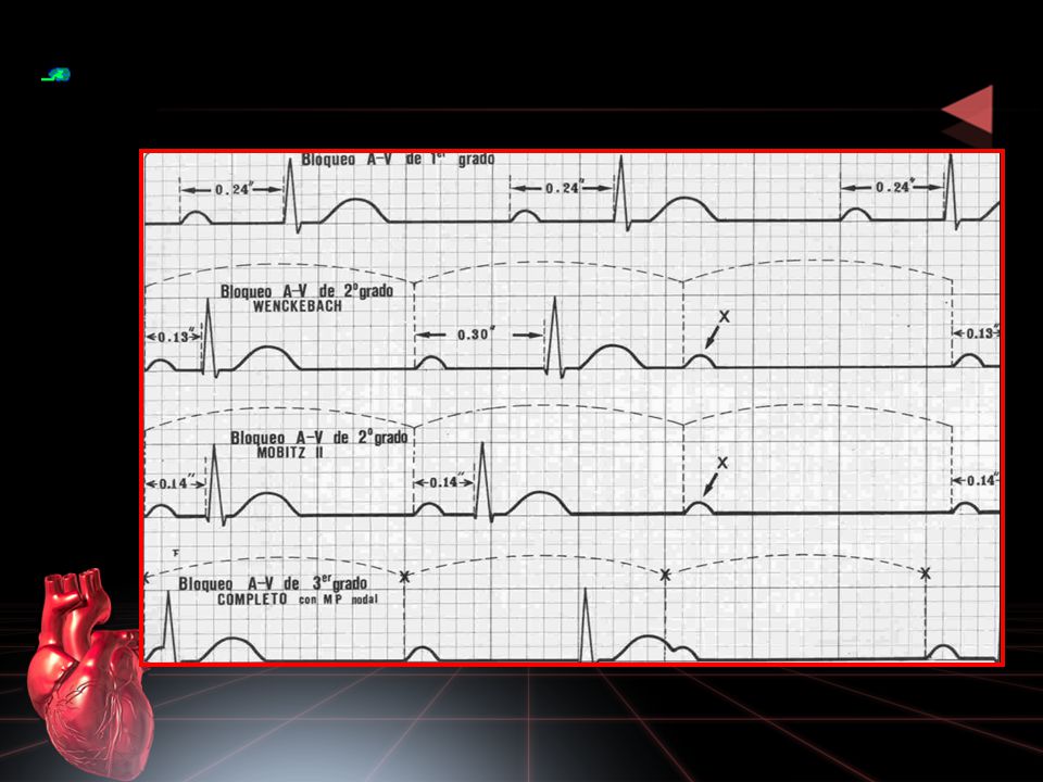
CLASIFICACIÓN DE LOS BLOQUEOS AV
Bloqueo AV de primer grado Bloqueo AV de segundo grado Mobitz I (Fenómeno de Wenckebach) Bloqueo AV de segundo grado – Mobitz II Bloqueo AV completo
 Bloqueo AV de segundo grado – Mobitz II. Bloqueo AV completo.")
18
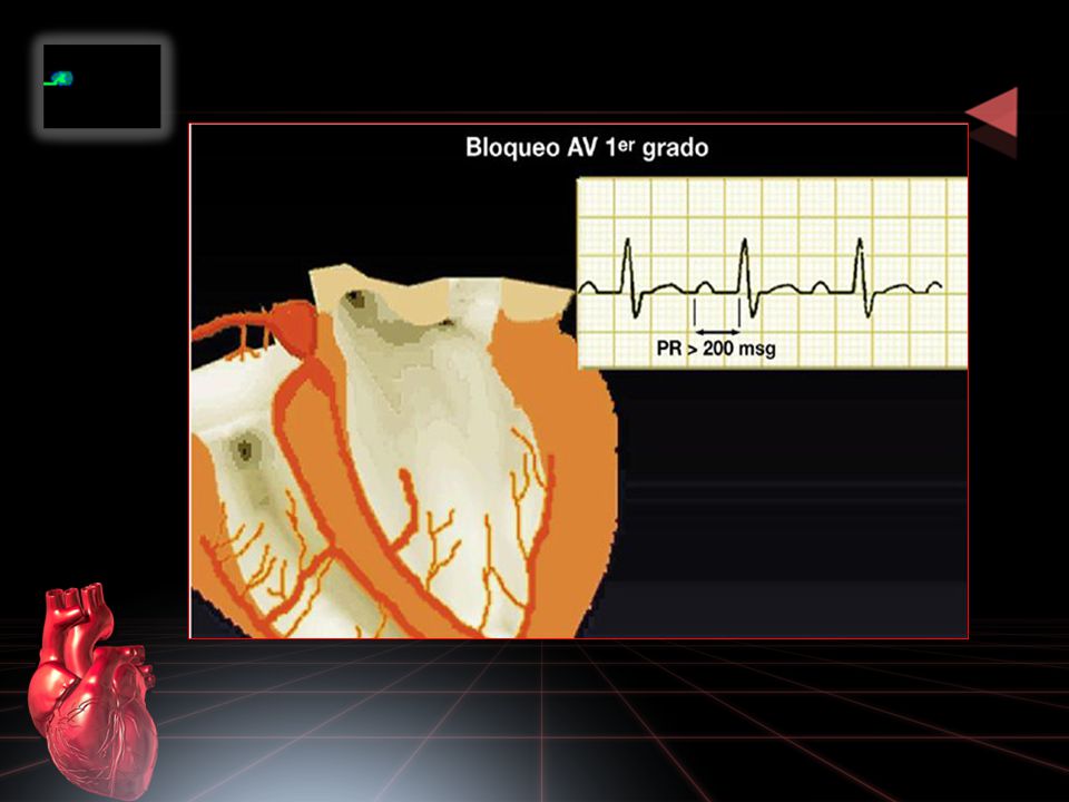
BLOQUEO AV DE PRIMER GRADO
Intervalo PR > 0.20 seg. ó 200 mseg. Intervalo PR prolongado es fijo. Siempre a la onda P le sigue un QRS. Ritmo es regular. The 10 rules for a normal ECG For an ECG to be determined as normal, Chamberlain has described 10 rules which must be met.1 The next ten slides will outline these rules. Chamberlain DAC. Personal communications.

19
BLOQUEO AV DE PRIMER GRADO
Rule 1 As described in Module 3, the PR interval is the time from initiation of depolarisation of the atria to initiation of the depolarisation of the ventricles. The PR interval should be 120 to 200 milliseconds, or 3 to 5 little squares. A longer PR may imply a block in conduction and a shorter interval indicates a vulnerability to arrhythmias.

21
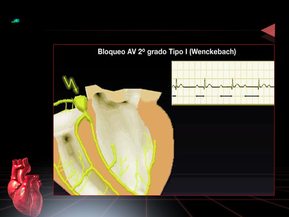
MOBITZ I (FENOMENO DE WENCKEBACH)
BAV 2DO GRADO MOBITZ I (FENOMENO DE WENCKEBACH) Prolongación del intervalo PR hasta que una P no se conduce. Complejo QRS de morfología normal. Ritmo es irregular. Rule 3 The QRS complex should be dominantly upright in leads I and II. Slight disparities are likely to be acceptable.
 Prolongación del intervalo PR hasta que una P no se conduce. Complejo QRS de morfología normal. Ritmo es irregular. Rule 3. The QRS complex should be dominantly upright in leads I and II. Slight disparities are likely to be acceptable.")
22
MOBITZ I (FENOMENO DE WENCKEBACH)
Rule 4 The QRS and T waves tend to have the same direction in the standard leads.

24
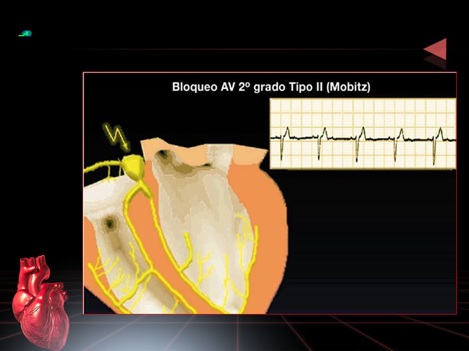
BLOQUEO AV DE SEGUNDO GRADO MOBITZ II
Intervalo PR es constante hasta que una P no conduce. Fijo: 2:1, 3:1, 4:1 Variable: 2:1, 4:1, 3:1 Avanzado: Dos ó mas P son bloqueadas. Rule 5 All waves are negative in lead aVR. This has to be so: aVR represents electrical activity as “seen” from the right shoulder. The sinus node is placed top right in the heart nearest the right shoulder, and the electrical activity is moving downwards and leftwards towards the left ventricle.

25
BLOQUEO AV DE SEGUNDO GRADO MOBITZ II (3:1)
Rule 6 The normality of QRS complexes recorded from the precordial leads is dependent on both morphological and dimensional criteria.

26
BLOQUEO AV DE SEGUNDO GRADO MOBITZ II (AVANZADO)
Location of infarction and its relation to the ECG: anterior infarction As was discussed in the previous module, the different leads look at different aspects of the heart, and so infarctions can be located by noting the changes that occur in different leads. The precordial leads (V1–6) each lie over part of the ventricular myocardium and can therefore give detailed information about this local area. aVL, I, V5 and V6 all reflect the anterolateral part of the heart and will therefore often show similar appearances to each other. II, aVF and III record the inferior part of the heart, and so will also show similar appearances to each other. Using these we can define where the changes will be seen for infarctions in different locations. Anterior infarctions usually occur due to occlusion of the left anterior descending coronary artery resulting in infarction of the anterior wall of the left ventricle and the intraventricular septum. It may result in pump failure due to loss of myocardium, ventricular septal defect, aneurysm or rupture and arrhythmias. ST elevation in I, aVL, and V2–6, with ST depression in II, III and aVF are indicative of an anterior (front) infarction. Extensive anterior infarctions show changes in V1–6 , I, and aVL.
 each lie over part of the ventricular myocardium and can therefore give detailed information about this local area. aVL, I, V5 and V6 all reflect the anterolateral part of the heart and will therefore often show similar appearances to each other. II, aVF and III record the inferior part of the heart, and so will also show similar appearances to each other. Using these we can define where the changes will be seen for infarctions in different locations. Anterior infarctions usually occur due to occlusion of the left anterior descending coronary artery resulting in infarction of the anterior wall of the left ventricle and the intraventricular septum. It may result in pump failure due to loss of myocardium, ventricular septal defect, aneurysm or rupture and arrhythmias. ST elevation in I, aVL, and V2–6, with ST depression in II, III and aVF are indicative of an anterior (front) infarction. Extensive anterior infarctions show changes in V1–6 , I, and aVL.")
28
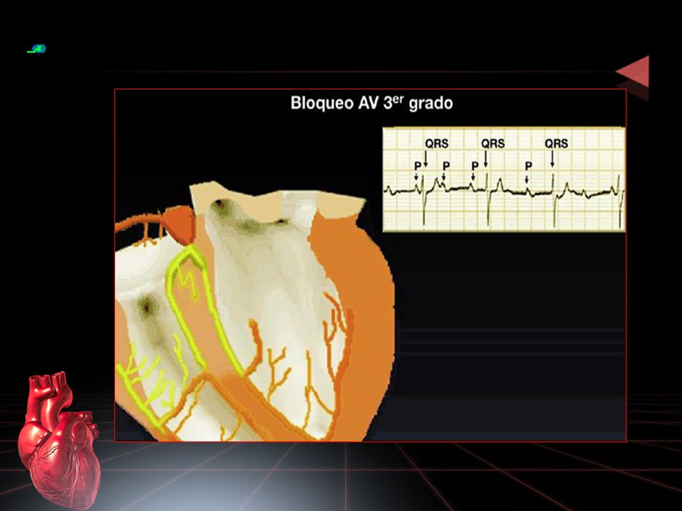
BLOQUEO AV COMPLETO Ó DE TERCER GRADO
Disociación entre aurículas y ventrículos PP es constante RR es constante Frecuencia es mayor en las P que en los complejos QRS. La morfología del QRS depende del marcapaso subsidiario. Rule 7 The ST segment should start isoelectric except in V1 and V2 where it may be elevated.

29
BLOQUEO AV COMPLETO Ó DE TERCER GRADO
Rule 8 In leads I, II, and V2 to V6 the P waves should be upright.

30
BLOQUEO AV COMPLETO Ó DE TERCER GRADO
Sequence of changes in evolving AMI The ECG changes that occur due to myocardial infarction do not all occur at the same time. There is a progression of changes correlating to the progression of infarction. Within minutes of the clinical onset of infarction, there are no changes in the QRS complexes and therefore no definitive evidence of infarction. However, there is ST elevation providing evidence of myocardial damage. The next stage is the development of a new pathological Q wave and loss of the r wave. These changes occur at variable times and so can occur within minutes or can be delayed. Development of a pathological Q wave is the only proof of infarction. As the Q wave forms the ST elevation is reduced and after 1 week the ST changes tend to revert to normal, but the reduction in R wave voltage and the abnormal Q waves usually persist. The late change is the inversion of the T wave and in a non-Q wave myocardial infarct, when there is no pathological Q wave, this T wave change may be the only sign of infarction. Months after an MI the T waves may gradually revert to normal, but the abnormal Q waves and reduced voltage R waves persist. In terms of diagnosing AMI in time to make thrombolysis a life-saving possibility, the main change to look for on the ECG is ST segment elevation.

32
TRATAMIENTO Bradicardia sintomática Atropina
Marcapaso transcutáneo ó transvenoso Dopamina Epinefrina Isoproterenol Deep Q wave The only diagnostic changes of acute myocardial infarction are changes in the QRS complexes and the development of abnormal Q waves. However, this may be a late change and so is not useful for the diagnosis of AMI in the pre-hospital situation. Remember that Q waves of more than 0.04 seconds , or 1 little square, are not generally seen in leads I, II or the precordial leads.

33
R E S U E L V E L O

34
R E S U E L V E L O

35
R E S U E L V E L O

36
R E S U E L V E L O

37
R E S U E L V E L O

38
R E S U E L V E L O

39
Bloqueos fasciculares: Hemibloqueo anterior (anterosuperior)
Clasificación de los bloqueos de rama y fasciculares Bloqueos fasciculares: Hemibloqueo anterior (anterosuperior) Hemibloqueo posterior (posteroinferior) Bloqueo bifasciculares: Bloqueo de rama derecha + hemibloqueo anterior. Bloqueo de rama derecha + hemibloqueo posterior. Bloqueos trifasciculares: Bloqueo de rama derecha + hemibloqueo anterior + hemibloqueo posterior. Bloqueo bifascicular + bloqueo atrioventricular de primer grado (PR largo).
 Hemibloqueo posterior (posteroinferior) Bloqueo bifasciculares: Bloqueo de rama derecha + hemibloqueo anterior. Bloqueo de rama derecha + hemibloqueo posterior. Bloqueos trifasciculares: Bloqueo de rama derecha + hemibloqueo anterior + hemibloqueo posterior. Bloqueo bifascicular + bloqueo atrioventricular de primer grado (PR largo).")
40
Hemibloqueo anterior de rama izquierda (HARI)
Se impide la entrada directa de los impulsos eléctricos en la pared anterior y lateral del VI. El tabique interventricular se despolariza en dirección normal: Izq a Der Despolarización del VD Despolarización de la pared posterior del VI Despolarización de la pared anterior y lateral del VI

41
HEMIBLOQUEO ANTERIOR QRS < 0.12 s
Hiperdesviación del QRS a la izquierda (-45º y -75º) Complejos qR empastados en D1 y Avl Complejos rS empastados en D2, D3 y aVF Retraso del tiempo de deflexión intrinsecoide en D1 y aVL
 Complejos qR empastados en D1 y Avl. Complejos rS empastados en D2, D3 y aVF. Retraso del tiempo de deflexión intrinsecoide. en D1 y aVL.")
42
HEMIBLOQUEO ANTERIOR

43
Hemibloqueo posterior de rama izquierda
En el se impide la entrada directa de los impulsos eléctricos en el tabique y la pared posterior y lateral del VI. El tabique interventricular se despolariza en dirección anormal: D a I Despolarización del VD Despolarización de la pared anterior y lateral del VI Despolarización de la pared posterior del VI

44
HEMIBLOQUEO POSTERIOR
Duración del QRS < 0.12 s Hiperdesviación del QRS a la derecha +90 y +120 Complejos rS empastados en D1 y aVL Complejos qR empastados en D2, D3 y aVF Retrazo del tiempo de deflexión intrisecoide en D2, D3 y aVF

45
HEMIBLOQUEO POSTERIOR

46
BRD + HEMIBLOQUEO ANTERIOR
Morfología característica BRD en V1-2 y V5-6 QRS desviación izquierda (-60) D1 y aVL retraso tiempo deflexión intrinsecoide D2, D3 y aVF onda S empastada. Ondas R altas y empastadas en aVR y aVL
 D1 y aVL retraso tiempo deflexión intrinsecoide. D2, D3 y aVF onda S empastada. Ondas R altas y empastadas en aVR y aVL.")
47
BRD + HEMIBLOQUEO ANTERIOR

48
BRD + HEMIBLOQUEO POSTERIOR
Morfología característica BRD en V1-2 y V5-6 D2, D3 y aVR retraso tiempo deflexión intrinsecoide Ondas R altas y empastadas en D2, D3 y aVR

49
BRD + HEMIBLOQUEO POSTERIOR

50
Para Información y Revisión de las presentaciones: www. cardiologica
Para Información y Revisión de las presentaciones:

Presentaciones similares
 es el registro gráfico, en función del tiempo, de las variaciones de potencial eléctrico.>")


