Descargar la presentación
La descarga está en progreso. Por favor, espere

1
Maduración del Linfocito B
U.T.I. Biología Tisular Depto. Inmunobiología Mayo 2008

2
Independiente del Antígeno Dependiente del Antígeno
Maduración del Linfocito B Independiente del Antígeno Médula ósea Rearreglo genes Ig Selección Dependiente del Antígeno Organos linfoides periféricos Diferenciación a plasmocitos Diferenciación a células B memoria Maduración de la afinidad Cambio de Clase

3
- contactos célula-célula
La maduración B en la médula ósea depende de la interacción de las células precursoras con las células estromales: - contactos célula-célula - factores de crecimiento (proliferación, diferenciación) The growth of early B-lineage cells is stimulated by stem-cell factor (SCF), a membrane-bound cytokine present on stromal cells, which interacts with the cell-surface receptor tyrosine kinase Kit on B-cell precursors. Developing B cells at later stages require the secreted cytokine interleukin-7 (IL-7). The chemokine stromal cell-derived factor 1 or pre-B cell growth-stimulating factor (SDF-1/PBSF) has an important role in the early stages of B-cell development, as shown by the failure of B-cell development in mice lacking the gene for this molecule. SDF-1 is produced constitutively by bone marrow stromal cells and one of its roles may be to retain developing B-cell precursors in the marrow microenvironment. Figure 4.5 The early stages of B-cell development are dependent on bone marrow stromal cells. The top panels show the interactions of developing B cells with bone marrow stromal cells. Stem cells and early pro-B cells use the integrin VLA-4 to bind to the adhesion molecule VCAM-1 on stromal cells. This and interactions between other cell adhesion molecules (CAMs) promote the binding of the receptor Kit on the B cell to stem-cell factor (SCF) on the stromal cell. Activation of Kit causes the B cell to proliferate. B cells at a later stage of maturation require interleukin-7 (IL-7) to stimulate their growth and proliferation. Panel a is a light micrograph of a tissue culture showing small round B-cell progenitors in intimate contact with stromal cells, which have extended processes fastening them to the plastic dish on which they are grown. Panel b is a high-magnification electron micrograph showing two lymphoid cells (L) adhering to a flattened stromal cell.
 The growth of early B-lineage cells is stimulated by stem-cell factor (SCF), a membrane-bound cytokine present on stromal cells, which interacts with the cell-surface receptor tyrosine kinase Kit on B-cell precursors. Developing B cells at later stages require the secreted cytokine interleukin-7 (IL-7). The chemokine stromal cell-derived factor 1 or pre-B cell growth-stimulating factor (SDF-1/PBSF) has an important role in the early stages of B-cell development, as shown by the failure of B-cell development in mice lacking the gene for this molecule. SDF-1 is produced constitutively by bone marrow stromal cells and one of its roles may be to retain developing B-cell precursors in the marrow microenvironment. Figure 4.5 The early stages of B-cell development are dependent on bone marrow stromal cells. The top panels show the interactions of developing B cells with bone marrow stromal cells. Stem cells and early pro-B cells use the integrin VLA-4 to bind to the adhesion molecule VCAM-1 on stromal cells. This and interactions between other cell adhesion molecules (CAMs) promote the binding of the receptor Kit on the B cell to stem-cell factor (SCF) on the stromal cell. Activation of Kit causes the B cell to proliferate. B cells at a later stage of maturation require interleukin-7 (IL-7) to stimulate their growth and proliferation. Panel a is a light micrograph of a tissue culture showing small round B-cell progenitors in intimate contact with stromal cells, which have extended processes fastening them to the plastic dish on which they are grown. Panel b is a high-magnification electron micrograph showing two lymphoid cells (L) adhering to a flattened stromal cell.")
4
El desarrollo de células B procede en etapas definidas por el rearreglo y expresión de genes de inmunoglobulinas

5
El desarrollo de células B procede en etapas definidas por el rearreglo y expresión de genes de inmunoglobulinas Figure 4.3 The development of B cells proceeds through stages defined by the rearrangement and expression of the immunoglobulin genes. In the stem cell, the immunoglobulin (Ig) genes are in the germline configuration. The first rearrangements are of the heavy-chain (H-chain) genes. Joining DH to JH defines the early pro-B cell, which becomes a late pro-B cell on joining VH to DJH. Expression of a functional μ chain and its expression at the cell surface as part of the pre-B receptor defines the large pre-B cell. Large pre-B cells proliferate, producing small pre-B cells in which rearrangement of the light-chain (L-chain) gene occurs. Successful light-chain gene rearrangement and expression of IgM on the cell surface define the immature B cell. The mature B cell is defined by the use of alternative splicing of heavy-chain mRNA to place IgD on the cell surface as well as IgM.
 genes are in the germline configuration. The first rearrangements are of the heavy-chain (H-chain) genes. Joining DH to JH defines the early pro-B cell, which becomes a late pro-B cell on joining VH to DJH. Expression of a functional μ chain and its expression at the cell surface as part of the pre-B receptor defines the large pre-B cell. Large pre-B cells proliferate, producing small pre-B cells in which rearrangement of the light-chain (L-chain) gene occurs. Successful light-chain gene rearrangement and expression of IgM on the cell surface define the immature B cell. The mature B cell is defined by the use of alternative splicing of heavy-chain mRNA to place IgD on the cell surface as well as IgM.")
6
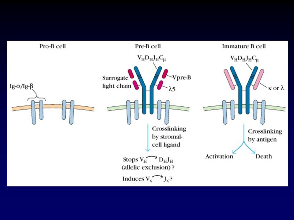
El receptor pre-B contiene una pseudo cadena liviana
Figure 4.4 The pre-B-cell receptor resembles the B-cell receptor except for the surrogate light chain.

9
Orden del rearreglo génico que conduce a la expresión de inmunoglobulinas de membrana
Figure 4.7 The order of gene rearrangements leading to the expression of cell-surface immunoglobulin. The heavy-chain genes are rearranged before the light-chain genes. Developing B cells are allowed to proceed to the next stage only when a productive rearrangement has been made. If an unproductive rearrangement is made on one chromosome of a homologous pair, then rearrangement is attempted on the second chromosome

10
La señalización vía el producto proteico funcional es usado para terminar el proceso de rearreglo de los genes de Ig (exclusión alélica) El proceso de exclusión alélica asegura que cada célula B exprese receptores B producto de un único rearreglo para cada cadena Figure 4.9 Signaling via the functional protein product is used to terminate immunoglobulin-gene rearrangement. First panel: in early pro-B cells, no functional μ protein is expressed and the biochemical machinery that performs gene rearrangement is active. Second panel: on completion of a productive heavy-chain gene rearrangement, μ heavy chains are synthesized and assembled into a complex with the λ5 and VpreB polypeptides that make up the surrogate light chain, and with Igα and Igβ. This complex, the pre-B-cell receptor, is transported to the cell surface, where it interacts with an unknown ligand that signals the cell to halt heavy-chain gene rearrangement and to proceed to the large pre-B-cell stage. Proliferation of the large pre-B cell yields small pre-B cells that commence immunoglobulin light-chain rearrangement. Note that λ5 and VpreB are coded by genes that are distinct from those in the immunoglobulin loci. Third panel: on completion of a productive light-chain gene rearrangement a light chain is made and assembles with μ to form IgM. This associates with Igα and Igβ and is transported to the cell surface. Its presence tells the cell to halt light-chain gene rearrangements

11
La expresión de proteínas involucradas en el rearreglo y expresión de genes de Ig cambian durante el desarrollo de las células B Figure 4.10 The expression of proteins involved in the rearrangement and expression of immunoglobulin genes changes during B-cell development. The rearrangement of immunoglobulin genes and the expression of the pre-B-cell receptor and IgM on the cell surface requires several categories of specialized proteins at different times during B-cell development. Examples of such proteins are listed here, with their expression during B-cell development shown with red shading. Of the proteins not discussed in the text, EBF regulates the transcription of the gene for Igα, and the B-lineage specific-activator protein (BSAP) regulates the expression of several of the other proteins listed. Oct-2 acts at the heavy-chain promoter and GATA-2 is a transcription factor active in several types of hematopoietic cells. CD45 is a cell-surface phosphotyrosine phosphatase that is restricted to hematopoietic cells. It is involved in regulating signal transduction from the pre-B-cell and B-cell receptors.
 regulates the expression of several of the other proteins listed. Oct-2 acts at the heavy-chain promoter and GATA-2 is a transcription factor active in several types of hematopoietic cells. CD45 is a cell-surface phosphotyrosine phosphatase that is restricted to hematopoietic cells. It is involved in regulating signal transduction from the pre-B-cell and B-cell receptors.")
12
La unión con antígenos propios en la médula ósea puede conducir a la deleción o inactivación de células B inmaduras Figure 4.13 Binding to self-antigens in the bone marrow can lead to the deletion or inactivation of immature B cells. Left panels: immature B cells that do not encounter a stimulatory self-antigen leave the bone marrow and enter the peripheral circulation, expressing both IgM and IgD on their surface. Middle panels: when immature B cells express receptors that recognize common cell-surface components of human cells, they are deleted from the repertoire by the induction of apoptosis. Right panels: immature B cells that bind soluble self-antigens are rendered unresponsive or anergic to the antigen, and, as a consequence, express low levels of IgM at the cell surface. They enter the peripheral circulation, where they express IgD but remain anergic.

13
Figure 2-21 Las células B vírgenes maduras que salen de la médula ósea co-expresan IgD e IgM en la membrana. Esto está regulado por splicing alternativo del ARNm. 13

14
Independiente del Antígeno Dependiente del Antígeno
Maduración del Linfocito B Independiente del Antígeno Médula ósea Rearreglo genes Ig Selección Dependiente del Antígeno Organos linfoides periféricos Diferenciación a plasmocitos Diferenciación a células B memoria Maduración de la afinidad Cambio de Clase

15
Ruta de circulación de células B maduras “naives” a través del ganglio linfático
Figure 4.16 The circulation route of mature, naive B cells through a lymph node. Having matured in the bone marrow, B cells migrate in the blood to lymph nodes and other secondary lymphoid tissues. B cells leave the blood and enter the cortex of the lymph node through the walls of specialized high endothelial venules (HEV). If they do not encounter their specific antigen, the B cells pass through the primary follicles and leave the node in the efferent lymph, which eventually joins the blood at veins in the neck.
. If they do not encounter their specific antigen, the B cells pass through the primary follicles and leave the node in the efferent lymph, which eventually joins the blood at veins in the neck.")
16
Las células B que encuentran al antígeno en los órganos linfoides secundarios forman centros germinales y se diferencian a células plasmáticas Figure 4.17 B cells encountering antigen in secondary lymphoid tissues form germinal centers and undergo differentiation to plasma cells. A lymph node is illustrated here. A B cell entering the lymph node through a HEV encounters antigen in the lymph node cortex. Antigen was delivered in the afferent lymph that drained from infected tissue. The B cell is activated by CD4 helper T cells (blue) in the T-cell areas to form a primary focus of dividing cells. From this, some B cells migrate directly to the medullary cords and differentiate into antibody-secreting plasma cells. Other B cells migrate into a primary follicle to form a germinal center. B cells continue to divide and differentiate within the germinal center. Activated B cells migrate from the germinal center to the medulla of the lymph node or to the bone marrow to complete their differentiation into plasma cells.
 in the T-cell areas to form a primary focus of dividing cells. From this, some B cells migrate directly to the medullary cords and differentiate into antibody-secreting plasma cells. Other B cells migrate into a primary follicle to form a germinal center. B cells continue to divide and differentiate within the germinal center. Activated B cells migrate from the germinal center to the medulla of the lymph node or to the bone marrow to complete their differentiation into plasma cells.")
17
Anatomía de los folículos linfoides
Figure 4.15 Anatomy of lymphoid follicles. This section through a human lymph node shows secondary lymphoid follicles, each of which has developed a germinal center (GC). Three zones can be recognized in a follicle, an outer mantle zone of resting B cells (M), a dark zone of proliferating blasts (D), and a light zone of differentiating cells (L). The areas between the follicles contains T cells, interdigitating cells (antigen-presenting), blood vessels, and sinuses.
. Three zones can be recognized in a follicle, an outer mantle zone of resting B cells (M), a dark zone of proliferating blasts (D), and a light zone of differentiating cells (L). The areas between the follicles contains T cells, interdigitating cells (antigen-presenting), blood vessels, and sinuses.")
18
Zona Clara (centrocitos)
Zona Oscura (centroblastos) IgD - CD38 + CD77 + HIPERMUTACION CFD T Manto Folicular IgD + CD38 - APOPTOSIS Zona Clara (centrocitos) IgD - CD38 + CD77 - SELECCION IgG IgE IgA plasmocito IgM plasmocytes CD38++ células B memoria IgD - CD38 -
 IgD - CD38 + CD77 + HIPERMUTACION. CFD. T. Manto Folicular. IgD + CD38 - APOPTOSIS. Zona Clara (centrocitos) IgD - CD38 + CD77 - SELECCION. IgG. IgE. IgA. plasmocito. IgM. plasmocytes. CD38++ células B memoria. IgD - CD38 -")
19
El proceso de mutación somática opera específicamente sobre los genes variables de Inmunoglobulinas

20
Maduración de la afinidad: hipermutación somática

21
El proceso de cambio de clase (switch) cambia el isotipo de una Ig manteniendo sus regiones variables (idiotipo).
 cambia el isotipo de una Ig manteniendo sus regiones variables (idiotipo).")
22
Los diferentes tumores de células B reflejan la heterogeneidad en las etapas del desarrollo y diferenciación de las células B normales Figure 4.19 The different B-cell tumors reflect the heterogeneity of developmental and differentiation states in the normal B-cell population. Each type of tumor corresponds to a normal state of B-cell development or differentiation. Tumor cells have similar properties to their normal cell equivalent, they migrate to the same sites in the lymphoid tissues, and have similar patterns of expression of cell-surface glycoproteins.

23
El Receptor de las células T

24
El receptor T es un heterodímero, cada cadena contiene un dominio constante y otro variable y se parece a un Fab unido a membrana. Figure 3.1 The T-cell receptor resembles a membrane-bound Fab fragment. Comparison of the T-cell receptor and an IgG antibody molecule. The T-cell receptor is a heterodimer composed of an α chain of kDa and a β chain of kDa in size. The extracellular portion of each chain consists of two immunoglobulin-like domains: the domain nearest to the membrane is a C region and the domain farthest from the membrane is a V region. Both the α and β chains span the cell membrane and have very short cytoplasmic tails. The three-dimensional structure formed by the four immunoglobulin-like domains of the T-cell receptor resembles that of the antigen-binding Fab fragment of antibody. Figure 3.2 Three-dimensional structure of the T-cell receptor showing the antigen-binding CDR loops. The ribbon diagram shows the α chain (in magenta) and the β chain (in blue). The receptor is viewed from the side as it would sit on a cell surface with the highly variable CDR loops, which bind the peptide:MHC molecule ligand, arrayed across its relatively flat top surface. The CDR loops are numbered 1-3 for each chain.
 and the β chain (in blue). The receptor is viewed from the side as it would sit on a cell surface with the highly variable CDR loops, which bind the peptide:MHC molecule ligand, arrayed across its relatively flat top surface. The CDR loops are numbered 1-3 for each chain.")
25
Estructura tridimensional del receptor T

26
Organización y rearreglos de los genes del receptor de células T
Figure 3.3 Organization and rearrangement of the T-cell receptor genes. The top and bottom rows of the figure show the germline arrangement of the variable (V), diversity (D), joining (J), and constant (C) gene segments at the T-cell receptor α- and β-chain loci. During T-cell development, a V-region sequence for each chain is assembled by DNA recombination. For the α chain (top), a Vα gene segment rearranges to a Jα gene segment to create a functional exon encoding the V domain. For the β chain (bottom), rearrangement of a Vβ, a Dβ, and a Jβ gene segment creates the functional V-domain exon. The assembled genes are transcribed and spliced to produce mRNA (not shown) encoding the α and β chains. Exons encoding the membrane-spanning regions are not shown. L, leader sequence.
, diversity (D), joining (J), and constant (C) gene segments at the T-cell receptor α- and β-chain loci. During T-cell development, a V-region sequence for each chain is assembled by DNA recombination. For the α chain (top), a Vα gene segment rearranges to a Jα gene segment to create a functional exon encoding the V domain. For the β chain (bottom), rearrangement of a Vβ, a Dβ, and a Jβ gene segment creates the functional V-domain exon. The assembled genes are transcribed and spliced to produce mRNA (not shown) encoding the α and β chains. Exons encoding the membrane-spanning regions are not shown. L, leader sequence.")
27
Composición polipeptídica del complejo-receptor de células T
Figure 3.6 Polypeptide composition of the T-cell receptor complex. The functional antigen receptor on the surface of T cells is composed of eight polypeptides and is called the T-cell receptor complex. The α and β chains bind antigen and form the core T-cell receptor (TCR). They associate with one copy each of CD3γ and CD3δ and two copies each of CD3ε and the ζ chain. These associated invariant polypeptides are necessary for transport of newly synthesized TCR to the cell surface and for transduction of signals to the cell's interior after the TCR has bound antigen. The transmembrane domains of the α and β chains contain positively charged amino acids (+), which form strong electrostatic interactions with negatively charged amino acids (-) in the transmembrane regions of the CD3γ, δ, and ε chains
. They associate with one copy each of CD3γ and CD3δ and two copies each of CD3ε and the ζ chain. These associated invariant polypeptides are necessary for transport of newly synthesized TCR to the cell surface and for transduction of signals to the cell s interior after the TCR has bound antigen. The transmembrane domains of the α and β chains contain positively charged amino acids (+), which form strong electrostatic interactions with negatively charged amino acids (-) in the transmembrane regions of the CD3γ, δ, and ε chains.")
28
Existen dos clases de receptores T: a:b y g:d
Tienen estructuras similares pero están codificados por grupos de genes diferentes Figure 3.7 There are two classes of T-cell receptors. The α:β T-cell receptor (left panel) and the γ:δ T-cell receptor (right panel) have similar structures, but they are encoded by different sets of rearranging gene segments and have different functions.
 and the γ:δ T-cell receptor (right panel) have similar structures, but they are encoded by different sets of rearranging gene segments and have different functions.")
29
¿Cómo reconoce el receptor T al antígeno?
Procesamiento del antígeno y su presentación Figure 3.9 Antigen processing and presentation. The antigens recognized by T cells are peptides that arise from the breakdown of macromolecular structures, the unfolding of individual proteins, and their cleavage into short fragments. These events constitute antigen processing. For a T-cell receptor to recognize a peptide antigen, the peptide must be bound by an MHC molecule and displayed at the cell surface, a process called antigen presentation

30
Existen dos compartimentos mayores separados por membranas en las células
Figure 3.16 There are two major compartments within cells, separated by membranes. One compartment is the cytosol, which is contiguous with the nucleus via the pores in the nuclear membrane. The other compartment is the vesicular system, which consists of the endoplasmic reticulum, the Golgi apparatus, endocytic vesicles, lysosomes, and other intracellular vesicles. The vesicular system is effectively contiguous with the extracellular fluid. Secretory vesicles bud off from the endoplasmic reticulum and by successive fusion and budding with the Golgi membranes move vesicular contents out of the cell. In contrast, endocytic vesicles take up extracellular material into the vesicular system

31
Procesamiento de antígenos presentados por moléculas MHC de clase I y de clase II ocurre en diferentes compartimentos celulares Figure 3.19 Processing of antigens presented by MHC class II and MHC class I molecules occurs in different cellular compartments. The left half of the figure shows the fate of peptides derived from extracellular antigens and pathogens. Extracellular material is taken up by endocytosis and phagocytosis into the vesicular system of the cell, in this case a macrophage. Proteases in these vesicles break down proteins to produce peptides that are bound by MHC class II molecules, which have been transported to the vesicles via the endoplasmic reticulum (ER) and the Golgi apparatus. The peptide:MHC class II complex is transported to the cell surface in outgoing vesicles. The right half of the figure shows the fate of peptides generated in the cytosol as a result of infection with viruses or intracytosolic bacteria. Proteins from such pathogens are broken down in the cytosol by the proteasome to peptides, which enter the ER. There the peptides are bound by MHC class I molecules. The peptide:MHC class I complex is transported to the cell surface via the Golgi apparatus.
 and the Golgi apparatus. The peptide:MHC class II complex is transported to the cell surface in outgoing vesicles. The right half of the figure shows the fate of peptides generated in the cytosol as a result of infection with viruses or intracytosolic bacteria. Proteins from such pathogens are broken down in the cytosol by the proteasome to peptides, which enter the ER. There the peptides are bound by MHC class I molecules. The peptide:MHC class I complex is transported to the cell surface via the Golgi apparatus.")
32
En el retículo endoplásmico existen proteínas de tipo chaperona que ayudan al ensamblado y carga peptídica de las moléculas MHC clase I Figure 3.18 Chaperone proteins aid the assembly and peptide loading of MHC class I molecules in the endoplasmic reticulum. MHC class I heavy chains assemble in the endoplasmic reticulum with the membrane-bound protein calnexin. When this complex binds β2-microglobulin (β2m) the partly folded MHC class I molecule is released from calnexin and then associates with the TAP-1 subunit of TAP by interacting with the TAP-associated protein tapasin and the chaperone protein calreticulin. The MHC class I molecule is retained in the endoplasmic reticulum until it binds a peptide, which completes the folding of the molecule. The peptide:MHC class I molecule complex is then released from tapasin and calreticulin, leaves the endoplasmic reticulum, and is transported to the cell surface
 the partly folded MHC class I molecule is released from calnexin and then associates with the TAP-1 subunit of TAP by interacting with the TAP-associated protein tapasin and the chaperone protein calreticulin. The MHC class I molecule is retained in the endoplasmic reticulum until it binds a peptide, which completes the folding of the molecule. The peptide:MHC class I molecule complex is then released from tapasin and calreticulin, leaves the endoplasmic reticulum, and is transported to the cell surface.")
33
La cadena invariante previene que péptidos se unan a moléculas MHC clase II hasta que alcancen el sitio de proteólisis extracelular Figure 3.20 The invariant chain prevents peptides from binding to a MHC class II molecule until it reaches the site of extracellular protein breakdown. MHC class II α and β chains are assembled with an invariant chain in the endoplasmic reticulum (ER); this complex is transported to the acidified vesicles of the endocytic system. The invariant chain is broken down, leaving just a small fragment called class II-associated invariant-chain peptide (CLIP) attached in the peptide-binding site. The vesicle membrane protein HLA-DM catalyzes the release of the CLIP fragment and its replacement by a peptide derived from endocytosed antigen that has been degraded within the acidic interior of the vesicles.
; this complex is transported to the acidified vesicles of the endocytic system. The invariant chain is broken down, leaving just a small fragment called class II-associated invariant-chain peptide (CLIP) attached in the peptide-binding site. The vesicle membrane protein HLA-DM catalyzes the release of the CLIP fragment and its replacement by a peptide derived from endocytosed antigen that has been degraded within the acidic interior of the vesicles.")
34
Estructuras tridimensionales de las moléculas MHC clase I y II.
Figure 3.13 The structures of MHC class I and MHC class II molecules are variations on a theme. An MHC class I molecule (left panels) is composed of one membrane-bound heavy (or α) chain and noncovalently bonded β2-microglobulin. The heavy chain has three extracellular domains, of which the amino-terminal α1 and α2 domains resemble each other in structure and form the peptide-binding site. An MHC class II molecule (right panels) is composed of two membrane-bound chains, an α chain (which is a different protein from MHC class I α) and a β chain. These have two extracellular domains each, the amino-terminal two (α1 and β1) resembling each other in structure and forming the peptide-binding site. The β2 domain of MHC class II molecules should not be confused with the β2-microglobulin of MHC class I molecules. The ribbon diagrams in the lower panels trace the paths of the polypeptide backbone chains.
 is composed of one membrane-bound heavy (or α) chain and noncovalently bonded β2-microglobulin. The heavy chain has three extracellular domains, of which the amino-terminal α1 and α2 domains resemble each other in structure and form the peptide-binding site. An MHC class II molecule (right panels) is composed of two membrane-bound chains, an α chain (which is a different protein from MHC class I α) and a β chain. These have two extracellular domains each, the amino-terminal two (α1 and β1) resembling each other in structure and forming the peptide-binding site. The β2 domain of MHC class II molecules should not be confused with the β2-microglobulin of MHC class I molecules. The ribbon diagrams in the lower panels trace the paths of the polypeptide backbone chains.")
35
Estructuras de las hendiduras de unión peptídica en las moléculas MHC clase I y II
Figure 3.15 The peptide-binding groove of MHC class I and MHC class II molecules. The T-cell receptor's view of the peptide-binding groove, with a peptide bound, is shown. In the MHC class I molecule (left panel) the groove is formed by the α1 and α2 domains of the MHC class I heavy chain; in the MHC class II molecule (right panel) it is formed by the α1 domain of the class II α chain and the β1 domain of the class II β chain. Amino-acid side-chains on the MHC molecule that are important for making interactions with the bound peptide are shown. The dotted blue lines indicate hydrogen bonds and ionic interactions made between the peptide and the MHC molecule. Peptides bind to MHC class I molecules by their ends (left panel), whereas in MHC class II molecules, the peptide extends beyond the peptide-binding groove and is held by interactions along its length (right panel).
 the groove is formed by the α1 and α2 domains of the MHC class I heavy chain; in the MHC class II molecule (right panel) it is formed by the α1 domain of the class II α chain and the β1 domain of the class II β chain. Amino-acid side-chains on the MHC molecule that are important for making interactions with the bound peptide are shown. The dotted blue lines indicate hydrogen bonds and ionic interactions made between the peptide and the MHC molecule. Peptides bind to MHC class I molecules by their ends (left panel), whereas in MHC class II molecules, the peptide extends beyond the peptide-binding groove and is held by interactions along its length (right panel).")
36
La mayoría de las células humanas expresan MHC clase I mientras que pocos tipos celulares expresan MHC clase II Figure 3.22 Most human cells express MHC class I, whereas only a few cell types express MHC class II. MHC class I molecules are expressed on almost all nucleated cells, although they are most highly expressed in hematopoietic cells. MHC class II molecules are normally expressed only by a subset of hematopoietic cells and by stromal cells in the thymus, although they can be produced by other cell types on exposure to the cytokine interferon-γ. *In humans, activated T cells express MHC class II molecules, whereas resting T cells do not. †In the brain, most cell types are MHC class II-negative, but microglia, which are related to macrophages, are MHC class II-positive.

37
Estructura del complejo MHC : péptido : receptor T
Figure 3.21 The MHC:peptide:T-cell receptor complex. Panel a shows a diagram of the polypeptide backbone of the complex of a T-cell receptor (TCR) bound to its peptide:MHC class I ligand. Panel b shows a schematic representation of this view of the receptor:ligand complex. In panel a the T-cell receptor's CDRs are colored: the α-chain CDR1 and CDR2 are light and dark blue respectively, while the β-chain CDR1 and CDR2 are light and dark purple respectively. The α chain CDR3 is yellow and the β-chain CDR3 is dark yellow. The eight amino-acid peptide is colored yellow and the positions of the first (P1) and last (P8) amino acids are indicated. Panel c is a view rotated 90o from that of panel a and shows the surface of the peptide:MHC class I ligand and the footprint made upon it by the T-cell receptor (outlined in black). Within this footprint the contributions of the CDRs are outlined in different colors and labeled. In panels d and e the diagonal orientation of the T-cell receptor with respect to the peptide-binding grooves of MHC class I and class II molecules, respectively, is shown in schematic diagrams. The T-cell receptor is represented by the black rectangle superimposed on the ribbon diagram (yellow) of the peptide-binding domains of the MHC molecules.
 bound to its peptide:MHC class I ligand. Panel b shows a schematic representation of this view of the receptor:ligand complex. In panel a the T-cell receptor s CDRs are colored: the α-chain CDR1 and CDR2 are light and dark blue respectively, while the β-chain CDR1 and CDR2 are light and dark purple respectively. The α chain CDR3 is yellow and the β-chain CDR3 is dark yellow. The eight amino-acid peptide is colored yellow and the positions of the first (P1) and last (P8) amino acids are indicated. Panel c is a view rotated 90o from that of panel a and shows the surface of the peptide:MHC class I ligand and the footprint made upon it by the T-cell receptor (outlined in black). Within this footprint the contributions of the CDRs are outlined in different colors and labeled. In panels d and e the diagonal orientation of the T-cell receptor with respect to the peptide-binding grooves of MHC class I and class II molecules, respectively, is shown in schematic diagrams. The T-cell receptor is represented by the black rectangle superimposed on the ribbon diagram (yellow) of the peptide-binding domains of the MHC molecules.")
38
Estructuras de los co-receptores CD4 y CD8
Presentan dominios tipo-inmunoglobulina CD8 se une a MHC- clase I CD4 se une a MHC- clase II Figure 3.10 The structures of the CD4 and CD8 glycoproteins. CD4 has four extracellular immunoglobulin-like domains (D1-D4) with a hinge between the D2 and D3 domains. CD8 consists of an α and a β chain, which both have an immunoglobulin-like domain that is connected to the membrane-spanning region by an extended stalk. C denotes the carboxy terminus
 with a hinge between the D2 and D3 domains. CD8 consists of an α and a β chain, which both have an immunoglobulin-like domain that is connected to the membrane-spanning region by an extended stalk. C denotes the carboxy terminus.")
39
Linfopoyesis T

40
Los precursores de células T migran desde la médula ósea al timo para madurar
Figure 5.1 T-cell precursors migrate from the bone marrow to the thymus to mature. T cells derive from bone marrow stem cells whose progeny migrate in the blood from the bone marrow to the thymus (left panel), where the development of T cells occurs. Mature T cells leave the thymus in the blood, from where they enter secondary lymphoid tissues (right panel) and then return to the blood in the lymph. In the absence of activation by specific antigen, mature T cells continue to recirculate between the blood, secondary lymphoid tissues, and lymph. GALT, gut-associated lymphoid tissue.
, where the development of T cells occurs. Mature T cells leave the thymus in the blood, from where they enter secondary lymphoid tissues (right panel) and then return to the blood in the lymph. In the absence of activation by specific antigen, mature T cells continue to recirculate between the blood, secondary lymphoid tissues, and lymph. GALT, gut-associated lymphoid tissue.")
41
Las células epiteliales del timo forman una red alrededor de los linfocitos que están en desarrollo
Figure 5.2 The epithelial cells of the thymus form a network surrounding developing thymocytes. In this scanning electron micrograph of the thymus, the developing thymocytes (the spherical cells) occupy the interstices of an extensive network of epithelial cells.
 occupy the interstices of an extensive network of epithelial cells.")
42
Organización celular del timo
Figure 5.3 The cellular organization of the thymus. The thymus is made up of several lobules. A section through a lobule stained with hematoxylin and eosin and viewed with the light microscope is shown in the left panel. The cells in this view are shown in diagrammatical form in the right panel. In the left panel, the darker staining of cortex compared with the medulla can be discerned. As shown in the right panel, the cortex consists of immature thymocytes (blue), branched cortical epithelial cells (light orange) and a few macrophages (yellow). The medulla consists of mature thymocytes (blue), medullary epithelial cells (orange), dendritic cells (yellow), and macrophages (yellow). One of the functions of the macrophages in both cortex and medulla is to remove the many thymocytes that fail to mature properly. A characteristic feature of the medulla is Hassall's corpuscles, which are believed to be sites of cell destruction.
, branched cortical epithelial cells (light orange) and a few macrophages (yellow). The medulla consists of mature thymocytes (blue), medullary epithelial cells (orange), dendritic cells (yellow), and macrophages (yellow). One of the functions of the macrophages in both cortex and medulla is to remove the many thymocytes that fail to mature properly. A characteristic feature of the medulla is Hassall s corpuscles, which are believed to be sites of cell destruction.")
43
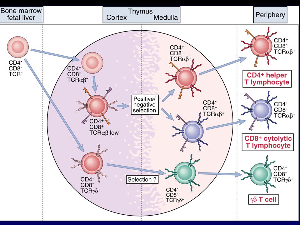
Poblaciones de linfocitos T en el timo
Figure 5.13 Positive and negative selection are mediated by different types of cell in the thymus. As thymocytes mature they move from the subcapsular region deeper into the thymus. Double-positive cells are found in the cortex where they undergo positive selection on cortical epithelial cells. The positively selected cells encounter dendritic cells and macrophages at the cortico-medullary junction; this is where most negative selection occurs. The surviving mature single-positive T cells leave the thymus and enter the blood circulation at venules in the medulla.

44
Etapas en el desarrollo tímico de células T a:b .
Figure 5.19 Stages of α:β T-cell development in the thymus correlate with T-cell receptor gene rearrangement and expression of cell-surface proteins by the developing T cell.

45
Rearreglos de los genes que codifican para el receptor T (I)
")
46
Rearreglos de los genes que codifican para el receptor T (II)
")
47
Rearreglos de los genes que codifican para el receptor T (III)
")
48
La interacción de una célula T doble-positiva con un complejo péptido propio : MHC propio durante la selección positiva determina si la célula T se convierte en CD4 o CD8. CD4 CD4 CD8 CD8 MHC I MHC II Figure 5.12 Interaction of a double-positive T cell with a self-peptide:self-MHC complex during positive selection determines whether the T cell will become a CD4 or a CD8 T cell. The left panels show the selection of a T cell whose T-cell receptor (TCR) interacts with peptide:MHC class I complexes on a thymic epithelial cell. The right panels show the outcome for a cell bearing a receptor that interacts with peptide:MHC class II complexes CD8 CD4
 interacts with peptide:MHC class I complexes on a thymic epithelial cell. The right panels show the outcome for a cell bearing a receptor that interacts with peptide:MHC class II complexes. CD8. CD4.")
49
La selección positiva y negativa está mediada por diferentes tipos de células en el timo
Figure 5.13 Positive and negative selection are mediated by different types of cell in the thymus. As thymocytes mature they move from the subcapsular region deeper into the thymus. Double-positive cells are found in the cortex where they undergo positive selection on cortical epithelial cells. The positively selected cells encounter dendritic cells and macrophages at the cortico-medullary junction; this is where most negative selection occurs. The surviving mature single-positive T cells leave the thymus and enter the blood circulation at venules in the medulla.

Presentaciones similares