Descargar la presentación
La descarga está en progreso. Por favor, espere

2
Fluidoterapia en el perioperatorio
Dr. Kwok Ho Sánchez Suen Agosto – 2012

3
“Y después de haber ayunado cuarenta días y cuarenta noches, tuvo hambre”
Mateo 4.2. Aspecto del desierto marciano visto por la sonda Spirit en 2004

4
Desierto de Sossusvlei, Namibia

5
El Desierto de Atacama es el desierto más árido del mundo; se extiende por el norte de Chile y la franja costera del Perú. Se sitúa, más o menos, sobre el Trópico de Capricornio, al igual que el Desierto del Kalahari, o que el Gran Desierto de Australia. La aridez, determinada por su latitud, se incrementa debido al efecto barrera de la Cordillera de los Andes, que bloquea la humedad procedente del Océano Atlántico a través de la Cuenca Amazónica. En el Desierto de Atacama se han registrado períodos de hasta 400 años sin lluvias en su sector central, delimitado por las ciudades de Antofagasta, Calama, Iquique y Arica. [editar] Historia El desierto ha estado poblado desde los comienzos de la colonización americana por los indígenas, especialmente por tribus como los atacameños, mientras que en su litoral vivían los changos. Dominado por el Imperio Inca, fue denominado el "despoblado de Atacama" por los españoles. Después de la Independencia, Bolivia ocupó la región más importante, incluyendo los valiosos depósitos de salitre que estuvieron en la génesis de la Guerra del Pacífico. Después de dicho conflicto, Chile se anexó la región y en ella se ha desarrollado una intensa actividad minera que incluye explotaciones de cobre y otros metales.

6
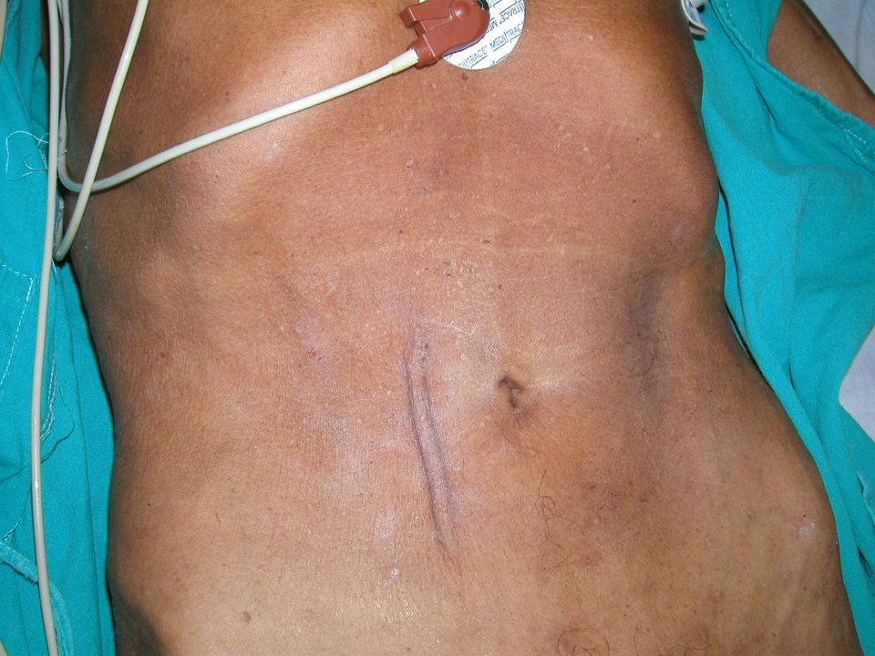
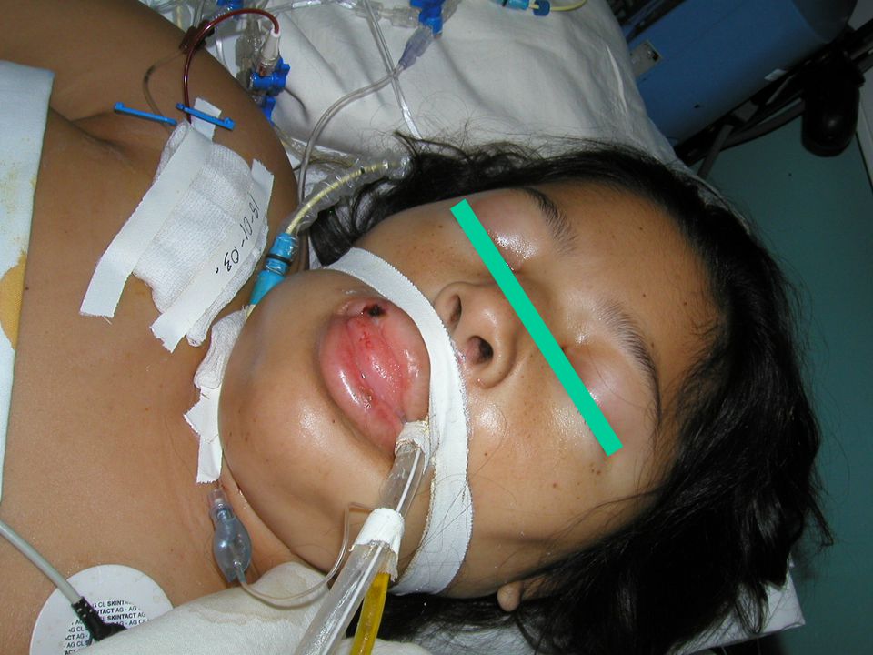
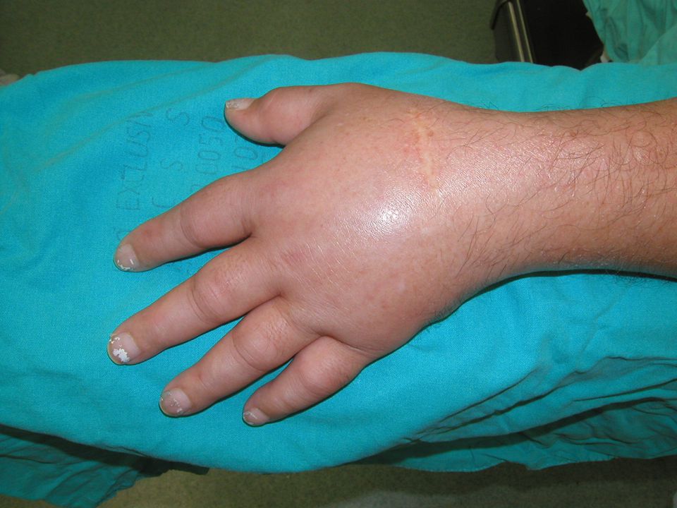
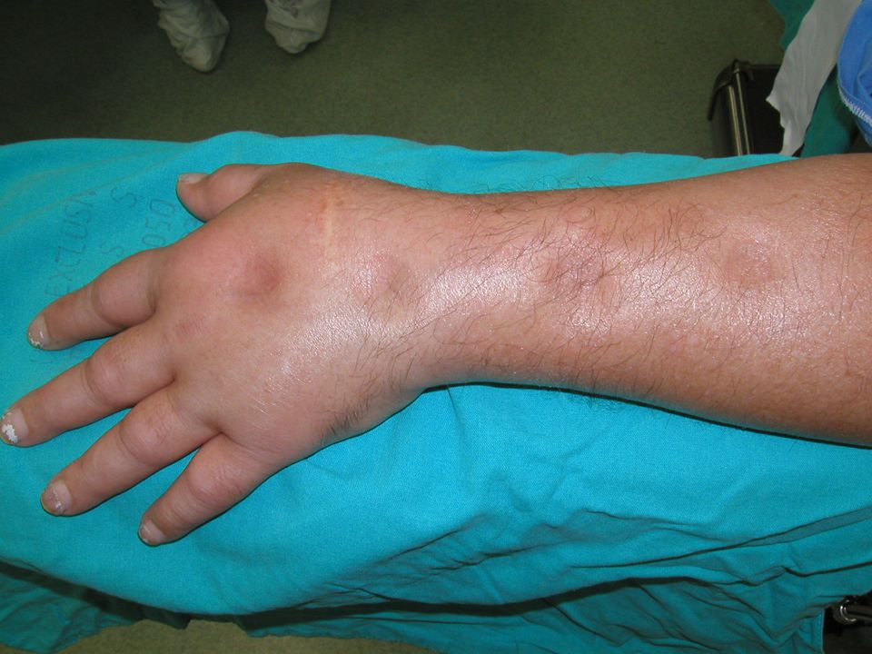
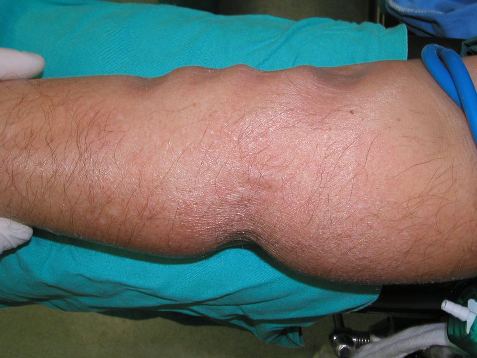
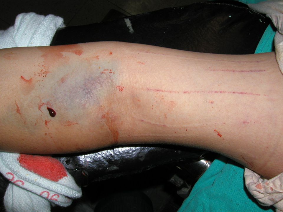
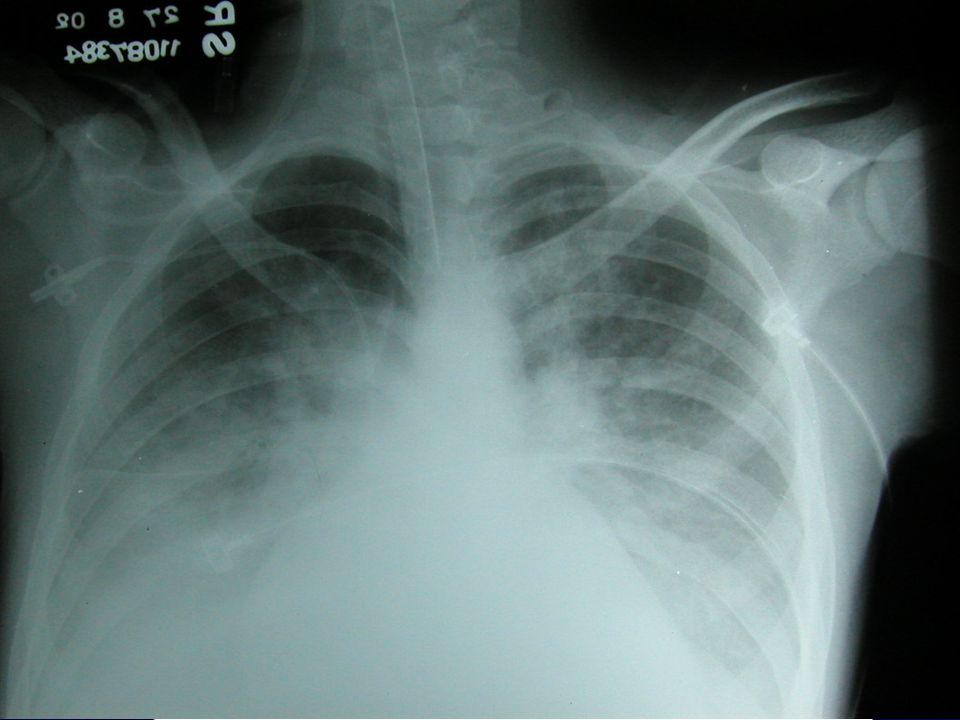
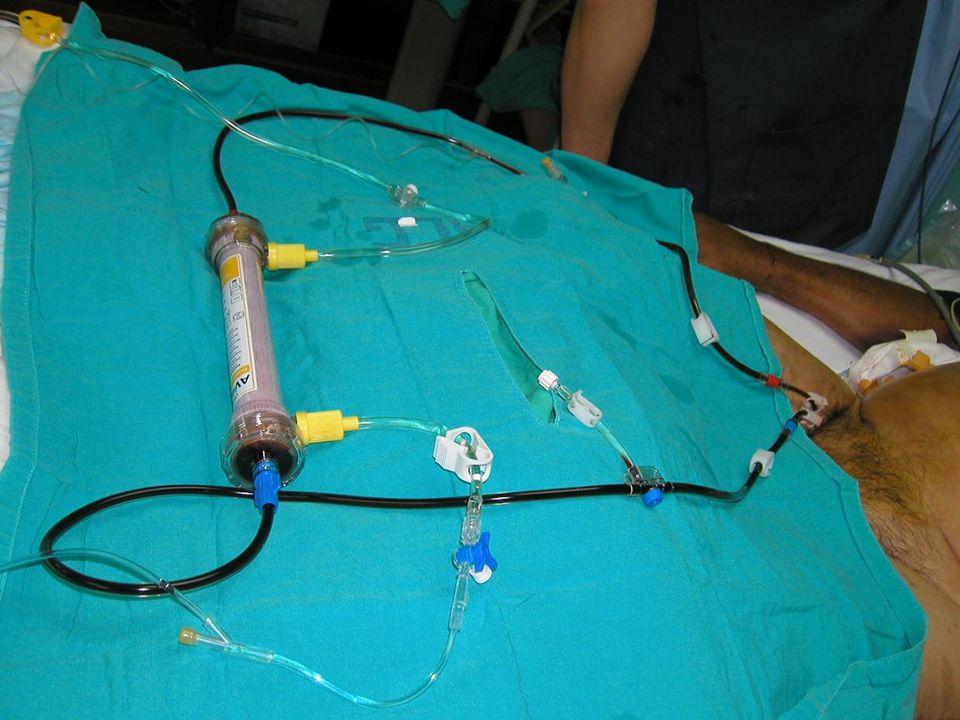
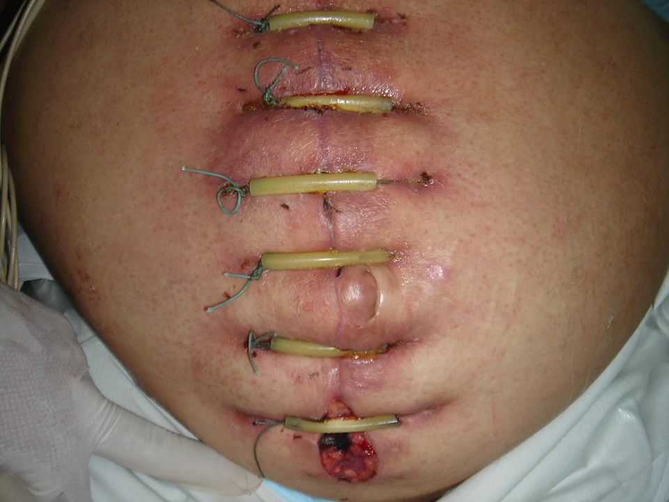
Caso clínico Paciente masculino de 55 años
Hipertenso de 10 años de evolución Diabetes mellitus tipo 2 de 8 años de evolución Referido al Servicio de ORL por cuadro de hipoacusia, tinnitus y vértigo Se internó para iniciarle estudios TAC con medio de contraste de oído medio Creatinina sérica 1,8 mg/dL → 4,5 mg/dL Fluidoterapia agresiva “para abrir el riñón” Anuria persistente

7
98.000 vidas perdidas/año por errores médicos …

9
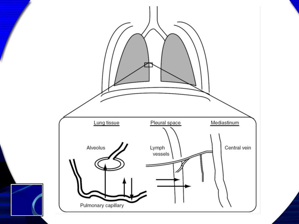
Diagrama de los líquidos corporales para un adulto promedio de 70 kilos
Compartimento Litros Volumen extracelular Volumen plasmático Volumen de glóbulos rojos Volumen intersticial 15 3 2 10 Volumen intracelular 25 Total 40

10
Fluidos corporales Agua corporal total (60%) LIC (40%) LEC (20%)
Na+ (10) K+ (156) Cl- (2) Proteína (55) Ca++ (3,3) Mg++ (26) HCO3- (8) HPO4= (95) SO4= (20) Na+ (145) K+ (4) Cl- (114) HCO3- (8) Na+ (142) K+ (4) Cl- (104) Proteína (13) Ca++ (5) Mg++ (2) HCO3- (27) HPO4= (2) SO4= (1) LIC (40%) LEC (20%)
 K+ (156) Cl- (2) Proteína (55) Ca++ (3,3) Mg++ (26) HCO3- (8) HPO4= (95) SO4= (20) Na+ (145) K+ (4) Cl- (114) HCO3- (8) Na+ (142) K+ (4) Cl- (104) Proteína (13) Ca++ (5) Mg++ (2) HCO3- (27) HPO4= (2) SO4= (1) LIC (40%) LEC (20%)")
11
LI 15% LP 5% LEC 20% LIC 40%

12
Sed LIC Turgencia LI Hemodinamia LP

13
Preguntas guía ¿Con qué se va a reponer? ¿Cuánto de volumen?
¿A qué velocidad? ¿Durante cuánto tiempo? ¿Por cuál ruta? ¿Qué se debe de vigilar?

14
Equilibrio hídrico Egresos Arrastre Ingresos

16
Pregunta … Si recibió ml de NaCl 0,9% y luego orinó ml, entonces el balance es: Neutro No es neutro Tiene balance positivo … de NaCl (6 g)
")
18
Resucitación líquida 1/3 2/3

19
V2 k23 ki kt V3 V1 kr Drobin D, Hahn RG. Anesthesiology; 2002; 96:

20
Propiedades volémicas
Fluido * LIC LEC LI LP Dextrosa 5% NaCl 0,9% Albúmina Sangre total 660 -100 340 1100 1000 255 825 500 85 275 500 1000 * Basada en infusión de volumen de 1 litro

21
¿ La solución NaCl 0,9% es fisiológica ?

22
Solución salina al 0,9% Solución fisiológica (!?)
9 g NaCl/1000 ml agua 154 mEq/l de Na+ 154 mEq/l de Cl- Osmolaridad 308 mOsml/l Eficaz para sustitución de LEC Acidosis hiperclorémica

25
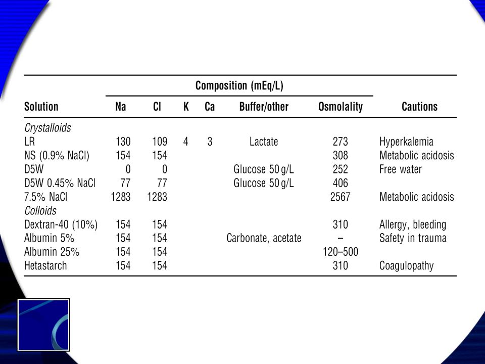
Opciones en la C.C.S.S. Solución NaCl 0,9%
Solución glucosada 5%, 10%, 50% Solución mixta Solución electrolítica balanceada Solución salina hipertónica Dextrán – 40 Poligelina al 3,5% Solución hiperoncótica – hipertónica

27
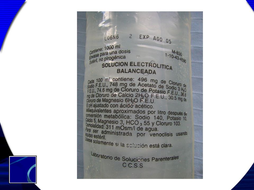
Se puede disolver en agua destilada ?
Disuelto en NaCl 0,9% Osmolaridad = 311 mOsm/L Se puede disolver en agua destilada ?

28
Osmolaridad aportada 311 – 308 = 3 mOsm/L

29
7-8 μm 1 μm 2 μm µm2 µm3

31
280 mOsm/L 200 mOsm/L

32
El puerto de Sadarghat en Dhaka
Dhaka ó Dacca

33
osmótico Efecto

34
Presión osmótica total
Oncótica Peso molecular → Osmolaridad calculada = 2 Na+ + glucosa/18 + BUN/2,3

35
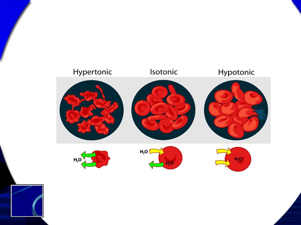
Presión osmótica ? Presión oncótica ?
B

36
Presión osmótica ? Presión oncótica ?
B

37
Presión osmótica ? Presión oncótica ?
B

38
Valores Presión osmótica coloidal: 1 mOsm = 19,6 mmHg Total: 28 mmHg
Albúmina: 21,8 mmHg Globulinas: 6 mmHg 1 mOsm = 19,6 mmHg

39
Todos los caminos conducen a Roma ?

41
Consideraciones ¿Secar al paciente como una pasa para luego correr con la fluidoterapia? ¿Administrar fluidoterapia masiva para luego intentar diuresis?

50
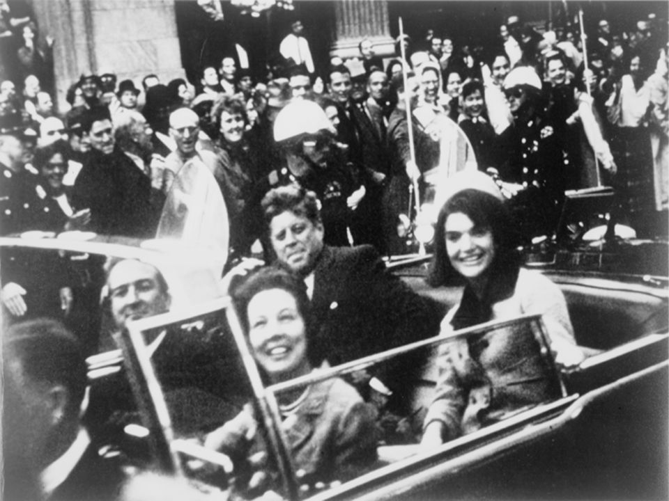
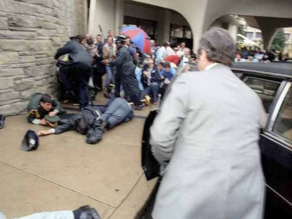
John Fitzgerald Kennedy
35° Presidente de EE.UU.

52
John Bowden Connally 1917-1993 Gobernador de Texas
John Bowden Connally, Jr. (February 27, 1917 – June 15, 1993) was an influential American politician, serving as Governor of Texas, Secretary of the Navy under President John F. Kennedy, and as Secretary of the Treasury under President Richard M. Nixon. While he was Governor in 1963, Connally was a passenger in the car in which President Kennedy was assassinated John Bowden Connally Gobernador de Texas
 was an influential American politician, serving as Governor of Texas, Secretary of the Navy under President John F. Kennedy, and as Secretary of the Treasury under President Richard M. Nixon. While he was Governor in 1963, Connally was a passenger in the car in which President Kennedy was assassinated. John Bowden Connally Gobernador de Texas.")
53
Supuesto asesino de JFK
Lee Harvey Oswald Supuesto asesino de JFK

54
Dallas County Administration Building in 2005, formerly the Texas School Book Depository

55
Oswald is shot by Jack Ruby

56
Asesino de Lee Harvey Oswald
Jacob Rubenstein (March 25, 1911 – January 3, 1967), who legally changed his name to Jack Leon Ruby in 1947, was an American nightclub operator in Dallas, Texas. He was convicted on March 14, 1964, of the murder of Lee Harvey Oswald on November 24, 1963, two days after Oswald was arrested for the assassination of President John F. Kennedy. He successfully appealed his conviction and death sentence. As a date for his new trial was being set,[1] he became ill and died of lung cancer on January 3, 1967. Conspiracy theorists claim that Ruby was involved with major figures in organized crime and killed Oswald as part of an overall plot surrounding the assassination of Kennedy. Others have disputed this, arguing that his connection with gangsters was minimal at best and that he was not the sort to be entrusted with such an act within a high-level conspiracy.[2] Ruby made a final statement from his hospital bed on December 19 that he and he alone had been responsible for the murder of Lee Harvey Oswald.[26] "There is nothing to hide," Ruby said. "There was no one else."[27] He died of a pulmonary embolism, secondary to bronchogenic carcinoma (lung cancer), on January 3, 1967 at Parkland Hospital, where Lee Harvey Oswald had died and President Kennedy had been pronounced dead after his assassination. Jack Ruby Asesino de Lee Harvey Oswald
, who legally changed his name to Jack Leon Ruby in 1947, was an American nightclub operator in Dallas, Texas. He was convicted on March 14, 1964, of the murder of Lee Harvey Oswald on November 24, 1963, two days after Oswald was arrested for the assassination of President John F. Kennedy. He successfully appealed his conviction and death sentence. As a date for his new trial was being set,[1] he became ill and died of lung cancer on January 3, Conspiracy theorists claim that Ruby was involved with major figures in organized crime and killed Oswald as part of an overall plot surrounding the assassination of Kennedy. Others have disputed this, arguing that his connection with gangsters was minimal at best and that he was not the sort to be entrusted with such an act within a high-level conspiracy.[2] Ruby made a final statement from his hospital bed on December 19 that he and he alone had been responsible for the murder of Lee Harvey Oswald.[26] There is nothing to hide, Ruby said. There was no one else. [27] He died of a pulmonary embolism, secondary to bronchogenic carcinoma (lung cancer), on January 3, 1967 at Parkland Hospital, where Lee Harvey Oswald had died and President Kennedy had been pronounced dead after his assassination. Jack Ruby Asesino de Lee Harvey Oswald.")
58
Dr. Tom Shires THREE PATIENTS at PARKLAND
Many Texas physicians have visited Parkland hospital; many have worked or trained there. Members of the Parkland staff are their acquaintances and friends. Many Texas physicians know personally the surviving gunshot victim, Gov. John Connally; some personally knew President John F. Kennedy, who died in Trauma Room 1; perhaps a few even knew Lee Harvey Oswald, the man charged by Dallas authorities with the assassination of the President and who was himself shot two days later. The assassination of President Kennedy, the wounding of Governor Connally, and the fatal shooting of Oswald are events of profound import to people everywhere, but they have special, personal meaning for Texans. So because a Texas hospital and Texas physicians figured prominently in this tragedy, the Texas State Journal of Medicine records for its readers of the medical profession a full account of treatment given a never-to-be-forgotten trio. When President John F. Kennedy in a moribund condition entered Parkland on Nov. 22, there was never opportunity for medical history taking. Such a history, had it been taken, would have shown that the patient "had survived several illnesses, the danger of war, the rigor of exposure in icy water, and had waged grueling electoral campaigns in spite of a serious and painful back injury." Parkland records show that the President arrived at the emergency room sometime after 12:30 p.m. (There is conflict as to the exact moment.) At 1 p.m. Dr. William Kemp Clark, associate professor and chairman of the Division of Neurosurgery of the University of Texas Southwestern Medical School, declared him dead. During the interim of less than 30 minutes, continuous resuscitative efforts were made. Later that day, several attending physicians filed reports. The following identifies these physicians and gives the gist of their reports: Charles J Carrico - Dr. Carrico was the first physician to see the President. A 1961 graduate of Southwestern Medical School, he is 28 and a resident in surgery at Parkland. He reported that when the patient entered the emergency room on an ambulance carriage he had slow agonal respiratory efforts and occasional cardiac beats detectable by auscultation. Two external wounds were noted; one a small wound of the anterior neck in the lower one third. The other wound had caused avulsion of the occipitoparietal calvarium and shredded brain tissue was present with profuse oozing. No pulse or blood pressure were present. Pupils were bilaterally dilated and fixed. A cuffed endotracheal tube was inserted through the laryngoscope. A ragged wound of the trachea was seen immediately below the larynx. The tube was advanced past the laceration and the cuff inflated. Respiration was instituted using a respirator assistor on automatic cycling. Concurrently, an intravenous infusion of lactated Ringer's solution was begun via catheter placed in the right leg. Blood was drawn for typing and crossmatching. Type 0 Rh negative blood was obtained immediately. In view of the tracheal injury and diminished breath sounds in the right chest, tracheostomy was performed by Dr. Malcolm 0. Perry and bilateral chest tubes inserted. A second intravenous infusion was begun in the left arm. In addition, Dr. M. T. Jenkins began respiration with the anesthesia machine, cardiac monitor and stimulator attached. Solu-Cortef (300 mg.) was given intravenously. Despite those measures, blood pressure never returned. Only brief electrocardiographic evidence of cardiac activity was obtained. Malcolm 0. Perry - Dr. Perry is an assistant professor of surgery at Southwestern Medical School from which he received his degree in He was 34 years old and was certified by the American Board of Surgery in 1963. At the time of initial examination of the President, Dr. Perry has stated, the patient was noted to be nonresponsive . His eyes were deviated and the pupils dilated. A considerable quantity of blood was noted on the patient, the carriage, and the floor. A small wound was noted in the midline of the neck in the lower third anteriorly. It was exuding blood slowly. A large wound of the right posterior cranium was noted, exposing severely lacerated brain. Brain tissue was noted in the blood at the head of the carriage. Pulse or heart beat were not detectable but slow spasmodic respiration was noted. An endotracheal tube was in place and respiration was being controlled. An intravenous infusion was being placed in the leg. While additional venesections were done to administer fluids and blood, a tracheostomy was effected. A right lateral injury to the trachea was noted. The cuffed tracheostomy tube was put in place as the endotracheal tube was withdrawn and respirations continued. Closed chest cardiac massage was instituted after placement of sealed-drainage chest tubes, but without benefit. When electrocardiogram evaluation revealed that no detectable electrical activity existed in the heart, resuscitative attempts were abandoned. The team of physicians determined that the patient had expired. Charles R. Baxter - Dr. Baxter is an assistant professor of surgery at Southwestern Medical School where he first arrived as a medical student in Except for two years away in the Army he has been at Southwestern and Parkland ever since, moving up from student to intern to resident to faculty member. He is 34 and was certified by the American Board of Surgery in 1963. Recalling his attendance to President Kennedy, he says he learned at approximately 12 :35 that the President was on the way to the emergency room and that he had been shot. When Dr. Baxter arrived in the emergency room, he found an endotracheal tube in place and respirations being assisted. A left chest tube was being inserted and cut-downs were functioning in one leg and in the left arm. The President had a wound in the midline of the neck. On first observation of the other wounds, portions of the right temporal and occipital bones were missing and some of the brain was lying on the table. The rest of the brain was extensively macerated and contused. The pupils were fixed and deviated laterally and were dilated. No pulse was detectable and ineffectual respirations were being assisted. A tracheostomy was performed by Dr. Perry and Dr. Baxter and a chest tube was inserted into the right chest (second interspace anteriorly). Meanwhile one pint of O negative blood was administered without response. When all of these measures were complete, no heart beat could be detected. Closed chest massage was performed until a cardioscope could be attached. Brief cardiac activity was obtained followed by no activity. Due to the extensive and irreparable brain damage which existed and since there were no signs of life, no further attempts were made at resuscitation. Robert N. McClelland - Dr. McClelland, 34, assistant professor of surgery at Southwestern Medical School, is a graduate of the University of Texas Medical Branch in Galveston. He has served with the Air Force in Germany and was certified by the American Board of Surgery in 1963. Regarding the assassination of President Kennedy, Dr. McClelland says that at approximately 12:35 p.m. he was called from the second floor of the hospital to the emergency room. When he arrived, President Kennedy was being attended by Drs. Perry, Baxter, Carrico, and Ronald Jones, chief resident in surgery. The President was at that time comatose from a massive gunshot wound of the head with a fragment wound of the trachea. An endotracheal tube had been placed and assisted respiration started by Dr. Carrico who was on duty in the emergency room when the President arrived. Drs. Perry, Baxter, and McClelland performed a tracheostomy for respiratory distress and tracheal injury. Dr. Jones and Dr. Paul Peters, assistant professor of surgery, ; inserted bilateral anterior chest tubes for pneumothoraces secondary to the tracheo-mediastinal injury. Dr. Jones and assistants had started three cutdowns, giving blood and fluids immediately. In spite of this, the President was pronounced dead at 1:00 p.m. by Dr. Clark, the neurosurgeon, who arrived immediately after Dr. McClelland. The cause of death, according to Dr. McClelland was the massive head and brain injury from a gunshot wound of the right side of the head. The President was pronounced dead after external cardiac massage failed and electrocardiographic activity was gone. Fouad A, Bashour - Dr. Bashour received his medical education at the University of Beirut School of Medicine in Lebanon. He is 39 and an associate professor of medicine in cardiology at Southwestern Medical School. At 12 :50 p.m. Dr. Bashour was called from the first floor of the hospital and told that President Kennedy had been shot. He and Dr. Donald Seldin, professor and chairman of the Department of Internal Medicine, went to the emergency room. Upon examination, they found that the President had no pulsations, no heart beats, no blood pressure. The oscilloscope showed a complete standstill. The President was declared dead at 1:00 p.m. William Kemp Clark - Dr. Clark is associate professor and chairman of the Division of Neurosurgery at Southwestern Medical School. The 38-year-old physician has done research on head injuries and has been at Southwestern since 1956. He reports this account of the President's treatment: The President arrived at the emergency room entrance in the back seat of his limousine. Governor Connally of Texas was also in this car. The first physician to see the President was Dr. Carrico. Dr. Carrico noted the President to have slow, agonal respiratory efforts. He could hear a heart beat but found no pulse or blood pressure. Two external wounds, one in the lower third of the anterior neck, the other in the occipital region of the skull, were noted. Through the head wound, blood and brain were extruding. Dr. Carrico inserted a cuffed endotracheal tube and while doing so, he noted a ragged wound of the trachea immediately below the larynx. At this time, Drs. Perry, Baxter, and Jones arrived. Immediately thereafter, Dr. Jenkins and Drs. A. H. Giesecke, Jr., and Jackie H. Hunt, two other staff anesthesiologists, arrived. The endotracheal tube had been connected to a respirator to assist the President's breathing. An anesthesia machine was substituted for this by Dr. Jenkins. Only 100 per cent oxygen was administered. A cutdown was performed in the right ankle, and a polyethylene catheter inserted in the vein. An infusion of lactated Ringer's solution was begun. Blood was drawn for typing and crossmatching, but unmatched type O Rh negative blood was immediately obtained and begun. Hydrocortisone (300 mg.) was added to the intravenous fluids. Dr. McClelland arrived to help in the President's care. Drs. Perry, Baxter, and McClelland did a tracheostomy. Considerable quantities of blood were present in the President's oral pharynx. At this time, Dr. Peters and Dr. Clark arrived. Dr. Clark noted that the President had bled profusely from the back of the head. There was a large (3 by 3 cm.) amount of cerebral tissue present on the cart. There was a smaller amount of cerebellar tissue present also. The tracheostomy was completed and the endotracheal tube was withdrawn. Suction was used to remove blood in the oral pharynx. A nasogastric tube was passed into the stomach. Because of the likelihood of mediastinal injury, anterior chest tubes were placed in both pleural spaces. These were connected to sealed underwater drainage. Neurological examination revealed the President's pupils to be widely dilated and fixed to light. His eyes were divergent, being deviated outward; a skew deviation from the horizontal was present. No deep tendon reflexes or spontaneous movements were found. When Dr. Clark noted that there was no carotid pulse, he began closed chest massage. A pulse was obtained at the carotid and femoral levels. Dr. Perry then took over the cardiac massage so that Dr. Clark could evaluate the head wound. There was a large wound beginning in the right occiput extending into the parietal region. Much of the right posterior skull, at brief examination, appeared gone. The previously described extruding brain was present. Profuse bleeding had occurred and 1500 cc. of blood was estimated to be on the drapes and floor of the emergency operating room. Both cerebral and cerebellar tissue were extruding from the wound. By this time an electrocardiograph was hooked up. There was brief electrical activity of the heart which soon stopped. The President was pronounced dead at 1:00 p.m. by Dr. Clark. M. T. Jenkins - Dr. Jenkins is professor and chairman of the Department of Anesthesiology at Southwestern Medical School. He is 46, a graduate of the University of Texas Medical Branch in Galveston, and was certified by the American Board of Anesthesiology in During World War II he served in the Navy as a lieutenant commander. When Dr. Jenkins was notified that the President was being brought to the emergency room at Parkland, he dispatched Drs. Giesecke and Hunt with an anesthesia machine and resuscitative equipment to the major surgical emergency room area. He ran downstairs to find upon his arrival in the emergency operating room that Dr. Carrico had begun resuscitative efforts by introducing an orotracheal tube, connecting it for controlled ventilation to a Bennett intermittent positive pressure breathing apparatus. Drs. Baxter, Perry, and McClelland arrived at the same time and began a tracheostomy and started the insertion of a right chest tube, since there was also obvious tracheal and chest damage. Drs. Peters and Clark arrived simultaneously and immediately thereafter assisted respectively with the insertion of the right chest tube and with manual closed chest cardiac compression to assure circulation. Dr. Jenkins believes it evidence of the clear thinking of the resuscitative team that the patient received 300 mg. hydrocortisone intravenously in the first few minutes. For better control of artificial ventilation, Dr. Jenkins exchanged the intermittent positive pressure breathing apparatus for an anesthesia machine and continued artificial ventilation. Dr. Gene Akin, a resident in anesthesiology, and Dr. Giesecke connected a cardioscope to determine cardiac activity. During the progress of these activities, the emergency room cart was elevated at the feet in order to provide a Trendelenburg position, a venous cutdown was performed on the right saphenous vein and additional fluids were begun in a vein in the left forearm while blood was ordered from the blood bank. All of these activities were completed by approximately 12:50 at which time external cardiac massage was still being carried out effectively by Dr. Clark as judged by a palpable peripheral pulse. Despite these measures there was only brief electrocardiographic evidence of cardiac activity. These described resuscitative activities were indicated as of first importance, and after they were carried out, attention was turned to other evidences of injury. There was a great laceration on the right side of the head (temporal and occipital), causing a great defect in the skull plate so that there was herniation and laceration of great areas of the brain, even to the extent that part of the right cerebellum had protruded from the wound. There were also fragmented sections of brain on the drapes of the emergency room cart. With the institution of adequate cardiac compression, there was a great flow of blood from the cranial cavity, indicating that there was much vascular damage as well as brain tissue damage. President Kennedy was pronounced dead at 1 p.m. It is Dr. Jenkins' personal feeling that all methods of resuscitation were instituted expeditiously and efficiently. However, he says, the cranial and intracranial damage was of such magnitude as to cause irreversible damage
 At 1 p.m. Dr. William Kemp Clark, associate professor and chairman of the Division of Neurosurgery of the University of Texas Southwestern Medical School, declared him dead. During the interim of less than 30 minutes, continuous resuscitative efforts were made. Later that day, several attending physicians filed reports. The following identifies these physicians and gives the gist of their reports: Charles J Carrico - Dr. Carrico was the first physician to see the President. A 1961 graduate of Southwestern Medical School, he is 28 and a resident in surgery at Parkland. He reported that when the patient entered the emergency room on an ambulance carriage he had slow agonal respiratory efforts and occasional cardiac beats detectable by auscultation. Two external wounds were noted; one a small wound of the anterior neck in the lower one third. The other wound had caused avulsion of the occipitoparietal calvarium and shredded brain tissue was present with profuse oozing. No pulse or blood pressure were present. Pupils were bilaterally dilated and fixed. A cuffed endotracheal tube was inserted through the laryngoscope. A ragged wound of the trachea was seen immediately below the larynx. The tube was advanced past the laceration and the cuff inflated. Respiration was instituted using a respirator assistor on automatic cycling. Concurrently, an intravenous infusion of lactated Ringer s solution was begun via catheter placed in the right leg. Blood was drawn for typing and crossmatching. Type 0 Rh negative blood was obtained immediately. In view of the tracheal injury and diminished breath sounds in the right chest, tracheostomy was performed by Dr. Malcolm 0. Perry and bilateral chest tubes inserted. A second intravenous infusion was begun in the left arm. In addition, Dr. M. T. Jenkins began respiration with the anesthesia machine, cardiac monitor and stimulator attached. Solu-Cortef (300 mg.) was given intravenously. Despite those measures, blood pressure never returned. Only brief electrocardiographic evidence of cardiac activity was obtained. Malcolm 0. Perry - Dr. Perry is an assistant professor of surgery at Southwestern Medical School from which he received his degree in He was 34 years old and was certified by the American Board of Surgery in At the time of initial examination of the President, Dr. Perry has stated, the patient was noted to be nonresponsive . His eyes were deviated and the pupils dilated. A considerable quantity of blood was noted on the patient, the carriage, and the floor. A small wound was noted in the midline of the neck in the lower third anteriorly. It was exuding blood slowly. A large wound of the right posterior cranium was noted, exposing severely lacerated brain. Brain tissue was noted in the blood at the head of the carriage. Pulse or heart beat were not detectable but slow spasmodic respiration was noted. An endotracheal tube was in place and respiration was being controlled. An intravenous infusion was being placed in the leg. While additional venesections were done to administer fluids and blood, a tracheostomy was effected. A right lateral injury to the trachea was noted. The cuffed tracheostomy tube was put in place as the endotracheal tube was withdrawn and respirations continued. Closed chest cardiac massage was instituted after placement of sealed-drainage chest tubes, but without benefit. When electrocardiogram evaluation revealed that no detectable electrical activity existed in the heart, resuscitative attempts were abandoned. The team of physicians determined that the patient had expired. Charles R. Baxter - Dr. Baxter is an assistant professor of surgery at Southwestern Medical School where he first arrived as a medical student in Except for two years away in the Army he has been at Southwestern and Parkland ever since, moving up from student to intern to resident to faculty member. He is 34 and was certified by the American Board of Surgery in Recalling his attendance to President Kennedy, he says he learned at approximately 12 :35 that the President was on the way to the emergency room and that he had been shot. When Dr. Baxter arrived in the emergency room, he found an endotracheal tube in place and respirations being assisted. A left chest tube was being inserted and cut-downs were functioning in one leg and in the left arm. The President had a wound in the midline of the neck. On first observation of the other wounds, portions of the right temporal and occipital bones were missing and some of the brain was lying on the table. The rest of the brain was extensively macerated and contused. The pupils were fixed and deviated laterally and were dilated. No pulse was detectable and ineffectual respirations were being assisted. A tracheostomy was performed by Dr. Perry and Dr. Baxter and a chest tube was inserted into the right chest (second interspace anteriorly). Meanwhile one pint of O negative blood was administered without response. When all of these measures were complete, no heart beat could be detected. Closed chest massage was performed until a cardioscope could be attached. Brief cardiac activity was obtained followed by no activity. Due to the extensive and irreparable brain damage which existed and since there were no signs of life, no further attempts were made at resuscitation. Robert N. McClelland - Dr. McClelland, 34, assistant professor of surgery at Southwestern Medical School, is a graduate of the University of Texas Medical Branch in Galveston. He has served with the Air Force in Germany and was certified by the American Board of Surgery in Regarding the assassination of President Kennedy, Dr. McClelland says that at approximately 12:35 p.m. he was called from the second floor of the hospital to the emergency room. When he arrived, President Kennedy was being attended by Drs. Perry, Baxter, Carrico, and Ronald Jones, chief resident in surgery. The President was at that time comatose from a massive gunshot wound of the head with a fragment wound of the trachea. An endotracheal tube had been placed and assisted respiration started by Dr. Carrico who was on duty in the emergency room when the President arrived. Drs. Perry, Baxter, and McClelland performed a tracheostomy for respiratory distress and tracheal injury. Dr. Jones and Dr. Paul Peters, assistant professor of surgery, ; inserted bilateral anterior chest tubes for pneumothoraces secondary to the tracheo-mediastinal injury. Dr. Jones and assistants had started three cutdowns, giving blood and fluids immediately. In spite of this, the President was pronounced dead at 1:00 p.m. by Dr. Clark, the neurosurgeon, who arrived immediately after Dr. McClelland. The cause of death, according to Dr. McClelland was the massive head and brain injury from a gunshot wound of the right side of the head. The President was pronounced dead after external cardiac massage failed and electrocardiographic activity was gone. Fouad A, Bashour - Dr. Bashour received his medical education at the University of Beirut School of Medicine in Lebanon. He is 39 and an associate professor of medicine in cardiology at Southwestern Medical School. At 12 :50 p.m. Dr. Bashour was called from the first floor of the hospital and told that President Kennedy had been shot. He and Dr. Donald Seldin, professor and chairman of the Department of Internal Medicine, went to the emergency room. Upon examination, they found that the President had no pulsations, no heart beats, no blood pressure. The oscilloscope showed a complete standstill. The President was declared dead at 1:00 p.m. William Kemp Clark - Dr. Clark is associate professor and chairman of the Division of Neurosurgery at Southwestern Medical School. The 38-year-old physician has done research on head injuries and has been at Southwestern since He reports this account of the President s treatment: The President arrived at the emergency room entrance in the back seat of his limousine. Governor Connally of Texas was also in this car. The first physician to see the President was Dr. Carrico. Dr. Carrico noted the President to have slow, agonal respiratory efforts. He could hear a heart beat but found no pulse or blood pressure. Two external wounds, one in the lower third of the anterior neck, the other in the occipital region of the skull, were noted. Through the head wound, blood and brain were extruding. Dr. Carrico inserted a cuffed endotracheal tube and while doing so, he noted a ragged wound of the trachea immediately below the larynx. At this time, Drs. Perry, Baxter, and Jones arrived. Immediately thereafter, Dr. Jenkins and Drs. A. H. Giesecke, Jr., and Jackie H. Hunt, two other staff anesthesiologists, arrived. The endotracheal tube had been connected to a respirator to assist the President s breathing. An anesthesia machine was substituted for this by Dr. Jenkins. Only 100 per cent oxygen was administered. A cutdown was performed in the right ankle, and a polyethylene catheter inserted in the vein. An infusion of lactated Ringer s solution was begun. Blood was drawn for typing and crossmatching, but unmatched type O Rh negative blood was immediately obtained and begun. Hydrocortisone (300 mg.) was added to the intravenous fluids. Dr. McClelland arrived to help in the President s care. Drs. Perry, Baxter, and McClelland did a tracheostomy. Considerable quantities of blood were present in the President s oral pharynx. At this time, Dr. Peters and Dr. Clark arrived. Dr. Clark noted that the President had bled profusely from the back of the head. There was a large (3 by 3 cm.) amount of cerebral tissue present on the cart. There was a smaller amount of cerebellar tissue present also. The tracheostomy was completed and the endotracheal tube was withdrawn. Suction was used to remove blood in the oral pharynx. A nasogastric tube was passed into the stomach. Because of the likelihood of mediastinal injury, anterior chest tubes were placed in both pleural spaces. These were connected to sealed underwater drainage. Neurological examination revealed the President s pupils to be widely dilated and fixed to light. His eyes were divergent, being deviated outward; a skew deviation from the horizontal was present. No deep tendon reflexes or spontaneous movements were found. When Dr. Clark noted that there was no carotid pulse, he began closed chest massage. A pulse was obtained at the carotid and femoral levels. Dr. Perry then took over the cardiac massage so that Dr. Clark could evaluate the head wound. There was a large wound beginning in the right occiput extending into the parietal region. Much of the right posterior skull, at brief examination, appeared gone. The previously described extruding brain was present. Profuse bleeding had occurred and 1500 cc. of blood was estimated to be on the drapes and floor of the emergency operating room. Both cerebral and cerebellar tissue were extruding from the wound. By this time an electrocardiograph was hooked up. There was brief electrical activity of the heart which soon stopped. The President was pronounced dead at 1:00 p.m. by Dr. Clark. M. T. Jenkins - Dr. Jenkins is professor and chairman of the Department of Anesthesiology at Southwestern Medical School. He is 46, a graduate of the University of Texas Medical Branch in Galveston, and was certified by the American Board of Anesthesiology in During World War II he served in the Navy as a lieutenant commander. When Dr. Jenkins was notified that the President was being brought to the emergency room at Parkland, he dispatched Drs. Giesecke and Hunt with an anesthesia machine and resuscitative equipment to the major surgical emergency room area. He ran downstairs to find upon his arrival in the emergency operating room that Dr. Carrico had begun resuscitative efforts by introducing an orotracheal tube, connecting it for controlled ventilation to a Bennett intermittent positive pressure breathing apparatus. Drs. Baxter, Perry, and McClelland arrived at the same time and began a tracheostomy and started the insertion of a right chest tube, since there was also obvious tracheal and chest damage. Drs. Peters and Clark arrived simultaneously and immediately thereafter assisted respectively with the insertion of the right chest tube and with manual closed chest cardiac compression to assure circulation. Dr. Jenkins believes it evidence of the clear thinking of the resuscitative team that the patient received 300 mg. hydrocortisone intravenously in the first few minutes. For better control of artificial ventilation, Dr. Jenkins exchanged the intermittent positive pressure breathing apparatus for an anesthesia machine and continued artificial ventilation. Dr. Gene Akin, a resident in anesthesiology, and Dr. Giesecke connected a cardioscope to determine cardiac activity. During the progress of these activities, the emergency room cart was elevated at the feet in order to provide a Trendelenburg position, a venous cutdown was performed on the right saphenous vein and additional fluids were begun in a vein in the left forearm while blood was ordered from the blood bank. All of these activities were completed by approximately 12:50 at which time external cardiac massage was still being carried out effectively by Dr. Clark as judged by a palpable peripheral pulse. Despite these measures there was only brief electrocardiographic evidence of cardiac activity. These described resuscitative activities were indicated as of first importance, and after they were carried out, attention was turned to other evidences of injury. There was a great laceration on the right side of the head (temporal and occipital), causing a great defect in the skull plate so that there was herniation and laceration of great areas of the brain, even to the extent that part of the right cerebellum had protruded from the wound. There were also fragmented sections of brain on the drapes of the emergency room cart. With the institution of adequate cardiac compression, there was a great flow of blood from the cranial cavity, indicating that there was much vascular damage as well as brain tissue damage. President Kennedy was pronounced dead at 1 p.m. It is Dr. Jenkins personal feeling that all methods of resuscitation were instituted expeditiously and efficiently. However, he says, the cranial and intracranial damage was of such magnitude as to cause irreversible damage.")
67
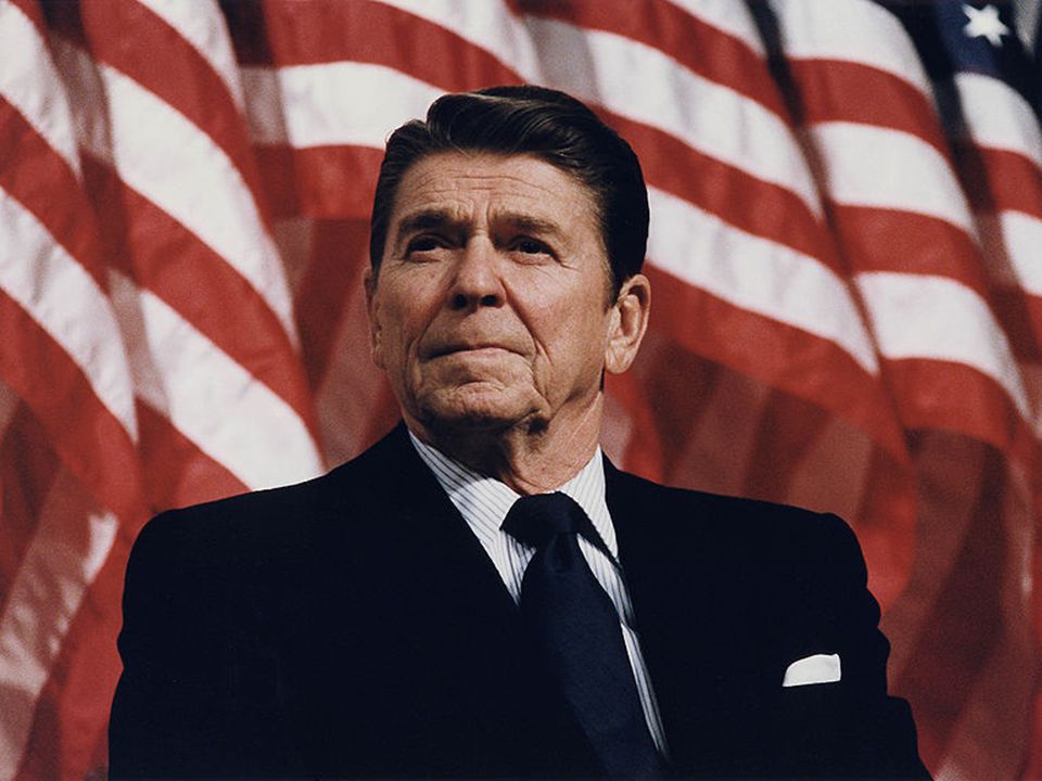
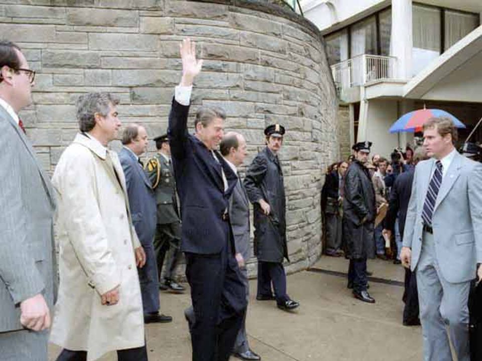
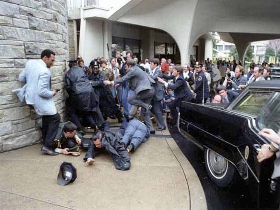
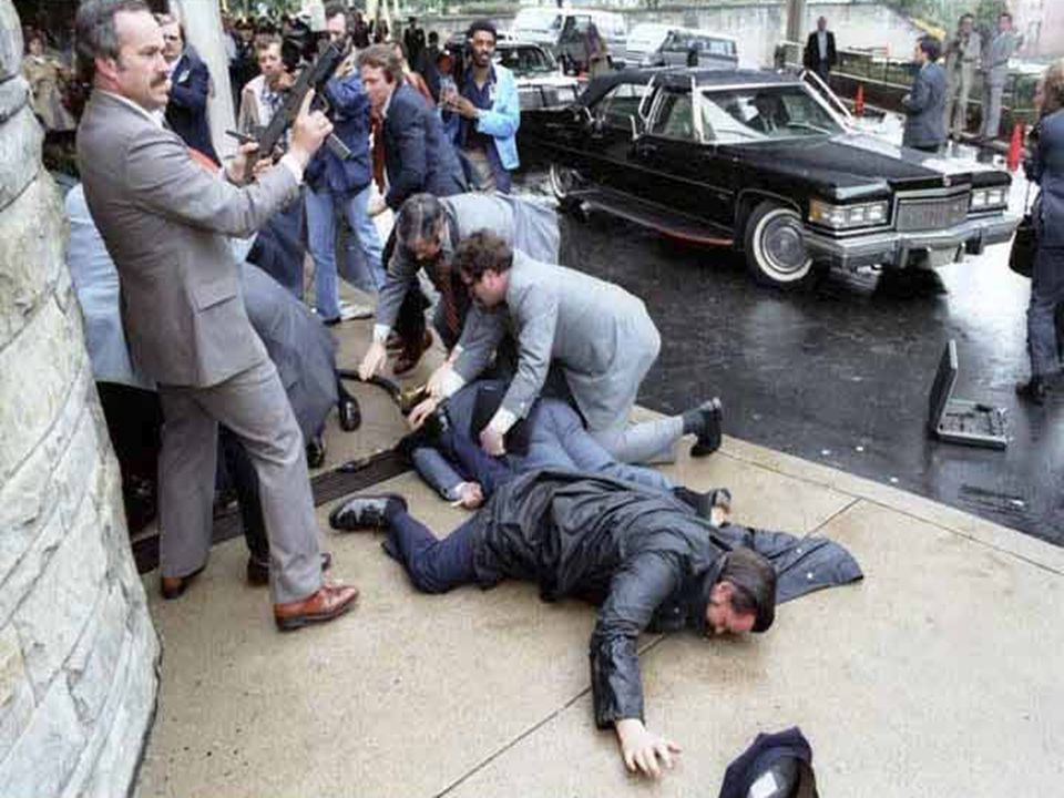
John Hinckley, Jr.

69
La verdad … está en el punto medio …

70
Estrategia liberal Estrategia conservadora Estrategia racional

72
Pregunta ¿ La diuresis es una guía confiable para la resucitación líquida? Sí No No necesariamente

73
Permeabilidad urinaria Perfusión renal Diuréticos Diuresis Aldosterona Función renal ADH

75
¿ El “suero fisiológico” es bueno para quitar la sed ?
Pregunta ¿ Un paciente con sed intensa es sinónimo de deshidratación ? ¿ El “suero fisiológico” es bueno para quitar la sed ?

76
el paciente está jadeando ?
Pregunta ¿ Un paciente con lengua seca, pero con edema periférico es sinónimo de deshidratación ? ¿ Has considerado si el paciente está jadeando ?

78
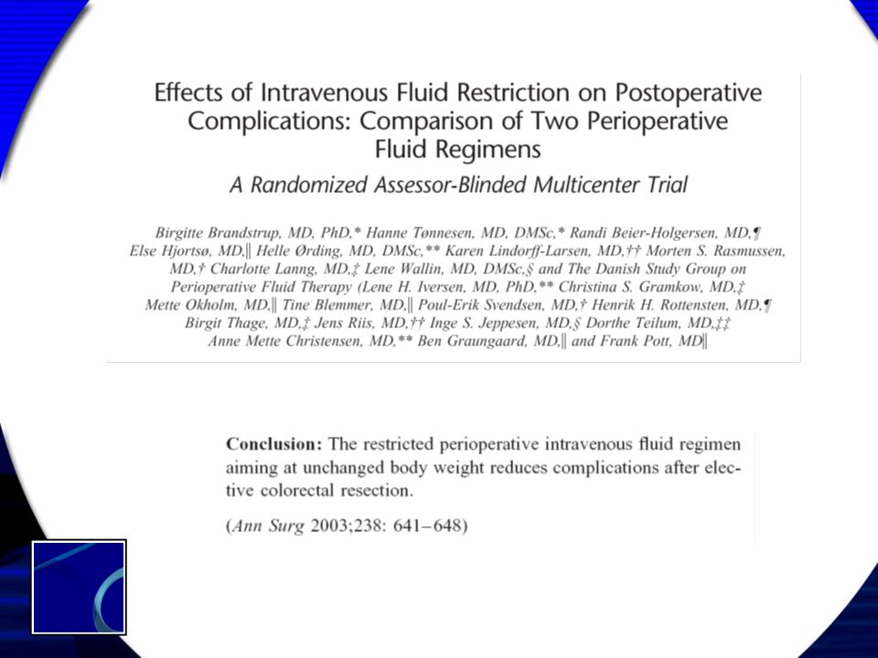
Régimen restricto No precarga para anestesia neuroaxial o máximo 10 ml/kg No reemplazo de tercer espacio Reposición de ayuno: 500 ml de SG 5% - ingreso oral durante ayuno Pérdida sanguínea: Volumen x volumen de coloide Transfusión sólo si sangra > 1500 ml Brandstrup et al. Ann Surg 2003; 238:641-8

79
Régimen estándar Ayuno Requerimientos basales horarios Diuresis
Sangrado Respiración Evaporación del campo quirúrgico Pérdidas por tercer espacio

80
Cálculos Paciente 45 años de 70 kg, para gastrectomía radical con ayuno de 10 h y cirugía que duró 4 horas: Ayuno = 1100 ml Req. basales = 110 ml/h x 4 = 440 ml Diuresis = 150 ml/h x 4 = 600 ml Sangrado = 1000 ml (3:1) = 3000 ml Evaporación = 10 ml/kg/h = 700 ml/h = 2800 ml Respiración = 1 ml/kg/h = 280 ml SNG = 250 ml Total = 8470
 = 3000 ml. Evaporación = 10 ml/kg/h = 700 ml/h = 2800 ml. Respiración = 1 ml/kg/h = 280 ml. SNG = 250 ml. Total =")
82
Régimen estándar vs restricto
Complicación Fuga anastomótica Sepsis Sangrado anormal Edema pulmonar Falla renal Restricto (n=69) 1 Estándar (n=72) 4 5 1 Brandstrup et al. Ann Surg 2003; 238:641-8
 1. Estándar (n=72) Brandstrup et al. Ann Surg 2003; 238:")
84
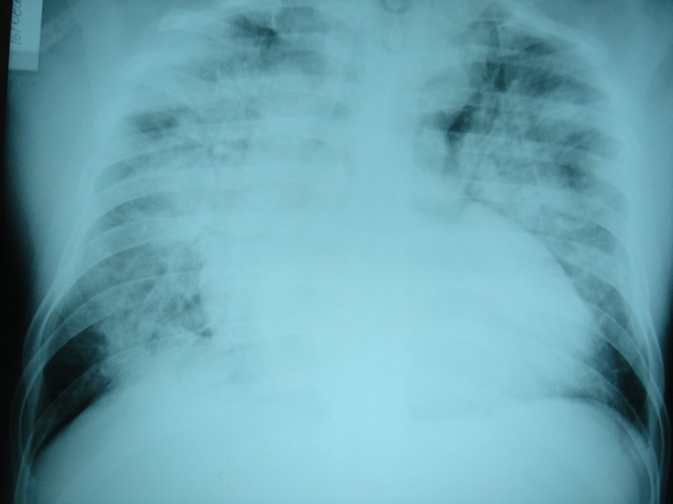
Fluidoterapia masiva Edema pulmonar Ileo adinámico
Hipertensión intraabdominal Coagulopatía dilucional Acidosis Deterioro de la función cardíaca Mala cicatrización Isquemia por disperfusión Scheingraber S. Anesthesiology 1999; 90:

91
La fluidoterapia no es tan inocua como pensábamos !

92
Pregunta ¿ Cuál de estos órganos funciona mal cuando presentan edema ?
Pulmones Cerebro Intestino Corazón

93
Isquemia por disperfusión

94
¿ Cuándo es beneficiosa la fluidoterapia ?

95
Fluidoterapia no mejora
función cardíaca Fluidoterapia mejora función cardíaca Gasto cardíaco Volumen de líquido administrado

96
Conclusiones Los fluidos son los medicamentos más utilizados en la práctica Su uso requiere de conocimientos profundos de la fisiología A veces, el no poner y no hacer nada es quizás mejor que poner y hacer … La vía más fisiológica es la oral Tortas

97
GRACIAS

98
¿ Dudas o preguntas ?

Presentaciones similares