Descargar la presentación
La descarga está en progreso. Por favor, espere

1
FISIOLOGIA DIGESTIVA (BCM II)
Clase 1: Sistema Nervioso Entérico Dr. Michel Baró Aliste

2
Requerimientos de la Función digestiva:
1-Tránsito de alimentos a lo largo del tubo digestivo 2-Secreción de jugos digestivos y digestión de los alimentos 3-Absorción de los productos digeridos 4-Circulación de la sangre y transporte de nutrientes 5-Control nervioso y hormonal del proceso

3
Funciones específicas de las partes del Sistema Digestivo:
Boca: Preparación del Bolo Faringe: Regulación del paso de alimentos y aire Esófago: Paso del bolo al estómago Estómago: Almacenamiento e inicio de la digestión Intestino delgado: Digestión y absorción Colon y recto: Excreción

5
Músculo liso intestinal características:
Longitud 100 a 500 micras Diámetro 2 a 10 micras Haces de hasta 1000 fibras paralelas, tramas Capa circular y longitudinal Uniones intercelulares (sincitio)
")
6
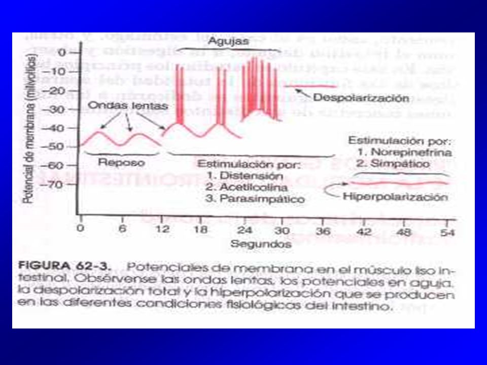
Actividad eléctrica del músculo liso
gastrointestinal 1-Ondas lentas: 3-12 /min estómago 3/min ¿bomba Na-K? 5-15 mV duodeno 12/min íleon 8-9/min 2-Potenciales en aguja canales Ca-Na umbral -40 mV reposo -50 a -60 mV frecuencia 1 a 10 /seg duración mseg (10 a 40 veces más que una neurona) 3-Potencial de reposo valor medio de -56 mV variable según estímulos mecánicos, nerviosos y humorales.
 3-Potencial de reposo. valor medio de -56 mV. variable según estímulos mecánicos, nerviosos y humorales.")
8
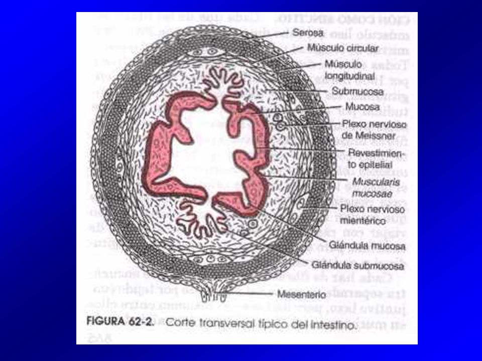
Sistema Nervioso Entérico
Control Nervioso gastrointestinal: Sistema Nervioso Entérico “Cerebro del Intestino” neuronas Derivado de las crestas neurales Interconectado con el SNC Funciones: Plexo mientérico: -Motilidad Plexo sub-mucoso: -Secreción endocrina y exocrina -Microcirculación Figure Cutaway view showing anatomy of the tubular esophagus. The esophagus is a muscular tube that is composed of longitudinal and circular muscle with extensive neural network in between. Auerbach's plexus (myenteric) lies between the longitudinal and circular muscle layers. Another nerve network, Meissner's plexus (submucosal), is situated between the muscularis mucosa and the circular muscle layer. Note that there is no serosa to the esophagus and that the lumen is collapsed and empty. In fact, activity of both esophageal sphincters preserves the vacuum of the esophagus; the upper esophageal sphincter acts to exclude air during respiration and the lower esophageal sphincter excludes gastric contents from refluxing back into the esophagus. (Adapted from Kahrilas [6].) References: [6]. Kahrilas PJ, The anatomy and physiology of dysphagia. In Dysphagia, Diagnosis, and Treatment. Edited by Gelfand DW, Richter JE. New York: Igaku-Shoin;
 lies between the longitudinal and circular muscle layers. Another nerve network, Meissner s plexus (submucosal), is situated between the muscularis mucosa and the circular muscle layer. Note that there is no serosa to the esophagus and that the lumen is collapsed and empty. In fact, activity of both esophageal sphincters preserves the vacuum of the esophagus; the upper esophageal sphincter acts to exclude air during respiration and the lower esophageal sphincter excludes gastric contents from refluxing back into the esophagus. (Adapted from Kahrilas [6].) References: [6]. Kahrilas PJ, The anatomy and physiology of dysphagia. In Dysphagia, Diagnosis, and Treatment. Edited by Gelfand DW, Richter JE. New York: Igaku-Shoin;")
9
Sistema Nervioso Entérico

10
Efectos de la estimulación del plexo mientérico:
Aumento de la contracción tónica (“tono”) de la pared intestinal aumento de la intensidad de las contracciones ligero aumento frecuencia de las contracciones aumento de la velocidad de conducción de las ondas excitatorias (peristalsis)
 de la pared intestinal. aumento de la intensidad de las contracciones. ligero aumento frecuencia de las contracciones. aumento de la velocidad de conducción de las ondas excitatorias (peristalsis)")
11
Efectos inhibitorios del plexo mientérico:
Neuronas inhibitorias (VIP, óxido nítrico) Relajación de esfínteres musculares píloro válvula íleo-cecal
 Relajación de esfínteres musculares. píloro. válvula íleo-cecal.")
12
Funciones del plexo submucoso:
origen de señales sensitivas control local de la secreción intestinal control local de la absorción contracción local de la muscularis mucosae

13
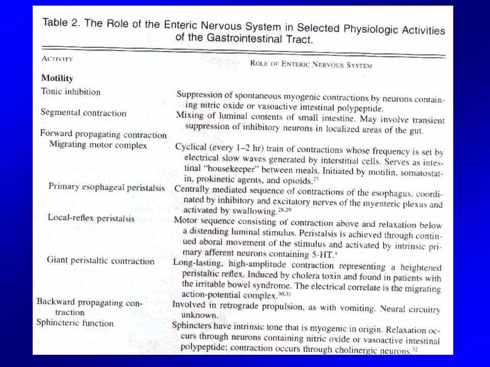
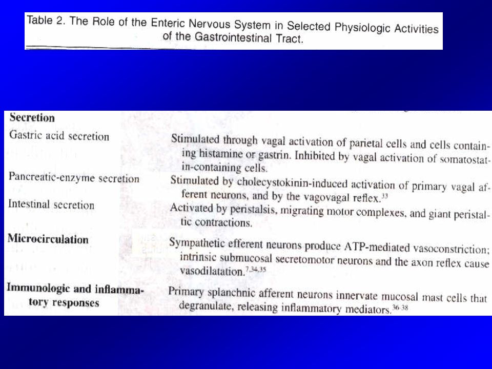
Sistema Nervioso Entérico
NEJM Vol334 Nº17, 1996.

14
Intrinsic nerves are more numerous than extrinsic nerves
Figure The intrinsic nerves are more numerous than the extrinsic nerves, and they communicate largely to modify local motor function through an extensive network of interconnected ganglion cells called plexuses [38]. These nerves communicate with sensory nerves from all layers of the intestine and coordinate the patterns of contraction in response to stimuli. This system is called the enteric nervous system (ENS) or gut brain. It is estimated that it contains one million nerves, about the same number as the spinal cord. ANSautonomic nervous system; DMVdorsal motor nucleus of the vagus; NTSnucleus of the solitary tract. (Adapted from Furness and Costa [38].) References: [38]. Furness JB, Costa M, Distribution of intrinsic nerve cell bodies and axons which take up aromatic amines and their precursors in the small intestine of the guinea pig. Cell Tissue Res
 or gut brain. It is estimated that it contains one million nerves, about the same number as the spinal cord. ANSautonomic nervous system; DMVdorsal motor nucleus of the vagus; NTSnucleus of the solitary tract. (Adapted from Furness and Costa [38].) References: [38]. Furness JB, Costa M, Distribution of intrinsic nerve cell bodies and axons which take up aromatic amines and their precursors in the small intestine of the guinea pig. Cell Tissue Res")
15
Neurotransmisores del
Sistema Nervioso Entérico

18
Control autónomo gastrointestinal
-Parasimpático: -craneal (vago): esófago, estómago, páncreas, intestino -sacro: (S2-4): I. grueso distal, sigmoides, recto, ano -Neuronas post-ganglionares en el plexo mientérico -Colinérgicas -Simpático: -(D5-L2), cadenas simpáticas, ganglios celíaco, mesentéricos -Distribución uniforme, efecto inhibitorio (noradrenalina) -Acción directa (músculo liso) e indirecta (neuronas entéricas)
: esófago, estómago, páncreas, intestino. -sacro: (S2-4): I. grueso distal, sigmoides, recto, ano. -Neuronas post-ganglionares en el plexo mientérico. -Colinérgicas. -Simpático: -(D5-L2), cadenas simpáticas, ganglios celíaco, mesentéricos. -Distribución uniforme, efecto inhibitorio (noradrenalina) -Acción directa (músculo liso) e indirecta (neuronas entéricas)")
19
* * *

20
* * *

21
Intestinal smooth muscle receives and sends neural messages
Núcleo Dorsal Motor 80%-90% de fibras aferentes (sensitivas) Figure The intestinal smooth muscle receives and sends neural messages that modulate its function [37]. The extrinsic nerves relay information to and from the brain, spinal cord, and the extraintestinal ganglia. The intestine can function quite well without the extrinsic nervous system; however, extrinsic nerves serve an important role to modify the activity of the intrinsic nervous system and the motor function of the intestine. In addition to modulating the local enteric reflexes, extrinsic nerves serve to integrate and coordinate widely separate regions of the gastrointestinal tract through its own reflexes. The extrinsic nervous system has three major subdivisions: the parasympathetic pathways, the sympathetic pathways, and the sensory pathways. The parasympathetic system to the intestine is provided entirely by the tenth cranial nerve, the vagus. The efferent nerve cell bodies arise in the dorsal motor nucleus of the vagus (DMV), and their axons pass directly to synapse mainly with the enteric ganglia. The vagus is made up of four types of nerve fibers: excitatory preganglionic cholinergic nerves, inhibitory preganglionic cholinergic nerves, sympathetic fibers from the cervical ganglia, and afferent fibers from the intestinal wall. In fact, 80% to 90% of the vagal fibers are afferent nerves that terminate on neurons of the nodose ganglia. The excitatory neurotransmitter released from the intramural nerves is acetylcholine; the inhibitory substance is probably nitric oxide. Bilateral section of the vagus has little long-lasting effect on the intestinal motility patterns whereas the response to vagal stimulation usually causes a mixture of excitation and inhibition. References: [37]. Gonella J, Bouvier-Blanquet F, Extrinsic nervous control of motility of small and large intestines and related sphincters. Physiol Rev
 Figure The intestinal smooth muscle receives and sends neural messages that modulate its function [37]. The extrinsic nerves relay information to and from the brain, spinal cord, and the extraintestinal ganglia. The intestine can function quite well without the extrinsic nervous system; however, extrinsic nerves serve an important role to modify the activity of the intrinsic nervous system and the motor function of the intestine. In addition to modulating the local enteric reflexes, extrinsic nerves serve to integrate and coordinate widely separate regions of the gastrointestinal tract through its own reflexes. The extrinsic nervous system has three major subdivisions: the parasympathetic pathways, the sympathetic pathways, and the sensory pathways. The parasympathetic system to the intestine is provided entirely by the tenth cranial nerve, the vagus. The efferent nerve cell bodies arise in the dorsal motor nucleus of the vagus (DMV), and their axons pass directly to synapse mainly with the enteric ganglia. The vagus is made up of four types of nerve fibers: excitatory preganglionic cholinergic nerves, inhibitory preganglionic cholinergic nerves, sympathetic fibers from the cervical ganglia, and afferent fibers from the intestinal wall. In fact, 80% to 90% of the vagal fibers are afferent nerves that terminate on neurons of the nodose ganglia. The excitatory neurotransmitter released from the intramural nerves is acetylcholine; the inhibitory substance is probably nitric oxide. Bilateral section of the vagus has little long-lasting effect on the intestinal motility patterns whereas the response to vagal stimulation usually causes a mixture of excitation and inhibition. References: [37]. Gonella J, Bouvier-Blanquet F, Extrinsic nervous control of motility of small and large intestines and related sphincters. Physiol Rev")
22
D5 L2 The thoracolumbar pathways form the sympathetic system
Figure The thoracolumbar pathways form the sympathetic system. Cholinergic preganglionic cell bodies emanate from the lower thoracic and upper lumbar segments of the cord. Spinal roots T9 and T10 provide the main sympathetic supply to the intestine. Their axons pass through the splanchnic nerves via the celiac, superior mesenteric, and inferior mesenteric ganglia; from there, they pass mainly to the intrinsic enteric ganglia. Noradrenergic postganglionic neurons originate in either the paravertebral ganglia or the prevertebral ganglia (celiac, superior mesenteric, inferior mesenteric); their postganglionic fibers synapse with enteric neurons. Stimulation of the efferent splanchnic nerves inhibits motility through release of norepinephrine. Section of the splanchnic nerves markedly increases motor activity. Thus, this system plays an important inhibitory role in the regulation of motor activity. L2
; their postganglionic fibers synapse with enteric neurons. Stimulation of the efferent splanchnic nerves inhibits motility through release of norepinephrine. Section of the splanchnic nerves markedly increases motor activity. Thus, this system plays an important inhibitory role in the regulation of motor activity. L2.")
23
Control autónomo gastrointestinal
Fibras sensitivas aferentes -Con cuerpo y axones en el plexo mientérico -Irritación mucosa intestinal -Distensión excesiva -Presencia de substancias específicas en el intestino -Con cuerpo en plexo mientérico y axones hacia los ganglios simpáticos pre-vertebrales (mesentéricos, celíacos e hipogástricos) -Con cuerpo celulares en ganglios de raíces dorsales y ganglios de nervios craneales (80% fibras del Vago): hacia la médula espinal y tronco encefálico
 -Con cuerpo celulares en ganglios de raíces. dorsales y ganglios de nervios craneales (80% fibras del Vago): hacia la médula espinal y tronco encefálico.")
24
Control autónomo gastrointestinal
Reflejos gastrointestinales -Reflejos integrados dentro del Sistema Nervioso Entérico -Secreción -Peristaltismo -Contracciones de mezclado -Inhibición local -Reflejos intestino - ganglios simpáticos prevertebrales- intestino -gastro-cólico -entero-gástricos -cólico-ileal -Reflejos intestino - médula o tronco - intestino -actividad motora y secretora gástrica (estómago, duodeno – tronco – estómago) -reflejos dolorosos - inhibición motora generalizada -reflejo defecatorio (colon, recto – médula – colon, recto, musc. abdominal)
 -reflejos dolorosos - inhibición motora generalizada. -reflejo defecatorio (colon, recto – médula – colon, recto, musc. abdominal)")
25
Control autónomo gastrointestinal
Control hormonal de la motilidad -Colecistoquinina: -Vaciamiento vesicular (cél “I” duodenales) -inhibición motilidad gástrica -Secretina: -inhibidor débil motilidad digestiva (cél. “S” duodenales) -Péptido inhibidor gástrico (intestino alto) -responde a los ac. grasos, aa, h. de c. -inhibidor leve de la motilidad gástrica -Otras: Motilina, Grelina
 -inhibición motilidad gástrica. -Secretina: -inhibidor débil motilidad digestiva. (cél. S duodenales) -Péptido inhibidor gástrico. (intestino alto) -responde a los ac. grasos, aa, h. de c. -inhibidor leve de la motilidad gástrica. -Otras: Motilina, Grelina.")
26
Cholecystokinin released from duodenal epithelium
CCK-A Vesícula Páncreas Cerebro CCK-B Estómago (compite con Gastrina) Figure Cholecystokinin (CCK) released from the duodenal epithelium (CCK enteroendocrine cells termed I cells) enters the bloodstream, and like a classic hormone, circulates to its target, the gallbladder and biliary tract [28]. CCK, a peptide synthesized in the duodenum and jejunum, consists of 33 amino acids (CCK-33) that contain its biologically active fragment, the 8 N-terminal amino acids (CCK-8). There are two types of receptors: CCK-A, found in the gallbladder, pancreas, and brain, and CCK-B, found in the stomach [13]. Gastrin and CCK are structurally similar and compete for CCK-B receptors. Several factors secreted into the proximal small intestine are capable of evoking the release of CCK; their own destruction by luminal proteases (eg, trypsin) provides a negative feedback loop regulating serum CCK levels and pancreatic secretion. Two factors are present in the intestinal lumen: (1) monitor protein is produced by acinar cells in the pancreas and secreted via the pancreatic duct into the duodenum, where it induces the CCK cells to secrete CCK; and (2) intestinal CCK releasing factor (CCK-RF), present in the duodenum under basal conditions, also stimulates CCK release from I cells. Dietary protein, the obvious substrate for trypsin, inhibits the destruction of these two releasing factors, resulting in a rise in CCK. CCK increases pancreatic secretions of monitor protein. The secreted proteases downregulate the whole process. References: [13]. Schjoldager BT, Role of CCK in gallbladder function. Ann N Y Acad Sci [28]. Walsh JH, Gastrointestinal hormones. In Physiology of the Gastrointestinal Tract. Edited by Johnso LR. New York: Raven Press;
 Figure Cholecystokinin (CCK) released from the duodenal epithelium (CCK enteroendocrine cells termed I cells) enters the bloodstream, and like a classic hormone, circulates to its target, the gallbladder and biliary tract [28]. CCK, a peptide synthesized in the duodenum and jejunum, consists of 33 amino acids (CCK-33) that contain its biologically active fragment, the 8 N-terminal amino acids (CCK-8). There are two types of receptors: CCK-A, found in the gallbladder, pancreas, and brain, and CCK-B, found in the stomach [13]. Gastrin and CCK are structurally similar and compete for CCK-B receptors. Several factors secreted into the proximal small intestine are capable of evoking the release of CCK; their own destruction by luminal proteases (eg, trypsin) provides a negative feedback loop regulating serum CCK levels and pancreatic secretion. Two factors are present in the intestinal lumen: (1) monitor protein is produced by acinar cells in the pancreas and secreted via the pancreatic duct into the duodenum, where it induces the CCK cells to secrete CCK; and (2) intestinal CCK releasing factor (CCK-RF), present in the duodenum under basal conditions, also stimulates CCK release from I cells. Dietary protein, the obvious substrate for trypsin, inhibits the destruction of these two releasing factors, resulting in a rise in CCK. CCK increases pancreatic secretions of monitor protein. The secreted proteases downregulate the whole process. References: [13]. Schjoldager BT, Role of CCK in gallbladder function. Ann N Y Acad Sci [28]. Walsh JH, Gastrointestinal hormones. In Physiology of the Gastrointestinal Tract. Edited by Johnso LR. New York: Raven Press;")
27
Meal composition determines cholecystokinin release
Table Meal composition determines cholecystokinin (CCK) release and hence gallbladder response. Meals with a high fat content, particularly polyunsaturated fats, are the most powerful stimuli for the release of CCK from the duodenal mucosa. Protein, or at least its constituent amino acids (particularly methionine, valine, phenylalanine, and tryptophan), is also a potent stimulus. Carbohydrates may cause gallbladder contraction but do not appear to release CCK. Calcium and magnesium also are potent stimulants for gallbladder contraction.
 release and hence gallbladder response. Meals with a high fat content, particularly polyunsaturated fats, are the most powerful stimuli for the release of CCK from the duodenal mucosa. Protein, or at least its constituent amino acids (particularly methionine, valine, phenylalanine, and tryptophan), is also a potent stimulus. Carbohydrates may cause gallbladder contraction but do not appear to release CCK. Calcium and magnesium also are potent stimulants for gallbladder contraction.")
28
Tipos de movimientos del tubo digestivo
Peristaltismo (propulsión) Segmentación (mezcla)
 Segmentación. (mezcla)")
30
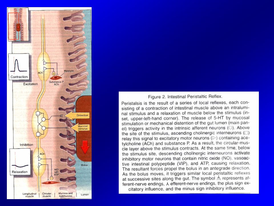
Mild distention at single site induces circular muscle contraction
5-HT Figure Mild distention at a single site induces circular muscle contraction proximal to the distention and inhibition distal to it. Excitatory neurons mostly run in a cephalad direction whereas inhibitory neurons are mainly directed in a caudad direction. This arrangement is responsible for the peristaltic reflex or "law of the intestine." Two predominant types account for approximately 80% of intrinsic motor neurons. Excitatory motor neurons contain acetylcholine (ACH) and tachykinins (neurokinin A and substance [SP]). They are responsible for the contractile phase of the peristaltic reflex. Inhibitory motor neurons contain nitric oxide synthase, vasoactive intestinal peptide (VIP), and PHM (peptide histidine methionine), which is derived from the same precursor as VIP. They are responsible for relaxation of the bowel wall distal to the distending bolus [40], [41]. The longitudinal muscle relaxes during circular muscle contraction and contracts during circular muscle relaxation. (Adapted from Grider [42].) References: [40]. Brooks SJ, Neuronal nitric oxide in the gut. J Gastroenterol Hepatol [41]. Daniel EE, Haugn C, Woskowski Z, et al. Role of nitric oxide-related inhibition in intestinal function: Relation to vasoactive intestinal peptide. Am J Physiol [42]. Grider JR, Peptidergic regulation of smooth muscle contractility. In Handbook of Experimental Pharmacology: Gastrointestinal Peptides. Edited by Brown DR. New York: Springer-Verlag;
 and tachykinins (neurokinin A and substance [SP]). They are responsible for the contractile phase of the peristaltic reflex. Inhibitory motor neurons contain nitric oxide synthase, vasoactive intestinal peptide (VIP), and PHM (peptide histidine methionine), which is derived from the same precursor as VIP. They are responsible for relaxation of the bowel wall distal to the distending bolus [40], [41]. The longitudinal muscle relaxes during circular muscle contraction and contracts during circular muscle relaxation. (Adapted from Grider [42].) References: [40]. Brooks SJ, Neuronal nitric oxide in the gut. J Gastroenterol Hepatol [41]. Daniel EE, Haugn C, Woskowski Z, et al. Role of nitric oxide-related inhibition in intestinal function: Relation to vasoactive intestinal peptide. Am J Physiol [42]. Grider JR, Peptidergic regulation of smooth muscle contractility. In Handbook of Experimental Pharmacology: Gastrointestinal Peptides. Edited by Brown DR. New York: Springer-Verlag;")
31
Intestinal contractions cause wall motion (a)
Figure Intestinal contractions cause wall motion, creating forces that increase intraluminal pressure and induce flow. Direct observation of wall motion is not feasible clinically. Highly sophisticated intraluminal sensors and modern recorders offer great accuracy, sensitivity, versatility, and portability. Two types of sensors are now common, both of which use transducers (converters of mechanical to electrical energy): perfused, open-tipped, side-hole, low-compliance catheters transmit forces to sensitive external transducers (panel A), or forces are transmitted directly to miniature transducers incorporated into small-diameter intraluminal catheters (panel B). Both of these approaches are sensitive and produce high-fidelity recordings of pressure waves within the lumen. Although the measurement of mechanical activity resulting from contractions is the more commonly performed study, the electromyogram yields additional important information about motor function. Electrical signals can be detected transmucosally using intraluminal electrodes. High-quality recordings from multiple bipolar electrodes can be made using a technique described by Coremans and associates [51]. Lead wires within the catheter are incorporated into collections of silver powder at opposite edges of holes in the catheter (panel C). The mucosa is aspirated into the hole by applying suction to the lumen of the catheter, creating an electrical contact between the intestine and the electrodes. This permits bipolar recordings and maintains a constant spatial relationships between the electrodes. References: [51]. Coremans G, Janssens H, Vantrappen G, Migrating action potential complexes in a patient with secretory diarrhea. Dig Dis Sci
: perfused, open-tipped, side-hole, low-compliance catheters transmit forces to sensitive external transducers (panel A), or forces are transmitted directly to miniature transducers incorporated into small-diameter intraluminal catheters (panel B). Both of these approaches are sensitive and produce high-fidelity recordings of pressure waves within the lumen. Although the measurement of mechanical activity resulting from contractions is the more commonly performed study, the electromyogram yields additional important information about motor function. Electrical signals can be detected transmucosally using intraluminal electrodes. High-quality recordings from multiple bipolar electrodes can be made using a technique described by Coremans and associates [51]. Lead wires within the catheter are incorporated into collections of silver powder at opposite edges of holes in the catheter (panel C). The mucosa is aspirated into the hole by applying suction to the lumen of the catheter, creating an electrical contact between the intestine and the electrodes. This permits bipolar recordings and maintains a constant spatial relationships between the electrodes. References: [51]. Coremans G, Janssens H, Vantrappen G, Migrating action potential complexes in a patient with secretory diarrhea. Dig Dis Sci")
32
Intestinal contractions cause wall motion (b)
Figure Intestinal contractions cause wall motion, creating forces that increase intraluminal pressure and induce flow. Direct observation of wall motion is not feasible clinically. Highly sophisticated intraluminal sensors and modern recorders offer great accuracy, sensitivity, versatility, and portability. Two types of sensors are now common, both of which use transducers (converters of mechanical to electrical energy): perfused, open-tipped, side-hole, low-compliance catheters transmit forces to sensitive external transducers (panel A), or forces are transmitted directly to miniature transducers incorporated into small-diameter intraluminal catheters (panel B). Both of these approaches are sensitive and produce high-fidelity recordings of pressure waves within the lumen. Although the measurement of mechanical activity resulting from contractions is the more commonly performed study, the electromyogram yields additional important information about motor function. Electrical signals can be detected transmucosally using intraluminal electrodes. High-quality recordings from multiple bipolar electrodes can be made using a technique described by Coremans and associates [51]. Lead wires within the catheter are incorporated into collections of silver powder at opposite edges of holes in the catheter (panel C). The mucosa is aspirated into the hole by applying suction to the lumen of the catheter, creating an electrical contact between the intestine and the electrodes. This permits bipolar recordings and maintains a constant spatial relationships between the electrodes. References: [51]. Coremans G, Janssens H, Vantrappen G, Migrating action potential complexes in a patient with secretory diarrhea. Dig Dis Sci
: perfused, open-tipped, side-hole, low-compliance catheters transmit forces to sensitive external transducers (panel A), or forces are transmitted directly to miniature transducers incorporated into small-diameter intraluminal catheters (panel B). Both of these approaches are sensitive and produce high-fidelity recordings of pressure waves within the lumen. Although the measurement of mechanical activity resulting from contractions is the more commonly performed study, the electromyogram yields additional important information about motor function. Electrical signals can be detected transmucosally using intraluminal electrodes. High-quality recordings from multiple bipolar electrodes can be made using a technique described by Coremans and associates [51]. Lead wires within the catheter are incorporated into collections of silver powder at opposite edges of holes in the catheter (panel C). The mucosa is aspirated into the hole by applying suction to the lumen of the catheter, creating an electrical contact between the intestine and the electrodes. This permits bipolar recordings and maintains a constant spatial relationships between the electrodes. References: [51]. Coremans G, Janssens H, Vantrappen G, Migrating action potential complexes in a patient with secretory diarrhea. Dig Dis Sci")
33
Most studied intestinal motility patterns: Migrating Motor Complex (MMC)
Figure The most studied intestinal motility patterns are the fasting migrating motor complex (equivalent to the migrating myoelectric complex, both of which are abbreviated MMC), and the fed or postprandial pattern [52]. The MMC is made up of three different phases: phase I lasts about 5 to 20 minutes and is characterized by absence of any spike bursts or contractions; phase II lasts from 10 to 40 minutes and exhibits intermittent contractions or spike bursts on about 50% of slow waves; and phase III (alternatively called the activity front) lasts from 3 to 6 minutes and consists of an intense burst of regular rhythmic contractions or spike bursts occurring at the same rate as the slow waves. The activity front begins in the stomach or the proximal jejunum and migrates along the small intestine. Only 10% of phase III activity fronts reach the ileocecal valve. When phase III begins in the stomach, it is preceded by an increase in serum motilin concentration. This complex repeats every 1 to 2 hours and recurs indefinitely until interrupted by a meal. In these recordings from normal subjects without gastrointestinal symptoms, the catheter (Gaeltec Limited, Dunvegan, Isle of Skye, Scotland) had strain gauge transducers spaced 15-cm apart with an outer diameter of 3.3 mm. Recordings were made using a 4 Mb solid-state datalogger at a sampling rate of 4 Hz (Synectics Medical, Irving, TX.) References: [52]. Szurszewski JH, A migrating electrical complex of the canine small intestine. Am J Physiol
, and the fed or postprandial pattern [52]. The MMC is made up of three different phases: phase I lasts about 5 to 20 minutes and is characterized by absence of any spike bursts or contractions; phase II lasts from 10 to 40 minutes and exhibits intermittent contractions or spike bursts on about 50% of slow waves; and phase III (alternatively called the activity front) lasts from 3 to 6 minutes and consists of an intense burst of regular rhythmic contractions or spike bursts occurring at the same rate as the slow waves. The activity front begins in the stomach or the proximal jejunum and migrates along the small intestine. Only 10% of phase III activity fronts reach the ileocecal valve. When phase III begins in the stomach, it is preceded by an increase in serum motilin concentration. This complex repeats every 1 to 2 hours and recurs indefinitely until interrupted by a meal. In these recordings from normal subjects without gastrointestinal symptoms, the catheter (Gaeltec Limited, Dunvegan, Isle of Skye, Scotland) had strain gauge transducers spaced 15-cm apart with an outer diameter of 3.3 mm. Recordings were made using a 4 Mb solid-state datalogger at a sampling rate of 4 Hz (Synectics Medical, Irving, TX.) References: [52]. Szurszewski JH, A migrating electrical complex of the canine small intestine. Am J Physiol")
34
Phase II activity varies considerably
Figure Phase II activity varies considerably. This recording is characterized by prominent "clustered" contractions. This type of activity during the fasting state has been reported to occur in the irritable bowel syndrome [53]. As can be seen in this recording, clustered activity during fasting can be very prominent in a normal subject with no abdominal pain. Thus, it is not an abnormal motor pattern in the fasting period. Clustered activity within an hour of a meal is abnormal and is characteristic of partial bowel obstruction, but also has been noted to occur after meals in pseudo-obstruction syndromes [54]. References: [53]. Kellow JE, Gill RC, Wingate DL, Prolonged ambulant recordings of small bowel motility demonstrate abnormalities in the irritable bowel syndrome. Gastroenterology [54]. Summers RW, Anuras S, Green J, Jejunal manometry patterns in health, partial intestinal obstruction, and pseudoobstruction. Gastroenterology

35
After a meal, a brisk increase in motor activity occurs
Figure After a meal, a brisk increase in motor activity occurs. The fed pattern lasts for 3 to 6 hours, depending on the size and content of the meal. During this time the fasting migrating motor complex (phases I, II, III) is not seen (ie, it is interrupted after meals). Meals high in fat and calories prolong the fed pattern. The fed pattern characterized by intermittent, irregular contractions or spike bursts that appear to occur randomly. All follow slow waves, but only about 30% to 60% of slow waves are followed by contractions. Spike bursts migrate from a few millimeters to 30 to 40 cm or more. Circumferential spike bursts are associated with lumen-occluding contractions. Spike bursts which do not occur in all quadrants around the bowel produce eccentric contractions and only partially occlude the lumen. The number of contractions at any one site is influenced by the content of the meal. Non-nutrient meals produce the greatest number of contractions, whereas meals mainly consisting of carbohydrates produce more contractions than meals which are high in protein and fat. Protein- and fat-rich meals not only produce fewer contractions, but the contractions also migrate shorter distances.
 is not seen (ie, it is interrupted after meals). Meals high in fat and calories prolong the fed pattern. The fed pattern characterized by intermittent, irregular contractions or spike bursts that appear to occur randomly. All follow slow waves, but only about 30% to 60% of slow waves are followed by contractions. Spike bursts migrate from a few millimeters to 30 to 40 cm or more. Circumferential spike bursts are associated with lumen-occluding contractions. Spike bursts which do not occur in all quadrants around the bowel produce eccentric contractions and only partially occlude the lumen. The number of contractions at any one site is influenced by the content of the meal. Non-nutrient meals produce the greatest number of contractions, whereas meals mainly consisting of carbohydrates produce more contractions than meals which are high in protein and fat. Protein- and fat-rich meals not only produce fewer contractions, but the contractions also migrate shorter distances.")
36
During sleep, fasting motor activity is often reduced
Figure During sleep, fasting motor activity is often reduced. The pattern resembles that of the awake fasting pattern, but the cycle duration is shorter. In most situations, phase II activity is markedly reduced and the duration of quiescent activity (phase I) is increased. Upon waking, a significant increase in motor activity occurs as the small bowel appears to awaken as well. It is important to recognize that a wide variety of contractile patterns occurs in normal persons.
 is increased. Upon waking, a significant increase in motor activity occurs as the small bowel appears to awaken as well. It is important to recognize that a wide variety of contractile patterns occurs in normal persons.")
37
Fin M.B.

Presentaciones similares


 * Mucosas * Secretoras * G Células ( Gastrina) Contracciones musculares del estomago.>")


