Descargar la presentación
La descarga está en progreso. Por favor, espere

1
Dr. Irving Rico Leal Servicio de Retina IVM 16/08/2010 THIS IS EALES!!
DR. BROWNING

2
HISTORIA 1880 Henry Eales, describió la enfermedad en un hombre joven el cual presentaba hemorragias vítreas recurrentes. No describió inflamación, ó neovasos. Angiopathia retinae juvenilis. Periphlebitis retinae. Primary perivasculitis of the retina. British ophthalmologist, Consultant in Birmingham and Midland Eye Hospital, Born 1852, Newton Abbot; died 1913.

3
DEFINICION Es una vasculopatía Obliterante, que afecta primariamente la retina periférica en adultos jóvenes.

4
EPIDEMIOLOGIA No hay diferencia racial.
Mayor prevalencia en India y Medio Este. Varones adultos (97.6%) > Mujeres. Murphy y Col. 55 px. = H y M. 80 – 90 % bilateral Edad predominante 20 – 35 años. Con un rango entre los 16 y 63 años. Middle East : asia, egipto, pakistan, afganistan, norte de africa. Murphy y col: in their study of 55 patients in the USA, found that men and women were equally affected
 > Mujeres. Murphy y Col. 55 px. = H y M. 80 – 90 % bilateral. Edad predominante 20 – 35 años. Con un rango entre los 16 y 63 años. Middle East : asia, egipto, pakistan, afganistan, norte de africa. Murphy y col: in their study of 55 patients in the USA, found that men and women were equally affected.")
5
MORBI/MORTALIDAD No mortalidad conocida.
AV con HV recurrentes 20/200 en 70% de los pacientes. Si la isquemia llega a Mácula, < 20/400.

6
ETIOLOGIA HLA-B5 : Behcet estomatitis aftosa (úlceras o llagas en la boca), úlceras genitales y uveítis DR1 is associated seronegative[3]-rheumatoid arthritis[ Dr4 LES AR.

7
FISIOPATOLOGIA Desconocida: se piensa que es un desorden primario de las paredes venosas de la retina (shunts), lo cual produce oclusiones, neovascularización periférica, y hemorragias vítreas. Se ha asociado a px con tuberculosis. De los shunts para ser específicos. Murphy et al.3 documented posterior vitreous detachment (PVD) in 27% of their patients with Eales disease. All these patients with PVD except one, experienced vitreous hemorrhage. Four of these patients had macular holes. The age range of the patients with PVD was from 13 to 63 years, with a mean age of 35 years. The PVD in the younger patients could have been due to a low-grade, chronic inflammation of the vitreous. The macular holes may represent a complication of posterior vitreous separation from the retina secondary to premature vitreous degeneration.Anteroposterior contraction of fibrovascular tissue adherent to both retina and posterior vitreous can cause both traction and rhegmatogenous retinal detachments, though the former are much more common Arteriolar y venular pueden estar afectados.
, lo cual produce oclusiones, neovascularización periférica, y hemorragias vítreas. Se ha asociado a px con tuberculosis. De los shunts para ser específicos. Murphy et al.3 documented posterior vitreous detachment (PVD) in 27% of their patients with Eales disease. All these patients with PVD except one, experienced vitreous hemorrhage. Four of these patients had macular holes. The age range of the patients with PVD was from 13 to 63 years, with a mean age of 35 years. The PVD in the younger patients could have been due to a low-grade, chronic inflammation of the vitreous. The macular holes may represent a complication of posterior vitreous separation from the retina secondary to premature vitreous degeneration.Anteroposterior contraction of fibrovascular tissue adherent to both retina and posterior vitreous can cause both traction and rhegmatogenous retinal detachments, though the former are much more common. Arteriolar y venular pueden estar afectados.")
8
SINTOMAS Flotadores “Telarañas” Vision Borrosa.
Disminución significativa de la visión. La visión puede ser normal, a MM y hasta PL. Disminución significativa de la visión. Secundaria a las multiples hemorragias.

9
SIGNOS CLINICOS En SA Inflamación ó Uveitis no granulomatosa en etapas tempranas. Cels y Flare, precipitados retro queráticos. SP: Vitreitis. Envainamiento vascular. Hemorragias en flama. Uveitis es muy rara en etapas tardías de la enfermedad. SP= segmento posterior: Envainamiento vascular =sheating.se envainan tanto los vass que pueden llegar a ocluirse. Hemorragias n flama se encuentran alrededor de los vasos envainados. A mild vitreous haze overlying the areas of vasculitis is more common

10
SIGNOS CLINICOS Edema macular cistoideo No perfusión periférica.
Vasos tortuosos con AMIRES Microanuerismas. Shunts arterio-venosos. Moldeado (beading) de los vasos. Cystoid macular edema can occur in patients with Eales disease due to increased capillary permeability, and inflamation. This can often be associated with significant vision loss. Beading: rectificar al parecer eso significa. Rubeosis que te puede llevar a GNV. Eales disease. Fundus photo of the peripheral retina, revealing vascular tortuosity and peripheral retinal neovascularization.
 de los vasos. Cystoid macular edema can occur in patients with Eales disease due to increased capillary permeability, and inflamation. This can often be associated with significant vision loss. Beading: rectificar al parecer eso significa. Rubeosis que te puede llevar a GNV. Eales disease. Fundus photo of the peripheral retina, revealing vascular tortuosity and peripheral retinal neovascularization.")
11
SIGNOS CLINICOS OVCR Exudadosduros y blandos.
Neovasos, en periferia 80%, en disco y rubeosis. Proliferación Fibro vascular → DR. Patients with Eales disease can also develop BVO, which can be either solitary or multiple. These patients can be differentiated from patients with primary BVO because the pathologic changes in BVO are usually confined to the affected quadrant of the retina. In contrast, Eales disease affects more extensive areas of the peripheral retina and does not respect either the anatomic distribution of venules in the retina or the horizontal midline. These two conditions can occur together, and Eales disease may predispose to the development of BVO The NVE is usually located at the junction between perfused and nonperfused retina

12
SIGNOS CLINICOS Figure 82-5 Hypovascular fibroproliferation emanating from the disc.

13
DIAGNOSTICO Por exclusión FAG: US: Los vasos envainados pueden fugar.
EMQ se observa en FAG US: Vitreosquisiss Tracción vitreo retinal DVP DR The cause of Eales disease is unknown. Eales disease is a diagnosis of exclusion and is thought to be idiopathic. No causative drugs, environmental factors, or infectious agents for Eales disease have been identified. Although a hypersensitivity to tuberculin protein has been reported, no clear relationship to tuberculosis has been found. An 88-kd acute phase reactant protein has been found in patients with Eales disease that is immunologically identical to that found in patients with posterior uveitis, tuberculosis, leprosy, and rheumatoid arthritis. The role of this protein is yet undetermined. Figure 82-2 Peripheral nonperfusion. Microaneurysms, venous beading, and arteriovenous shunting can be noted at the margin of perfused and nonperfused retina.

14
DIAGNOSTICO Figure 82-3 An example of severe nonperfusion which began peripherally and extended posteriorly to involve the macula and peripapillary retina.

15
DIAGNOSTICOS DIFERECNIALES
Enf inflamatorias. Enf que producen neovascularización. OVCR. RD. Sickle retinopathy. Sarcoidosis. LES. Enf vasculares del colágeno.

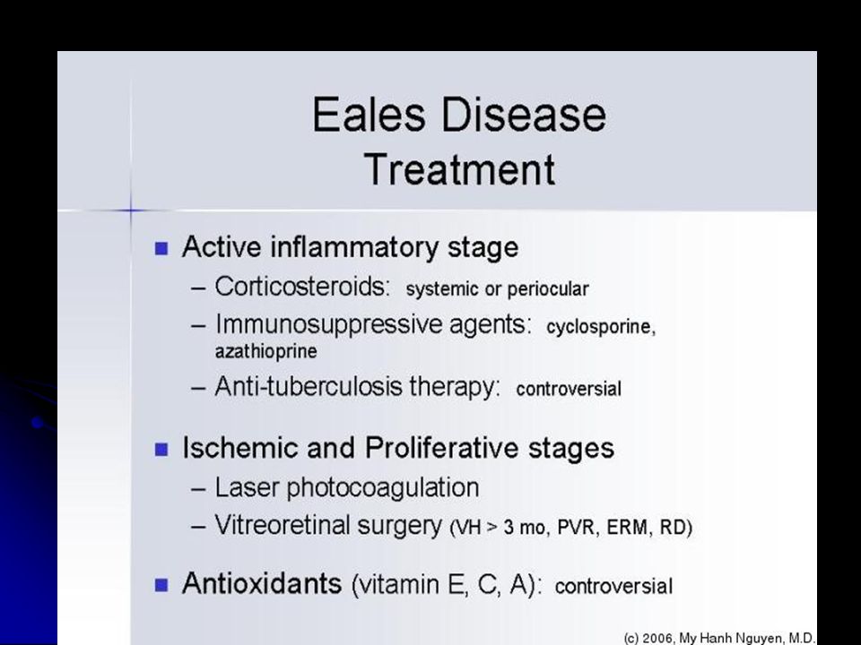
17
TRATAMIENTO Figure 82.6 A, Neovascularization of the disc. Note the segmental exudative arteriolar sheathing

18
TRATAMIETO B, Approximately 2½ years later the patient has had total regression of the neovascularization after treatment with full-scatter photocoagulation. Note that the former area of the arteriolar sheathing has resorbed.

19
GRACIAS…

Presentaciones similares