Descargar la presentación
La descarga está en progreso. Por favor, espere

1
MUJER DE 26 AÑOS CON UNA MASA OVÁRICA Dra. PI González Márquez Dra. I González-Rodilla CI REUNIÓN DE LA SEAP NOROESTE Asturias, Cantabria, Castilla y León, Galicia, Norte y Centro de Portugal.

2
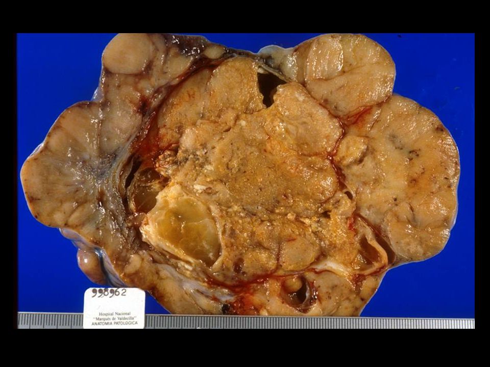
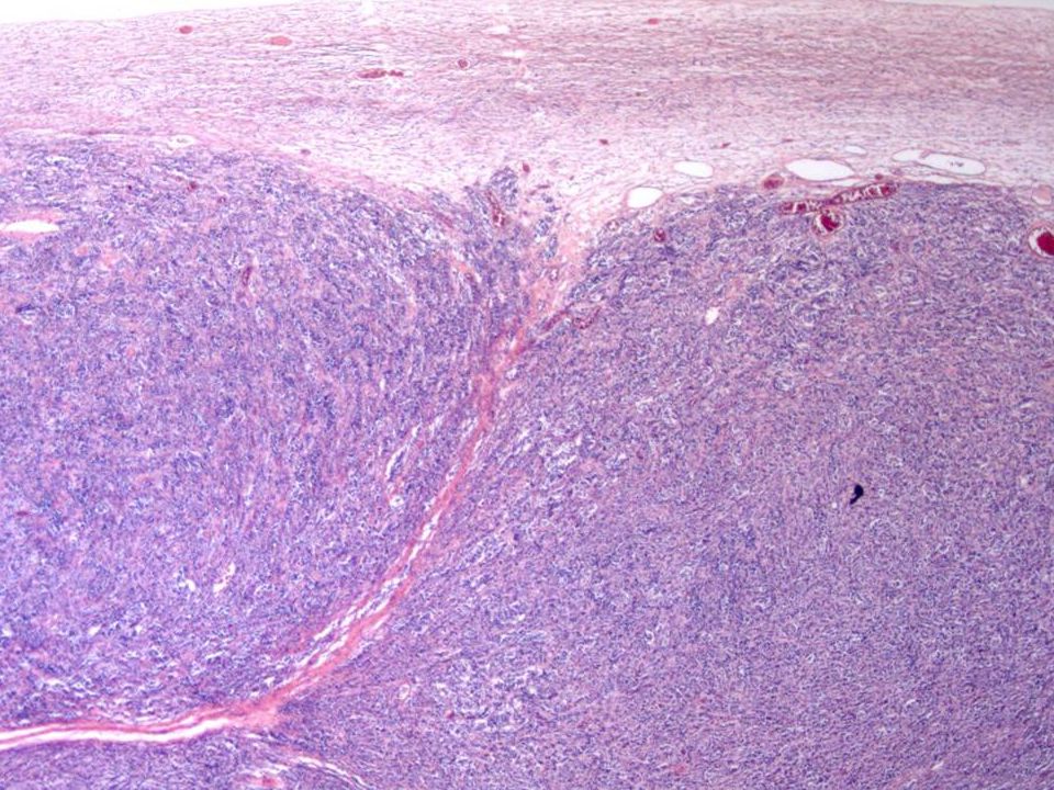
HISTORIA CLÍNICA Mujer de 26 años de edad Distensión abdominal Dificultad para la micción Masa palpable en fosa ilíaca derecha Anexectomía derecha y cuña ovárica izquierda.

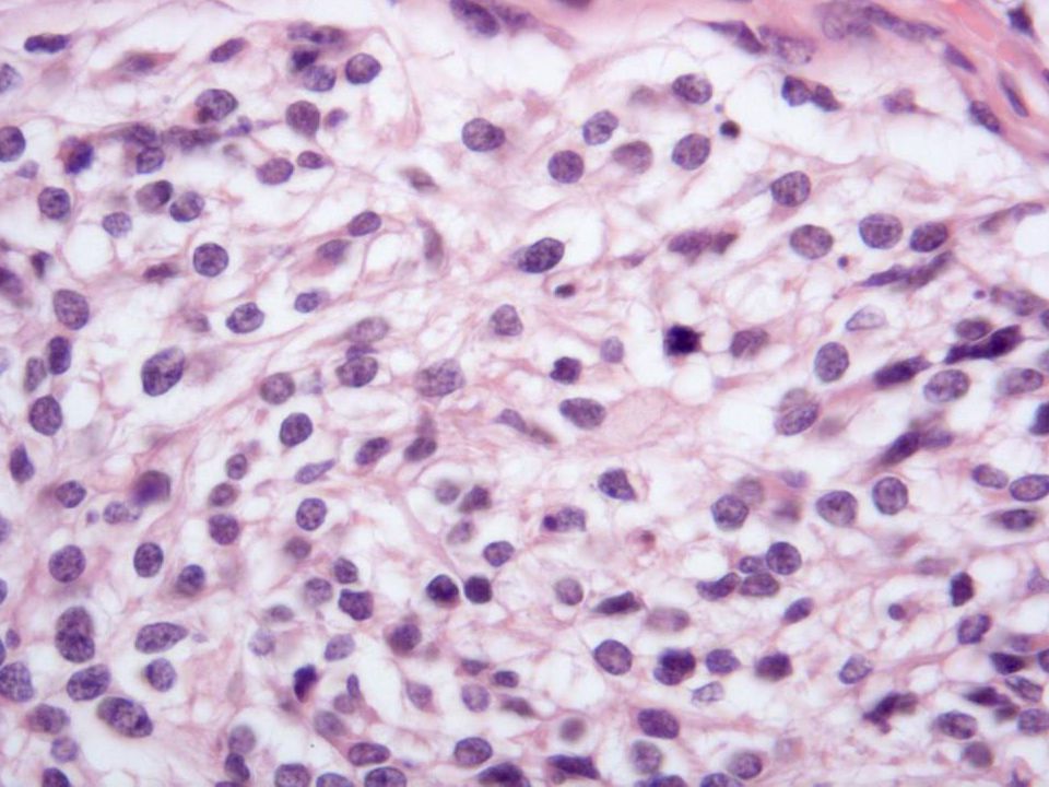
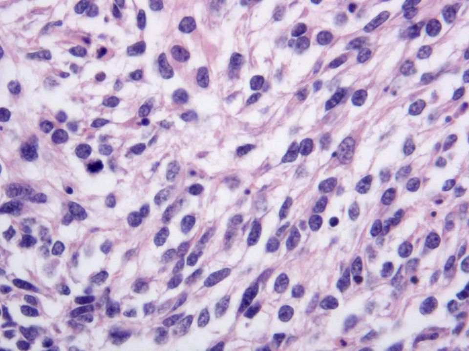
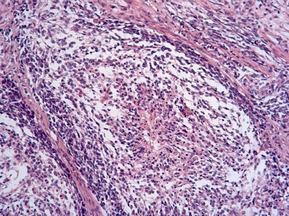
24
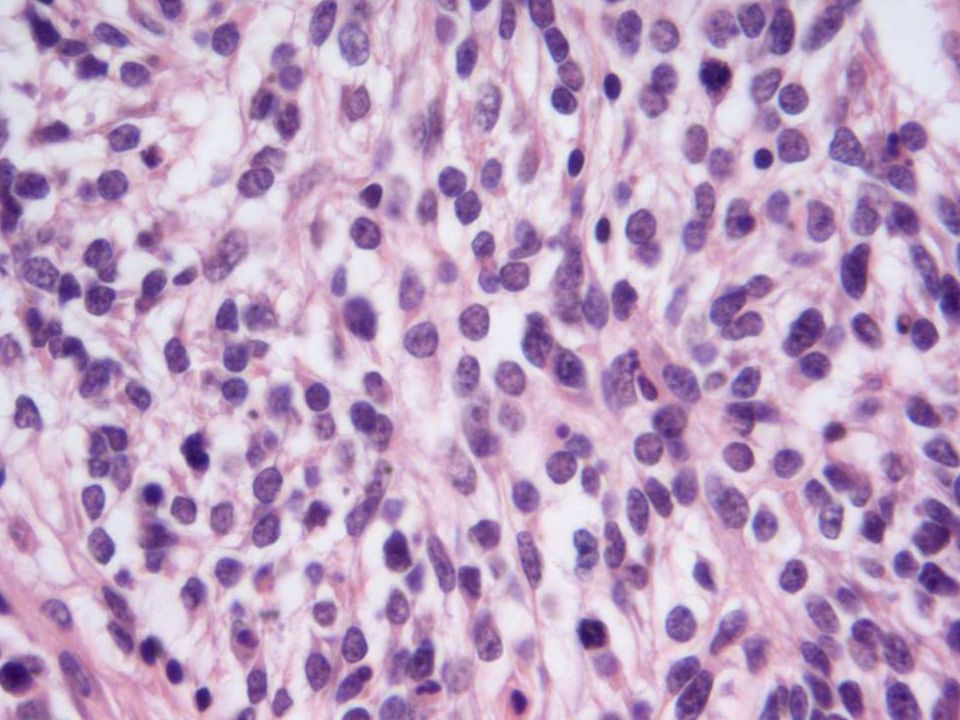
DIAGNÓSTICO DIFERENCIAL Tumor intraabominal desmoplásico de células pequeñas redondas y azules Carcinoma de células pequeñas –Hipercalcemia –Neuroendocrino (tipo pulmonar) Sarcoma del estroma endometrial Rabdomiosarcoma embrionario Linfoma indiferenciado Melanoma
 Sarcoma del estroma endometrial Rabdomiosarcoma embrionario Linfoma indiferenciado Melanoma")
25
DIAGNÓSTICO DIFERENCIAL TUMOR NEUROECTODÉRMICO PRIMITIVO DEL OVARIO TERATOMA INMADURO

26
NSE

27
GFAP

28
S-100

29
SINAPTOFISINA

30
CD99

31
VIMENTINA

32
DIAGNÓSTICO ANEJO UTERINO DERECHO CON: –TUMOR NEUROECTODERMICO PRIMITIVO del ovario. –Trompa de Falopio sin particularidades. Cuña del ovario izquierdo sin lesiones.

33
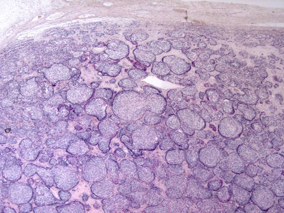
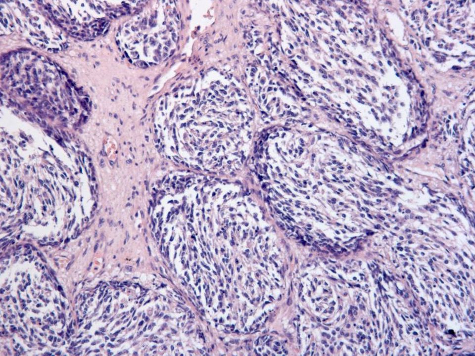
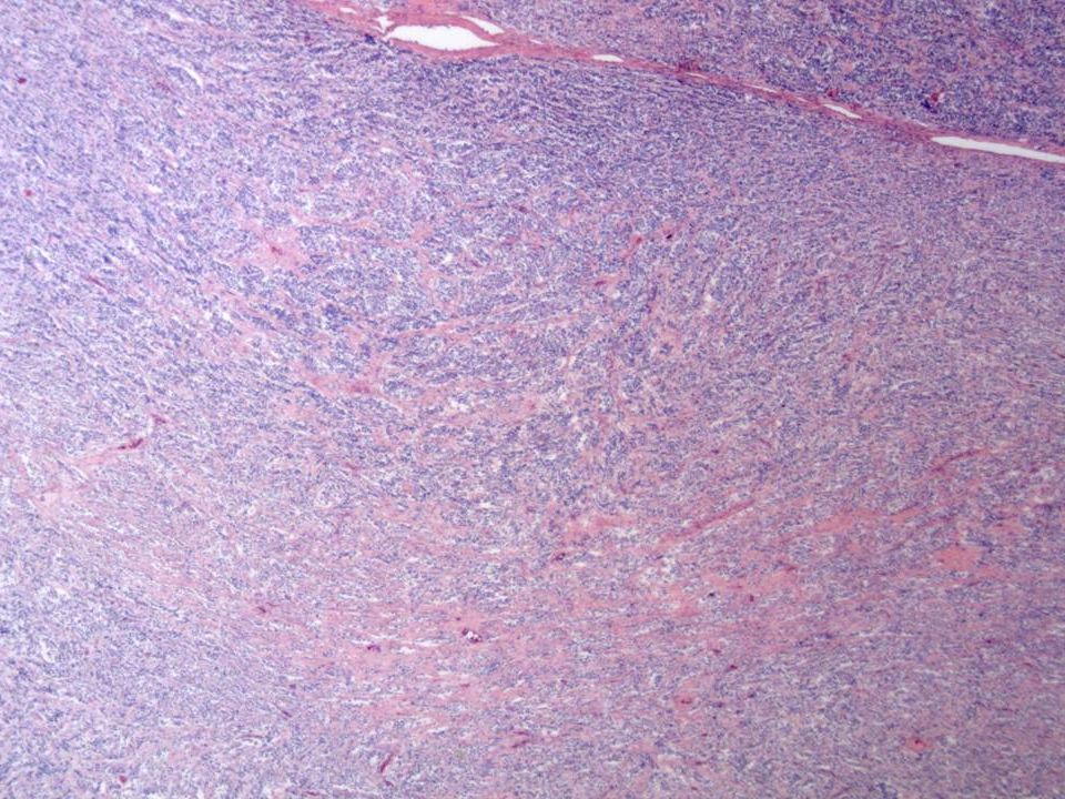
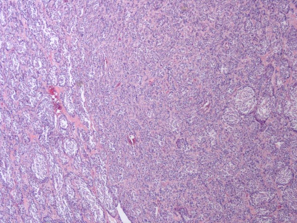
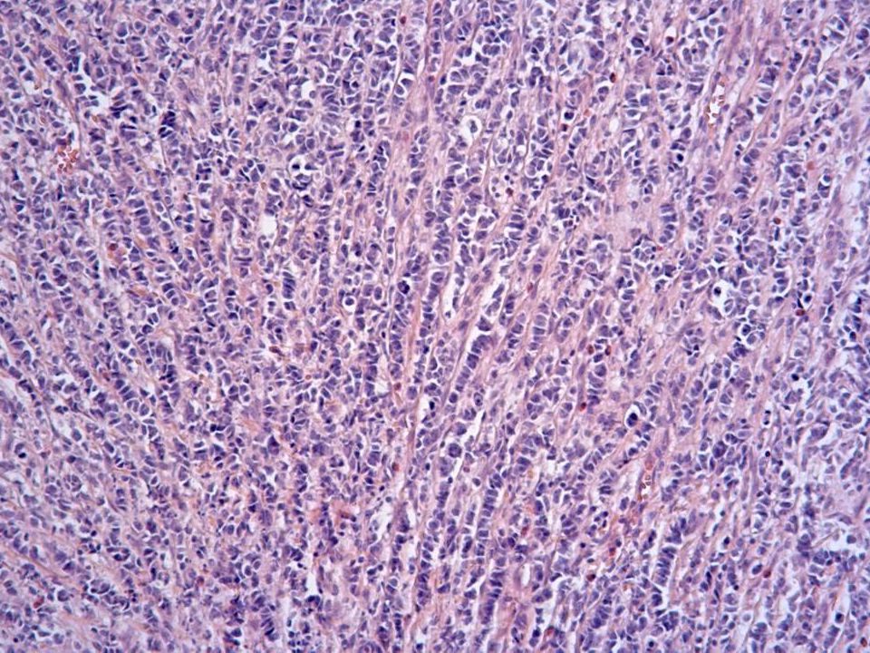
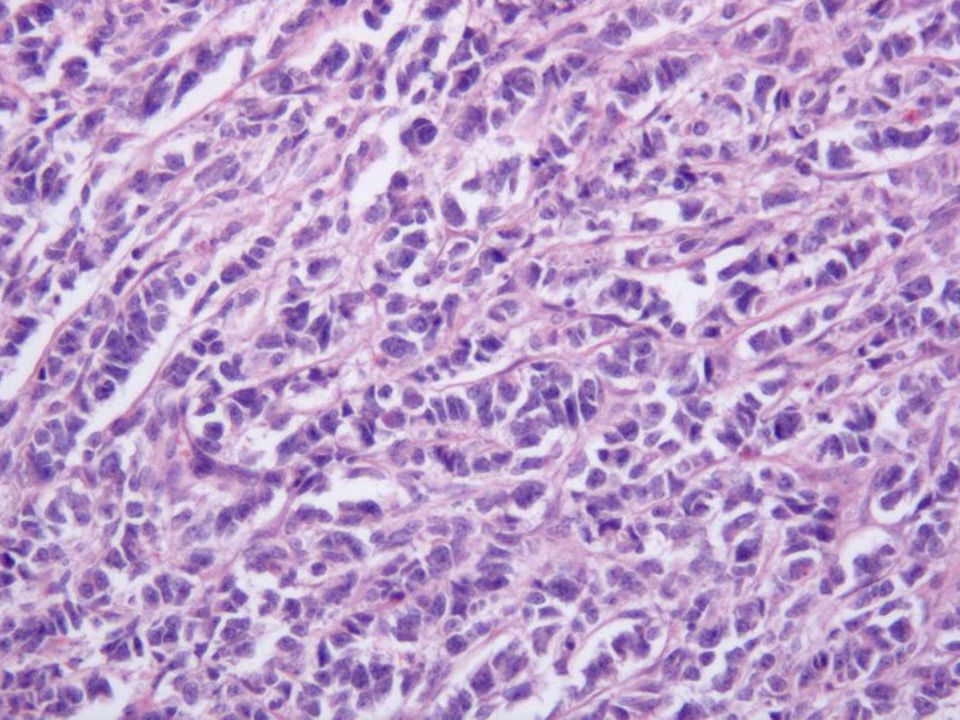
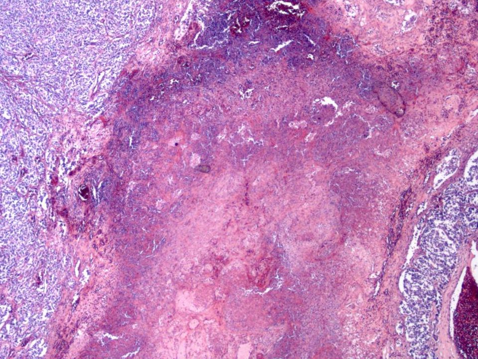
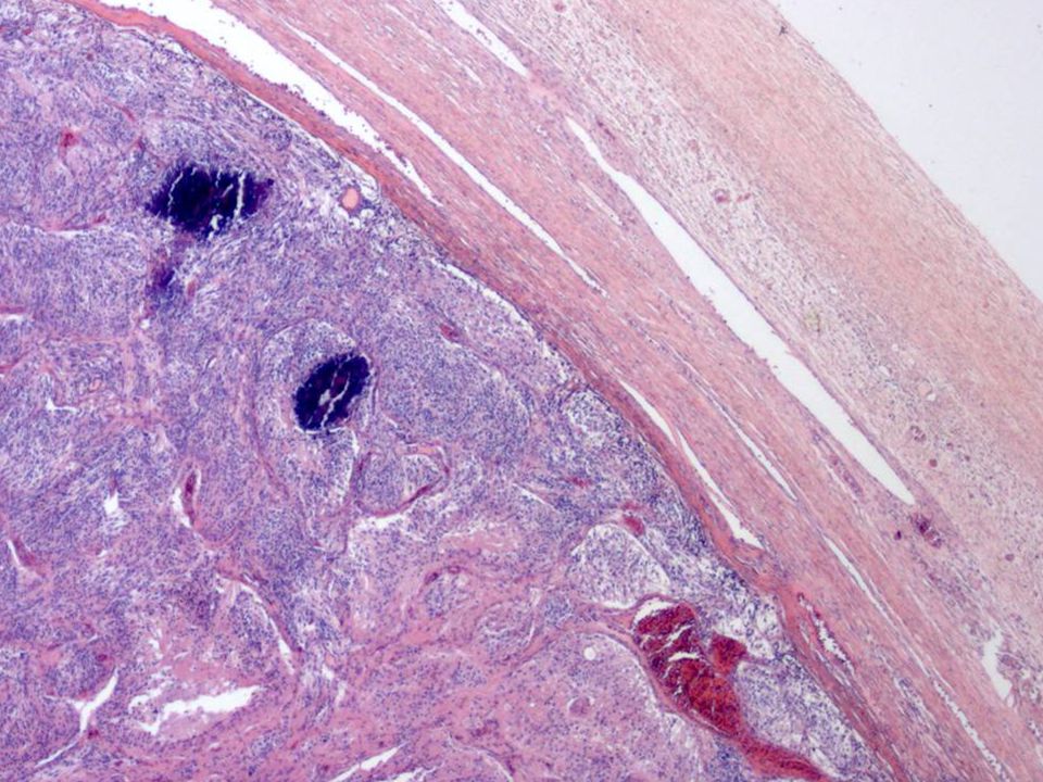
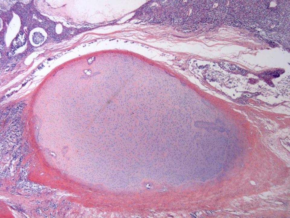
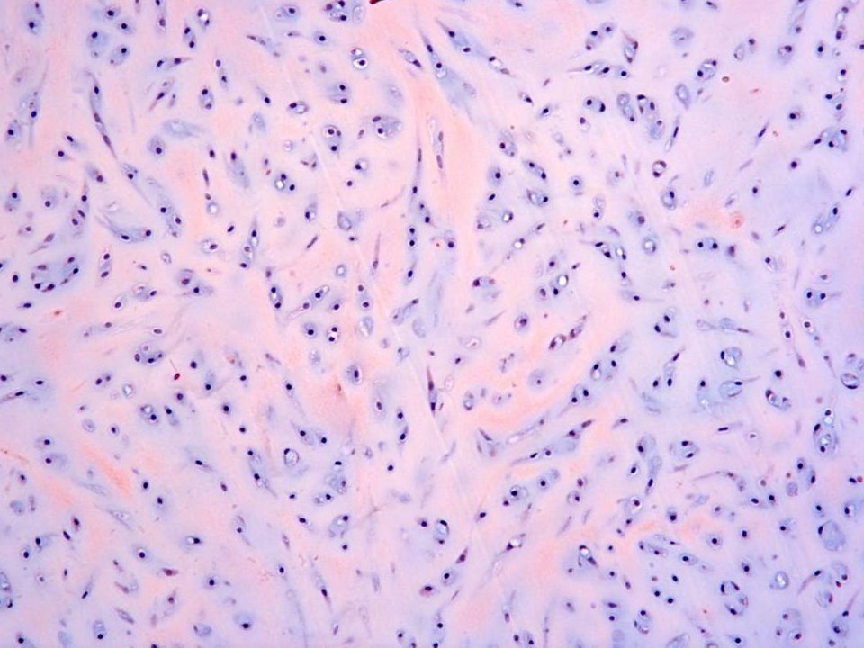
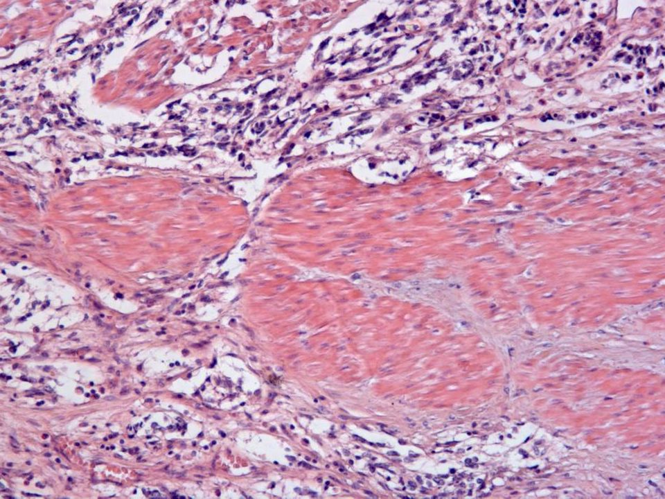
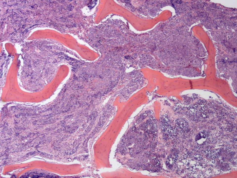
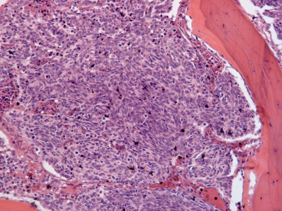
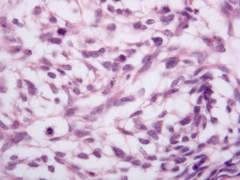
Neoplasias constituidas por células pequeñas Remedan arquitectural y citológicamente neoplasias del S.N.C. Subtipos histológicos: 1.Bien diferenciadas 2.Primitivos 3.Anaplásicos TUMOR NEUROECTODÉRMICO PRIMARIO de OVARIO EPENDIMOMANEUROBLASTOMA MEDULOEPITELIOMA MEDULOBLASTOMA GLIOBLASTOMA MUTIFORME

34
Pueden contener elementos teratomatosos Rosetas o pseudorosetas PNET se considera la forma mejor diferenciada de los tumores pertenecientes a la familia PNET/Sarcoma de Ewing. Traslocación (11;22) Hueso y partes blandas Muy raro en el ovario TUMOR NEUROECTODÉRMICO PRIMARIO de OVARIO
 Hueso y partes blandas Muy raro en el ovario TUMOR NEUROECTODÉRMICO PRIMARIO de OVARIO.")
35
Clínica: –Dolor abdominal o masa palpable Diagnóstico: –Simula neoplasias del SNC –GFAP, NSE, Sinaptofisina –CD 99 (MIC2) Tratamiento multidisciplinar Pronóstico infausto. TUMOR NEUROECTODÉRMICO PRIMARIO de OVARIO

39
BIBLIOGRAFÍA 1.Kleinman GM, Young RH, Scully RE. Primary neuroectodermal tumors of the ovary. A report of 25 cases. Am J Surg Pathol. 1993 Aug;17(8):764-78 2.Kim KJ, Jang BW, Lee SK, Kim BK, Nam SL. A case of peripheral primitive neuroectodermal tumor of the ovary. Int J Gynecol. Cancer 2004 Mar-Apr;14(2):370-2 3.Kawauchi S, Fuduka T, Miyamoto S, Yoshioka J, Shirahama S, Saito T, Tsukamoto N. Peripheral primitive neuroectodermal tumor of the ovary confirmed by CD99 immunostaining, Karyotypic analysis, and RT-PCR for EWS/FLI-1 chimeric mRNA. Am J Surg Pathol. 1998 Nov;22(11):1417-22 4.Aguirre P, Scully RE. Malignant neuroectodermal tumor of the ovary, a distinctive form of monodermal teratoma: report of five cases. Am J Surg Pathol. 1982 Jun;6(4):283-92 5.McCluggage WG. Ovarian neoplasms composed of small round cells: a review. Adv Anat Pathol. 2004 Nov;11(6):288-96
: Kim KJ, Jang BW, Lee SK, Kim BK, Nam SL. A case of peripheral primitive neuroectodermal tumor of the ovary. Int J Gynecol. Cancer 2004 Mar-Apr;14(2): Kawauchi S, Fuduka T, Miyamoto S, Yoshioka J, Shirahama S, Saito T, Tsukamoto N. Peripheral primitive neuroectodermal tumor of the ovary confirmed by CD99 immunostaining, Karyotypic analysis, and RT-PCR for EWS/FLI-1 chimeric mRNA. Am J Surg Pathol Nov;22(11): Aguirre P, Scully RE. Malignant neuroectodermal tumor of the ovary, a distinctive form of monodermal teratoma: report of five cases. Am J Surg Pathol Jun;6(4): McCluggage WG. Ovarian neoplasms composed of small round cells: a review. Adv Anat Pathol Nov;11(6):")
Presentaciones similares