Descargar la presentación
La descarga está en progreso. Por favor, espere

1
TRAUMA DE COLUMNA CERVICAL EN NIÑOS
ALVARO TORO POSADA FUSVP U de A

2
HISTORIA CLÍNICA TRAUMA CERVICAL INFANTIL
Torticolis Limitada movilidad Espasmo nucal Reflejos anormales Clonus Rigidez Hipotensiòn <frecuencia cardíaca Respiración diafragmática Priapismo Alteración temperatura Clinical Clearing the Cervical Spine NEXUS Low-Risk CriteriaNo midline cervical tenderness No evidence of intoxication No altered level of alertness No focal neurological deficit No distracting painful injury No CSI was identified in the pediatric group without at least one NEXUS risk factor About 20% less radiographs would have been performed However there were few pediatric patients with CSI. Lower end of CI: 87.8 Only 4 injured patients were younger than 9 yrs. NEXUS criteria can be used with caution in pediatric patients ≥ 8 years.

3
JUSTIFICACION Porque diferente del adulto? Como lo evalúo?
RX, TAC o RM y cuando? Manejo inicial Excepciones

4
TRAMPAS EN EL TRAUMA CERVICAL INFANTIL Haizlip JA; Scherrer PD
“no creo que necesite un collar cervical, está caminando por donde se accidentó” “hay que transportarlo en una camilla rígida como un adulto, con correas, está seguro si está quieto“ “ tiene 5 años, como no le duele el cuello, pienso que no necesita inmovilización con collar”

5
TRAMPAS DEL TRAUMA CERVICAL
“estoy casi seguro que la línea que veo en los RX es una placa de crecimiento, pues es pequeña y las fracturas a esta edad son muy raras” “el técnico de RX no podrá tomar en este niño unas placas pues no se queda quieto, menos una boca abierta para odontoides” “el niño enviado para placas en flexión y extensión tiene dolor, las hago?”

6
TRAMPAS TRAUMA CERVICAL
“para mayor seguridad todo niño con trauma cervical debo realizar una TAC” “ todas las radiografías son normales y ella parece estar bien, puedo decirle a los padres que no se preocupen de nada” “como el niño que está inconciente tiene RX y TAC cervical normal, puedo dejarlo sin collar”

7
Trauma de columna cervical Bollini
35-45% de Tx de columna 0.34% de traumas infantiles Cervicalgia Torticolis fija Dolor Deficit neurológico HC por el paciente o acompañantes Mecanismo Signos y síntomas Nx Intoxicación o medicamentos Antecedentes de trauma, enfermedades o síndromes medular anterior, central, Brown Sequard

8
LESIONES DE LA COLUMNA CEVICAL EN NIÑOS EPIDEMIOLOGÍA
Kokoska E. Patel J. Martin B. Fuente NPTR NPTR TARN Edad 0 - 20 0 - 15 No niños 24,740 75,172 19,538 Lesiones C C 408 (1.6) 1098 (1.5) 662 (3.4) % hombres 59 61 58 % lesión medular 35 29 21.9 % mortalidad 17 13 We all know that cervical spine injuries can produce severe and permanent neurological deficit. Fortunately CSI in children are uncommon. Three large studies, 2 in USA using data from the National Pediatric Trauma Registry and one in the United Kingdom with data from the Trauma Audit and Research Network
 1098 (1.5) 662 (3.4) % hombres % lesión medular % mortalidad We all know that cervical spine injuries can produce severe and permanent neurological deficit. Fortunately CSI in children are uncommon. Three large studies, 2 in USA using data from the National Pediatric Trauma Registry and one in the United Kingdom with data from the Trauma Audit and Research Network.")
9
TIPOS DE LESIÓN CERVICAL NIÑOS
lesiones Kokoska E. % Patel J. Fractura 56 luxación 25 22 Fracture/luxación - 5 SCIWORA Spinal cord injury without radiological abnormality 19 17 The most common types of CSI injuries in children are … but spinal cord injuries without radiological abnormalities account for almost 1/5 of CSI

10
LESIONES DE ACUERDO CON LA EDAD KOKOSKA
Tipo 0 - 10 p Fracturas (%) 42 65 < .01 Luxaciones (%) 31 20 SCIWORA (%) 27 15 The anatomical differences also account for a higher proportion of dislocations and sciwora in younger children
 < .01. Luxaciones (%) SCIWORA (%) The anatomical differences also account for a higher proportion of dislocations and sciwora in younger children.")
11
RX AP, lat, boca abierta, flexiòn extensiòn ?
Trauma cervical 65% H 35 %F RX AP, lat, boca abierta, flexiòn extensiòn ? TAC si hay imagen de fractura RM Acc tto 48% Deportes 35% futbol, clavados, fútbol americano, lucha libre Caídas 15% 52% cervical superior 40% TEC y 60% C0C1C2 30% Cx cervical spine injuries are rare, constituting between 0.65% and 9.47% of all cervical spine injuries. Between 1986 and 1997 we treated 102 pediatric patients (65% boys, 35% girls birth to 9 years of age (38 patients; Group 1) and 10 to 16 years of age (64 patients; Group 2). radiographic studies, including plain cervical radiographs (anteroposterior, lateral, swimmer’s, and openmouth odontoid views), were obtained in all patients. Flexion– extension radiographs were also obtained to assess spinal stability when plain radiography and computerized tomography studies demonstrated normal results and in patients with SCI without radiographic evidence of abnormality. Fine-slice computerized tomography scans were obtained if the presence of a fracture was uncertain. Magnetic resonance imaging was performed in 88 patients Clinical summary of management and outcome in 102 pediatric patients with cervical injuries* Surgical Outcome Nonsurgical Age Anterior Odontoid Complete Partial Partially No Range OC Posterior Disectomy/ Corp/ Screw Halo Collar/ Recov- Recov- Nonfunc- Recov- (yrs) Fusion Fusion Fusion Fusion Fixation Vest Other ery ery tional ery Death birth–9 5 2† 10–16 2 3‡ * OC = occipitocervical; corp = corpectomy; other = custom-molded brace. † These two patients underwent wiring fixation and fusion procedures. ‡ One of these patients underwent fusion with lateral mass plating, one with posterior wiring, and one with placement of a posterior cervical spine injuries, the mortality rate was 10%, whereas it was 20% in patients with upper cervical Pediatric cervical spine injuries: report of 102 cases and review of the literature MOHAMMED A. ELERAKY, M.D., NICHOLAS THEODORE, Neurosurg (Spine 1) 92:12–17, 2000
 and 10 to 16 years of age (64 patients; Group 2). radiographic studies, including plain cervical. radiographs (anteroposterior, lateral, swimmer’s, and openmouth. odontoid views), were obtained in all patients. Flexion– extension radiographs were also obtained to assess. spinal stability when plain radiography and computerized. tomography studies demonstrated normal results and in patients. with SCI without radiographic evidence of abnormality. Fine-slice computerized tomography scans were. obtained if the presence of a fracture was uncertain. Magnetic. resonance imaging was performed in 88 patients. Clinical summary of management and outcome in 102 pediatric patients with cervical injuries* Surgical Outcome. Nonsurgical. Age Anterior Odontoid Complete Partial Partially No. Range OC Posterior Disectomy/ Corp/ Screw Halo Collar/ Recov- Recov- Nonfunc- Recov- (yrs) Fusion Fusion Fusion Fusion Fixation Vest Other ery ery tional ery Death. birth–9 5 2† –16 2 3‡ * OC = occipitocervical; corp = corpectomy; other = custom-molded brace. † These two patients underwent wiring fixation and fusion procedures. ‡ One of these patients underwent fusion with lateral mass plating, one with posterior wiring, and one with placement of a posterior. cervical spine injuries, the mortality rate was 10%, whereas. it was 20% in patients with upper cervical. Pediatric cervical spine injuries: report of 102 cases and review of the literature MOHAMMED A. ELERAKY, M.D., NICHOLAS THEODORE, Neurosurg (Spine 1) 92:12–17,")
12
no familiarizado con patología, anatomía, variantes de lo normal
Errores DX <8a 24% ≥9 a 15%. > unión C0C2 Predisponen: no familiarizado con patología, anatomía, variantes de lo normal Error Patients (N=37) n % Misdiagnosed on initial imaging evaluation 7 19 True ″missed″ diagnosis 2 5.4 Called A-O dislocation, but was type I hangman’s fracture Missed segmental fracture of left C1 anterior tubercle ″Normal and/or developmental variants″ read as fractures or dislocation 5 13.5 Occiptal condyle fracture Jefferson fracture C4 vertebral body fracture C7 spinous process fracture A-O dislocation 123 Table 3 Error rates by cervical level Number at injury level (%)a Number of missed (%) Totals (%) Occiput–C /18 (28) C3–C /21 (10) aThere were 39 injury levels as two patients had injuries at two different cervical spine levels 124 The misdiagnosis of acute cervical spine injuries and fractures in infants and children: the 12-year experience of a level I pediatric and adult trauma center, Anthony M., Childs Nerv Syst (2005) 21: 122–127 Niño 2ª Sciwora
 n % Misdiagnosed on initial imaging evaluation True ″missed″ diagnosis Called A-O dislocation, but was type I. hangman’s fracture. Missed segmental fracture of left C1. anterior tubercle. ″Normal and/or developmental variants″ read. as fractures or dislocation Occiptal condyle fracture. Jefferson fracture. C4 vertebral body fracture. C7 spinous process fracture. A-O dislocation Table 3 Error rates by cervical level. Number at injury. level (%)a. Number of. missed (%) Totals (%) Occiput–C /18 (28) C3–C /21 (10) aThere were 39 injury levels as two patients had injuries at two. different cervical spine levels The misdiagnosis of acute cervical spine injuries and fractures in infants and children: the 12-year experience of a level I pediatric and adult trauma center, Anthony M., Childs Nerv Syst (2005) 21: 122–127. Niño 2ª Sciwora.")
13
El fulcro está C5C6 en adolescentes, C2C3 en niños menores
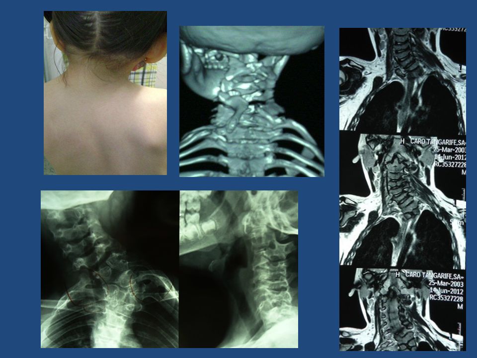
Predisponentes : anomalias , ciertas patologìas Down, Klippel Feil, Chiari, neoplasias, infecciones, ttno metabòlico El fulcro está C5C6 en adolescentes, C2C3 en niños menores La incidencia de SCIWORA en niños va de 4% a 67% Cervical Spine Injuries in Children: A Review of 103 Patients Treated Consecutively at a Level 1 Pediatric Trauma Center, Rebeccah L. Brown of Pediatric Surgery, Vol 36, No 8 (August), 2001:
, 2001:")
14
A Prospective Multicenter Study of Cervical Spine Injury in Children Peter Viccellio, MD*; Harold Simon, MD‡; Barry D. Pressman, MD§; Manish N. Shah, for the NEXUS Group PEDIATRICS Vol. 108 No. 2 August 2001 Diferencias anatòmicas entre niños y adultos hasta los 8a y un poco hasta los 12a– Nexus estudio prospectivo observacional >% niños grandes caracterìsticas como adultos SCIWORA es muy raro en niños centromedular, Brown Sequard, sìndrome medular posterior Maltrato infantil

15
POSNA 2012 Orientaciòn de las facetas màs horizontal
Fusiòn de los centros de osificaciòn de la odontoides entre 3-6ª A menor edad mayor movilidad de la columna que la mèdula Hipermovilidad y > tamaño de la cabeza, > vulnerabilidad cervical superior. TEC es la lesiòn más frecuente asociada en niños pequeños hasta 50% RX en flexiòn extensiòn cervical si hay hallazgos sospechosos Aufdermaur lesiones placa de crecimiento y SCIWORA y estudios recientes de RM muestran además lesiones ligamentarias As expected age plays a role in the mechanism of injury. MVA account for > 40% of injuries in young and older children, falls and pedestrian accidents are more common in younger children while bicycle and sports related accidents are more common in older children. Radiography of Cervical SpineInjury in Children: Are Flexion–Extension Radiographs Useful forAcute Trauma? Jerry Raphael Dwek Christine B. Chung AJR:174, June 2000

16
DIFERENCIAS NIÑOS ADULTOS
Here you can see wedge shaped vertebral bodies and more horizontal articulating facets as compare with the adult.

17
RX DE COLUMNA CERVICAL

18
COLUMNA CERVICAL EN NIÑOS
Reconocer centros de osificación y las fisis La sedoluxación C2/3 común en <8a, Siga la línea espinolaminar Swischuk Evaluación tejidos blandos anterior

19
INDICACIONES DE TAC RM Estado mental alterado
TAC 98% sensibilidad y especificidad (irradia x 10) IRM SCIWORA RM edema, lesiones ligamentarias C0C2, subluxación, distracción, contusión medular, hemorragia, protrusión discal lesiones ligamento longitudinal, fracturas ocultas
 IRM SCIWORA. RM edema, lesiones ligamentarias C0C2, subluxación, distracción, contusión medular, hemorragia, protrusión discal lesiones ligamento longitudinal, fracturas ocultas.")
20
Se solicitarà una RM cuando se tenga un niño con trauma cervical, o fractura de esta zona, cuando haya disociación entre los hallazgos clìnicos y RX MRI in the assessment of the supportive soft tissues of the cervical spine in acute trauma in children M. D.Keiper R. A.Zimmerman Neuroradiology (1998) 40: 359±363 A retrospective analysis of 52 young patients Although it is clear from this study that MRI is useful in patients requiring immediate surgical stabilization, the benefit of MRI in those patients with relatively minor, radiographically stable injury is less apparent. Although apparent minor injury may be identified on MRI, treatment of such injury is not determined. Fig.1a±c A 4-year-old child with severe head trauma and high suspicion of cervical softtissue injury. a Lateral plain radiograph demonstrates atlanto- occipital and C1±2 distraction but no osseous injury. b, c Sagittal spin-echo 500/12 and fast spin-echo 3500/90 images the same day reveal extensive soft tissue edema, ligamentous disruption, and osseous distraction. MRI demonstrates definitive soft tissue injury at the C3 level, indicating the extent of posterior fusion required for stabilization. Note also the unsuspected spinal cord contusion at the C1 level in c (arrow) 4a, TEC, trauma cervical C0C1C2 distracción sin lesión ósea. RM Sciwora
 40: 359±363. A retrospective analysis of 52 young patients. Although it is clear from this study that MRI is useful. in patients requiring immediate surgical stabilization, the benefit of MRI in those patients with relatively. minor, radiographically stable injury is less apparent. Although apparent minor injury may be identified on. MRI, treatment of such injury is not determined. Fig.1a±c A 4-year-old child. with severe head trauma and. high suspicion of cervical softtissue. injury. a Lateral plain. radiograph demonstrates atlanto- occipital and C1±2 distraction. but no osseous injury. b, c Sagittal spin-echo 500/12. and fast spin-echo 3500/90 images. the same day reveal extensive. soft tissue edema, ligamentous. disruption, and osseous. distraction. MRI demonstrates. definitive soft tissue injury. at the C3 level, indicating. the extent of posterior fusion. required for stabilization. Note. also the unsuspected spinal. cord contusion at the C1 level. in c (arrow) 4a, TEC, trauma cervical C0C1C2 distracción sin lesión ósea. RM Sciwora.")
21
MANEJO DEL TRAUMA CERVICAL
ABC Proteger la columna cervical, todo niño con trauma cervical o de cràneo, politraumatizado o con lesión neurológica, debe ser tratado como si tuviera un trauma de columna cervical Objetivos: Estabilizar las lesiones primarias y prevenir las lesiones secundarias entre 3% - 25% de lesiones se presentan durante el transporte o manejo inicial El tratamiento definitivo no es un objetivo inicial

22
TRATAMIENTO DEL TRAUMA CERVICAL
Estabilizar la lesión primaria y prevenir las secundarias No hay una guía universal Evaluación con neurocirugía Reducción cerrada y halo Cirugía en algunos casos, ligamentos Esteroides no hay guías en los niños

23
INMOVILIZACIÓN DE LA COLUMNA CERVICAL PARA EL TRANSPORTE DE NIÑOS
La cabeza con mayor tamaño incrementa la flexión en una tabla común Levantar los hombros o plano deprimido en la cabeza columna en neutro

24
INMOVILIZACIÓN DE LA COLUMNA CERVICAL
Collar duro Evitar collar grande pues hiperextiende Receso en la mesa de transporte para la cabeza, o realce de los hombros Correas en la frente, el mentón, hombros caderas, muslos y tobillos Pendiente si se presentan vómitos

29
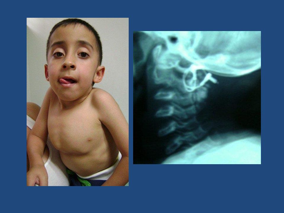
Fractura tubérculo anterior C1
Hagman

30
Radiographic studies obtained in a 2-year-old child
Radiographic studies obtained in a 2-year-old child. Left: Preoperative radiograph of the patient in a halo brace, demonstrating atlantooccipital dislocation. Center and Right: Postoperative flexion (center) and extension (right) radiographs obtained 6 months after occipitocervical fusion in which a Steinmann pin and bone graft were used, demonstrating an absence of pathological motion and an increase
 and extension (right) radiographs obtained 6 months after. occipitocervical fusion in which a Steinmann pin and bone graft were used, demonstrating an absence of pathological motion and an increase.")
31
Odontoid Process (Dens) Fracture
Fracture through base of dens. Dens and C1 posterior to C2

32
Head and Cervical Spine Differences Children vs. Adults
Characteristics Children Adults Head/Body Large Small Fulcrum C2 – C3 C5 – C6 Neck muscles, spine ligaments Weak, lax and elastic Strong, stiff, ↓ elasticity Vertebral bodies Anterior wedging Cartilaginous No wedging ossified Articulating facets More horizontally oriented Vertically oriented There are anatomical differences that influence the type of injuries. Children have

33
The ABCS of Radiographic Cervical Spine Evaluation
A. Alignment: Lordotic curves, malalignment, subluxation, distraction. B. Bones: Fractures, anterior and posterior cervical columns, ossification centers C. Cartilage: Intervertebral disk spaces, ossification centers S. Soft Tissues: Prevertebral, predental spaces.

34
Os Odontoideum

35
Cervical Spine Injuries in Children
Arturo S. Gastañaduy M.D. Associate Professor of Pediatrics Louisiana State University Health Sciences Center July 2010

36
Head and Cervical Spine Differences Children vs. Adults
Characteristics Children Adults Head/Body Large Small Fulcrum C2 – C3 C5 – C6 Neck muscles, spine ligaments Weak, lax and elastic Strong, stiff, ↓ elasticity Vertebral bodies Anterior wedging Cartilaginous No wedging ossified Articulating facets More horizontally oriented Vertically oriented There are anatomical differences that influence the type of injuries. Children have

37
Odontoid Fractures Better seen in open mouth views.
Type I: fracture at the tip of the odontoid. Type II: Fracture at the base of the odontoid. Type III: Fracture extends to the body of the odontoid

38
Posterior Cervical Line (PCL) of Swischuk
PCL connects the anterior aspect of the spinous processes of C1 and C3 If subluxation of C2 on C3, draw PCL (A) No subluxation. PCL cannot be applied (B) Subluxation: Anterior aspect of C2 spinous process misses PCL >2 mm (hangman’s fracture) (C) Pseudosubluxation: Anterior aspect of C2 spinous process <2 mm or touches PCL Now that we are talking of subluxations, there is a common apparent subluxation of C2 on C3 in children
 No subluxation. PCL cannot be applied. (B) Subluxation: Anterior aspect of C2 spinous process misses PCL >2 mm (hangman’s fracture) (C) Pseudosubluxation: Anterior aspect of C2 spinous process <2 mm or touches PCL. Now that we are talking of subluxations, there is a common apparent subluxation of C2 on C3 in children.")
39
Same patient MRI Diagnosis: SCIWORA

40
Limitations for the routine use of the CT and MRI in the evaluation of cervical spine in children
Cervical spine injuries are rare in children CT radiation dose is 10 times > plain films CT is more costly MRI availability is limited MRI difficult for critically ill child

41
Anatomy – C1 3 ossification centers at birth – body and 2 neurocentral arches Neurocentral synchondroses (F) fuse at about 7 years of age
 fuse at about 7 years of age.")
42
Anatomy – C2 4 ossification centers at birth – body, 2 neural arches, dens Neurocentral synchondroses (F) fuse at age 3-6 years Synchondrosis between body and dens (L) fuses age 3 – 6 years Thus no physis / synchondrosis should be visible on open mouth odontoid view in child older than 6 years
 fuses age 3 – 6 years. Thus no physis / synchondrosis should be visible on open mouth odontoid view in child older than 6 years.")
43
Anatomy – C2 Summit ossification center (H) appears at age 3 – 6 and fuses around age 12 Do not confuse with os odontoideum

Presentaciones similares


 y con quienes lo realizan.>")

