Descargar la presentación
La descarga está en progreso. Por favor, espere

1
PATOLOGIAS MAS FRECUENTES QUE CONDUCEN A LA BAJA VISION:
Dra. Alicia Balier.

2
ANATOMIA OCULAR El ojo es una extensión del sistema nervioso central, siendo la función visual un fenómeno complejo producido a nivel de la corteza cerebral. El ojo es el órgano receptor. El nervio óptico y la vía visual el conductor.

3
ANATOMIA OCULAR RELACIONADA A LAS PATOLOGIAS GLOBO OCULAR
Capa externa: Fibrosa esclerótica córnea Capa media: Vascular iris cuerpo ciliar coroides Capa interna: Neurosensorial retina

4
GLOBO OCULAR En su interior se limitan 3 compartimientos: Camaras: anterior posterior vítrea

5
GLOBO OCULAR

6
CORNEA Estructura transparente Poder refractivo 42 dioptrias
Diametro: 12 mm horizontal 11 mm vertical Radio de curvatura 7,8 mm Espesor: 0,6 mm en el centro 1,0 mm en la periferia

7
TRACTO UVEAL IRIS: actúa como diafragma (pupila) capas: estroma
capa muscular -m.dilatador esfínter epitelio pigmentario

8
CUERPO CILIAR Funciones: acomodación nutrición del seg. anterior
secreción de humor acuoso Porciones: pars plana pars plicata - procesos ciliares Musculo ciliar – longitudinal circular

9
COROIDES Abundante pigmento Capa vascular nutre 2/3 externos de retina
vasos mayores coriocapilar Se adhiere a EP de retina por membrana de Bruch

10
RETINA Función: transformar estímulo luminoso en impulso eléctrico
3 porciones: polo posterior – macula papila óptica retina intermedia retina periférica

11
POLO POSTERIOR Papila óptica Inicio del nervio optico
Macula Mancha oscura en el centro dela retina1 Elevada concentración de fotorreceptores (principalmente conos) Responsable de la vision de colores (fotópica) Papila óptica Inicio del nervio optico Carece de fotorreceptores Punto ciego
 Responsable de la vision de colores (fotópica) Papila óptica. Inicio del nervio optico. Carece de fotorreceptores. Punto ciego.")
12
COMPLEJO RETINA, EPR, COROIDES
Light-sensitive tissue lining the inside of the eye Converts light to neural signals Ganglion nerve cells overlie the photoreceptor layer and retinal pigment epithelium (RPE)1,2 Bruch’s membrane and the choroid sit behind the RPE
1,2. Bruch’s membrane and the choroid sit behind the RPE.")
13
Highly pigmented layer of the retina
Removes waste products from photoreceptors1 Nourishes photoreceptors2 Acts as a barrier to choroidal blood vessels2

14
Vascular layer behind the RPE
Provides oxygen and nourishment to the RPE1 Divided into five layers:1 Suprachoroidea Haller’s layer Sattler’s layer Choriocapillaris Bruch’s membrane

15
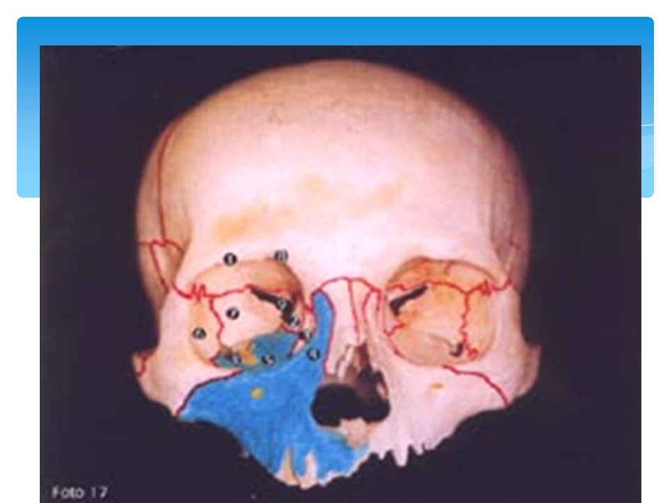
GLOBO OCULAR

17
VIA VISUAL

18
PREVALENCIA EN EL MUNDO DE BAJA VISION Y CEGUERA
Discapacidad visual: 285 million1 Baja Visión: 246 milliones ciegos: 39 milliones Ceguera legal visión 20/200

19
CAUSAS DE CEGUERA Y B.V.

20
CAUSAS GLOBALES DE CEGUERA
Ceguera evitable % Tratables y/o prevenibles 90% de las personas ciegas viven en países en desa rrollo

21
EVALUACION RAPIDA DE LA CEGUERA EVITABLE (RAAB)
El Raab se llevó a cabo en Uruguay en 2011, a nivel nacional La encuesta poblacional fue organizada por la Universidad de la República, el MSP (Minsa), IAPB, ORBIS y Visión 20/20 Latinoamérica. El objetivo era evaluar la situación actual sobre la ceguera y la discapacidadvisual en Uruguay. En este informe los datos se refieren a las personas mayores de 50 anos.
, IAPB, ORBIS y Visión 20/20 Latinoamérica. El objetivo era evaluar la situación actual sobre la ceguera y la discapacidadvisual en Uruguay. En este informe los datos se refieren a las personas mayores de 50 anos.")
22
RESULTADOS La prevalencia de la ceguera y la baja visión aumentan fuertemente con la edad, como en todas las comunidades. El ajuste de la prevalencia en baja visión de personas que requieren de servicios especializados se estimó en 0,8%: personas, hombres y mujeres. La causa más frecuente fue la catarata bilateral; 48,6%, glaucoma, 14,3%; degeneración macular senil 8,6%; otras enfermedades de polo posterior 8,6%

23
EPIDEMIOLOGIA Global prevalence of AMD 30 million1,2
AMD-related blindness >3 million (8.7% of all causes)2 Increasing prevalence as population ages3,4
2. Increasing prevalence as population ages3,4.")
24
EPIDEMIOLOGIA

25
PATOLOGIAS PRINCIPALES EN BAJA VISION Fundacion Braille del Uruguay

26
DEGENERACION MACULAR RELACIONADA A LA EDAD
Enfermedad degenerativa de la retina. Principal causa de ceguera en paises desarrollados Edad de inicio 65 años. Incidencia anual de afección del otro ojo es 12%. Primariamente asociada a fallo del epitelio pigmentario de la retina

27
DEGENERACION MACULAR RELACIONADA A LA EDAD
Tipos: Seca (80% a 90%) Humeda: (10% a 20%) Sintomas: Metamorfopsias Escotomas centrales o paracentrales Disminución de la A.V. lenta o brusca Disminución de la sensibilidad de contraste
 Humeda: (10% a 20%) Sintomas: Metamorfopsias. Escotomas centrales o paracentrales. Disminución de la A.V. lenta o brusca. Disminución de la sensibilidad de contraste.")
28
FACTORES DE RIESGO DE DMRE
Increased risk of AMD Smoking,1,2 diet3 Genetics1 Family history1,2 Cardiovascular disease2,3 Age,1,2 race1

29
ESCOTOMA CENTRAL EN DMRE

30
DISFUNCION DE EPR EN DMRE
Drusen Choroidal neovascularization (CNV) Fovea
 Fovea.")
31
Pre-symptomatic AMD1,2 Clearance of waste starts to falter
At stages 1 and 2 (‘no AMD’ or AREDS category 1)1 there are no or few small drusen due to a small amount of retinal waste build-up (<63 μm in diameter), but no other visual signs or symptoms.2 Approximately 90% of the Caucasian population aged ≥40 years will have one or two of these drusen, and there is very little risk of progression to advanced AMD in these subjects.3 Age-Related Eye Disease Study (AREDS) research group. Arch Opthalmol 2001;119: Ambati J, et al. Survey of Ophthalmology 2003;48: Cook H, et al. Br Med Bull 2008;85: 20/20 vision No signs or symptoms 1. Age-Related Eye Disease Study (AREDS) research group. Arch Opthalmol 2001;119: ; 2. Ambati J, et al. Survey of Ophthalmology 2003;48:
1 there are no or few small drusen due to a small amount of retinal waste build-up (<63 μm in diameter), but no other visual signs or symptoms.2 Approximately 90% of the Caucasian population aged ≥40 years will have one or two of these drusen, and there is very little risk of progression to advanced AMD in these subjects.3. Age-Related Eye Disease Study (AREDS) research group. Arch Opthalmol 2001;119: Ambati J, et al. Survey of Ophthalmology 2003;48: Cook H, et al. Br Med Bull 2008;85: /20 vision. No signs or symptoms. 1. Age-Related Eye Disease Study (AREDS) research group. Arch Opthalmol 2001;119: ; 2. Ambati J, et al. Survey of Ophthalmology 2003;48:")
32
Early AMD1,2 The RPE fails to clear waste from Bruch’s membrane
Pigment clumping At stages 3 and 4 (‘early AMD’ or AREDS category 2)1 more small drusen and some intermediate drusen ( μm; 125 μm is the approximate diameter of a retinal vein at the optic nerve head)2 develop, as well as minimal RPE abnormalities (e.g. changes in pigmentation).1 The larger and more prevalent the drusen, the higher the risk of progression to advanced AMD.2 However, patients with ‘early AMD’ have only a 1.3% risk of progressing to advanced AMD in 5 years.1 At these stages of disease progression, vision is likely to be unaffected, although some vision impairment may appear towards the end of stage 4. Age-Related Eye Disease Study (AREDS) research group. Arch Ophthalmol 2001;119: Cook H, et al. Br Med Bull 2008;85: More and larger drusen Minimal RPE abnormalities Vision likely to be unaffected 20/50 vision 1. Age-Related Eye Disease Study (AREDS) research group. Arch Ophthalmol 2001;119: ; 2. Cook H, et al. Br Med Bull 2008;85:127-49
1 more small drusen and some intermediate drusen ( μm; 125 μm is the approximate diameter of a retinal vein at the optic nerve head)2 develop, as well as minimal RPE abnormalities (e.g. changes in pigmentation).1 The larger and more prevalent the drusen, the higher the risk of progression to advanced AMD.2 However, patients with ‘early AMD’ have only a 1.3% risk of progressing to advanced AMD in 5 years.1. At these stages of disease progression, vision is likely to be unaffected, although some vision impairment may appear towards the end of stage 4. Age-Related Eye Disease Study (AREDS) research group. Arch Ophthalmol 2001;119: Cook H, et al. Br Med Bull 2008;85: More and larger drusen. Minimal RPE abnormalities. Vision likely to be unaffected. 20/50 vision. 1. Age-Related Eye Disease Study (AREDS) research group. Arch Ophthalmol 2001;119: ; 2. Cook H, et al. Br Med Bull 2008;85:")
33
Intermediate AMD1 The RPE fails to adequately nourish the retina
At stage 5 (‘Intermediate/early advanced AMD’ or AREDS category 3/early 4)1, signs include extensive medium–large-sized drusen (≥125 μm) and areas of geographical atrophy (including loss of RPE, choriocapillaris and photoreceptor cells) not involving the center of the fovea.1, A substantial decrease in visual acuity is generally noticeable at this stage due to the destruction of the photoreceptors, involving distortion, blurring or a black or gray patch in central vision, known as a scotoma.2 The 5-year progression from intermediate to advanced AMD was reported to be 18% in the AREDS study.1 Age-Related Eye Disease Study (AREDS) research group. Arch Ophthalmol 2001;119: Cook H, et al. Br Med Bull 2008:85; Extensive medium–large drusen Geographical atrophy (not central fovea) Gradual loss of vision 20/200 vision 1. Age-Related Eye Disease Study (AREDS) research group. Arch Ophthalmol 2001;119:
1, signs include extensive medium–large-sized drusen (≥125 μm) and areas of geographical atrophy (including loss of RPE, choriocapillaris and photoreceptor cells) not involving the center of the fovea.1, A substantial decrease in visual acuity is generally noticeable at this stage due to the destruction of the photoreceptors, involving distortion, blurring or a black or gray patch in central vision, known as a scotoma.2 The 5-year progression from intermediate to advanced AMD was reported to be 18% in the AREDS study.1. Age-Related Eye Disease Study (AREDS) research group. Arch Ophthalmol 2001;119: Cook H, et al. Br Med Bull 2008:85; Extensive medium–large drusen Geographical atrophy. (not central fovea) Gradual loss of vision. 20/200 vision. 1. Age-Related Eye Disease Study (AREDS) research group. Arch Ophthalmol 2001;119:")
34
Severe and rapid vision loss
Advanced AMD1,2 Growth of abnormal new blood vessels throughout the RPE and retina results in bleeding and exudation into the macula At stage 6 (‘Advanced AMD’ or AREDS category 4), pathological changes in Bruch’s membrane cause retinal hypoxia, which leads to the upregulation of vascular endothelial growth factor (VEGF).1 As a result, more fragile blood vessels are formed though angiogenesis.1 At this stage the individual is likely to experience severe and rapid vision loss. One or more of the following signs may also be present:2 Geographic atrophy (advanced dry AMD) of the RPE and choriocapillaris involving the central fovea, with multiple large drusen surrounding the atrophic site. Macular damage as a result of new blood vessel growth through the RPE and retina (neovascular maculopathy), including: CNV, leading to bleeding and exudation into the macula Serous fluid and/or haemorrhagic detachment of the sensory retina or RPE Retinal hard exudates (as a result of leakage) Subretinal and sub-RPE fibrovascular proliferation Permanent retinal scarring as a result of damage/bleeding Salminen A, et al. Mol Med 2010;16:53542. Age-Related Eye Disease Study (AREDS) research group. Arch Ophthalmol 2001;119: Geographical atrophy of the central fovea Advanced dry AMD Neovascular maculopathy, including CNV Wet AMD Severe and rapid vision loss 20/400 vision 1. Salminen A, et al. Mol Med 2010;16:53542; 2. Age-Related Eye Disease Study (AREDS) research group. Arch Ophthalmol 2001;119:
, pathological changes in Bruch’s membrane cause retinal hypoxia, which leads to the upregulation of vascular endothelial growth factor (VEGF).1 As a result, more fragile blood vessels are formed though angiogenesis.1 At this stage the individual is likely to experience severe and rapid vision loss. One or more of the following signs may also be present:2. Geographic atrophy (advanced dry AMD) of the RPE and choriocapillaris involving the central fovea, with multiple large drusen surrounding the atrophic site. Macular damage as a result of new blood vessel growth through the RPE and retina (neovascular maculopathy), including: CNV, leading to bleeding and exudation into the macula. Serous fluid and/or haemorrhagic detachment of the sensory retina or RPE. Retinal hard exudates (as a result of leakage) Subretinal and sub-RPE fibrovascular proliferation. Permanent retinal scarring as a result of damage/bleeding. Salminen A, et al. Mol Med 2010;16: Age-Related Eye Disease Study (AREDS) research group. Arch Ophthalmol 2001;119: Geographical atrophy of the central fovea. Advanced dry AMD. Neovascular maculopathy, including CNV. Wet AMD. Severe and rapid vision loss. 20/400 vision. 1. Salminen A, et al. Mol Med 2010;16:53542; 2. Age-Related Eye Disease Study (AREDS) research group. Arch Ophthalmol 2001;119:")
35
TRATAMAIENTO DE LA DMRE HUMEDA
Los VEGF-A (factores de crecimiento del endotelio vascuclar) estimulan la angiogénesis y aumentan la permeabilidad vascular. El standard actual de tratamiento es la inyección intravítrea de anti VEGF (ranibizumab, bevacizumab)
 estimulan la angiogénesis y aumentan la permeabilidad vascular. El standard actual de tratamiento es la inyección intravítrea de anti VEGF (ranibizumab, bevacizumab)")
36
GLAUCOMA Neuropatía óptica Principal factor de riesgo: PIO elevada
Tipos: Cronico primario de ángulo abierto De ángulo cerrado Secundario Sintomas principales: Perdida de campo visual Perdida de agudeza visual tardía Ceguera

37
GLAUCOMA

38
GLAUCOMA

39
MIOPIA PATOLOGICA Patológica cuando el eje axial es > 26mm o el error refractivo es > de 5 dioptrías

40
MIOPIA PATOLOGICA Degeneración retiniana progresiva Tipos de lesión:
Placas de atrofia corioretiniana Atrofia peripapilar cono miópico Neovascularización macular Mancha de Fuchs Lesiones de retina periférica predisponentes al desprendimiento de retina Estafiloma posterior

41
MIOPIA PATOLOGICA Disminución progresiva o brusca de agudeza visual Asociación con glaucoma Licuefacción vítrea Desprendimiento de retina Mayor frecuencia de catarata subcapsular posterior

42
MIOPIA PATOLOGICA ≥3 años 20% de ojos 29% de ojos ≥3 años NVC miopica

43
MIOPIA PATOLOGICA

44
EPIDEMIOLOGIA DEL EDEMA MACULAR DIABETICO
Prevalence of diabetes worldwide1 346 million Diabetic retinopathy2 ~23% of patients with diabetes DME3 ~7% of patients with diabetes

45
RETINOPATIA DIABETICA CAUSA 4,8% DE LA CEGUERA GLOGAL

46
RETINOPATIA DIABETICA
Tipos: Tipo 1 Insulino dependiente Enfermedad autoinmune Tipo 2 No insulino dependiente Resistencia a la insulina Déficit en la producción 46% historia familiar

47
EMD: FACTORES DE RIESGO PRIMARIOS
Increased risk of DME Glycemic control Diabetic retinopathy severity Age, gender, smoking Diabetes duration Hypertension The primary risk factor for the development of DME is diabetes mellitus. The type, duration of disease and age of onset have been associated with the incidence of DME:1,2 A US study showed that, over a period of 10 years, DME occurred in 20.1% of younger-onset patients, 25.4% of older-onset patients receiving insulin, and 13.9% of older-onset patients not receiving insulin.1 Additional factors associated with DME include:3 Having more severe diabetic retinopathy Gender (higher incidence in males) Higher glycosylated hemoglobin (poor glycemic control) Higher systolic and diastolic blood pressure Smoking Klein R, et al. Ophthalmology 1995;102:7-16. Chen E, et al. Curr Med Res Opin 2010;26: Klein R, et al. Ophthalmology 2009;116: 1. Klein R, et al. Ophthalmology 1995;102:7-16; 2. Chen E, et al. Curr Med Res Opin 2010;26: ; 3. Klein R, et al. Ophthalmology 2009;116:
 Higher glycosylated hemoglobin (poor glycemic control) Higher systolic and diastolic blood pressure. Smoking. Klein R, et al. Ophthalmology 1995;102:7-16. Chen E, et al. Curr Med Res Opin 2010;26: Klein R, et al. Ophthalmology 2009;116: Klein R, et al. Ophthalmology 1995;102:7-16; 2. Chen E, et al. Curr Med Res Opin 2010;26: ; 3. Klein R, et al. Ophthalmology 2009;116:")
48
RETINOPATIA DIABEICA

49
RETINOPATIA DIABETICA
MANEJO Control de factores de riesgo Control de glicemia: hemoglobina glicosilada Controles oftalmológicos seriados

50
RETINOPATIA DIABETICA
Tratamiento: Corticoides Láser Antiangiogénicos

51
RETINOPATIA DIABETICA

52
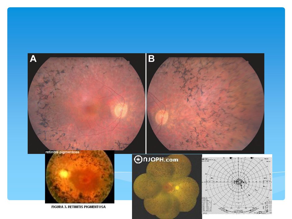
Retinosis Pigmentaria
Distrofia retiniana mas frecuente Hereditaria Autosómica recesiva (la más frecuente) Autosómica dominante Ligada al sexo En el fondo de ojo: Atrofia papila Estrechamiento de las arterias Alteración de la distribución del pigmento retiniano en la periferia media (espículas óseas) Maculopatía Sintomas Ceguera nocturna (ERG escotópico muy alterado) Alteración de campo visual periférico (visión tubular) Ceguera
 Autosómica dominante. Ligada al sexo. En el fondo de ojo: Atrofia papila. Estrechamiento de las arterias. Alteración de la distribución del pigmento retiniano en la periferia media (espículas óseas) Maculopatía. Sintomas. Ceguera nocturna (ERG escotópico muy alterado) Alteración de campo visual periférico (visión tubular) Ceguera.")
54
RETINOSIS PIGMENTARIA

55
FIN…

Presentaciones similares


>")


