Descargar la presentación
La descarga está en progreso. Por favor, espere

1
SISTEMA RESPIRATORIO HUMANO
Depto. de Biología Instituto Nacional Prof. Valentina Olivares Fredes.

2
Respiración: Respiración celular y Ventilación pulmonar
Figura 1. Relación existente entre la ventilación pulmonar y la respiración celular.

3
Las funciones del sistema respiratorio son:
Intercambiar gases respiratorios (O2 y CO2) entre la atmósfera y la sangre, a nivel de los alvéolos pulmonares (hematosis). Controlar el grado de acidez sanguínea (pH), mediante la regulación de la concentración de CO2 en la sangre. Posee receptores para el sentido del olfato, filtra el aire inspirado. Produce sonidos (fonación). Excretar H2O, lo que ayuda a la regulación de la temperatura corporal.
 entre la atmósfera y la sangre, a nivel de los alvéolos pulmonares (hematosis). Controlar el grado de acidez sanguínea (pH), mediante la regulación de la concentración de CO2 en la sangre. Posee receptores para el sentido del olfato, filtra el aire inspirado. Produce sonidos (fonación). Excretar H2O, lo que ayuda a la regulación de la temperatura corporal.")
4
Estructuras anatómicas involucradas en la respiración:
Vías respiratorias Porción respiratoria Porción conductora Bronquiolos respiratorios y alvéolos Bronquiolos terminales Laringe Tráquea Bronquios Nariz Faringe Transporte del aire Hematosis vías aéreas Figura 2. Porciones conductora y respiratoria de las vías respiratorias.

5
(a) nasal cavity pharynx epiglottis larynx esophagus trachea bronchi
Figure: 29-7 part a Title: The human respiratory system part a Caption: (a) Air enters through the nasal cavity and mouth and passes through the pharynx and the larynx into the trachea. The epiglottis prevents food from going down the trachea. The trachea splits into two large branches, the bronchi, which lead into the two lungs. The smaller branches of the bronchi, the bronchioles, lead to the microscopic alveoli (which are enmeshed in capillaries), where gas exchange occurs. The pulmonary artery carries deoxygenated blood (in blue) to the lungs; the pulmonary vein carries oxygenated blood (in red) back to the heart. bronchioles pulmonary artery pulmonary vein
 Air enters through the nasal cavity and mouth and passes through the pharynx and the larynx into the trachea. The epiglottis prevents food from going down the trachea. The trachea splits into two large branches, the bronchi, which lead into the two lungs. The smaller branches of the bronchi, the bronchioles, lead to the microscopic alveoli (which are enmeshed in capillaries), where gas exchange occurs. The pulmonary artery carries deoxygenated blood (in blue) to the lungs; the pulmonary vein carries oxygenated blood (in red) back to the heart. bronchioles. pulmonary artery. pulmonary vein.")
6
branch of pulmonary artery
bronchiole branch of pulmonary artery smooth muscle arteriole branch of pulmonary vein lymphatic vessel Figure: 29-7 part b Title: The human respiratory system part b Caption: (b) Close-up of alveoli (their interiors are shown in the cut-away section) and their surrounding capillaries. Question The nasal passages of mammals tend to follow long, complicated, winding pathways. Why are nasal passages so convoluted? alveoli capillary network
 Close-up of alveoli (their interiors are shown in the cut-away section) and their surrounding capillaries. Question The nasal passages of mammals tend to follow long, complicated, winding pathways. Why are nasal passages so convoluted alveoli. capillary network.")
7
Alvéolos pulmonares Por sus delgadas paredes es posible el intercambio de gases respiratorios. Sus delgadas paredes que separan a alvéolos vecinos poseen poros colaterales, que previenen colapso pulmonar. Están recubiertos por un epitelio constituido por dos tipos de células: neumocitos tipo I y neumocitos tipo II. Estos últimos producen el surfactante pulmonar.

8
Cada pulmón posee 300 millones de alvéolos, que aportan una superficie de 70 metros cuadrados aprox. Tamaño de una cancha de tenis.

9
Mecánica respiratoria
(a) Inhalation (b) Exhalation air moves in air moves out ribcage contracts ribcage expands lungs compress lungs expand Figure: 29-11 Title: The mechanics of breathing Caption: (a) During inhalation, rhythmic nerve impulses from the brain stimulate the diaphragm to contract (pulling it downward) and the muscles surrounding the ribs to contract (moving them up and outward). The result is an increase in the size of the chest cavity, causing air to rush in. (b) Relaxation of these muscles (exhalation) allows the diaphragm to dome upward and the rib cage to collapse, forcing air out of the lungs. Question Imagine that a person living at sea level travels to a high mountaintop, and there inhales with a muscular contraction of exactly the same strength as had been her habit while at rest in her sea level home. Would the resulting inhalation contain a volume of air that was larger, smaller, or the same as at sea level? Why? diaphragm contracts downward diaphragm relaxes upward Mecánica respiratoria
 Inhalation. (b) Exhalation. air moves in. air moves out. ribcage contracts. ribcage expands. lungs compress. lungs expand. Figure: Title: The mechanics of breathing. Caption: (a) During inhalation, rhythmic nerve impulses from the brain stimulate the diaphragm to contract (pulling it downward) and the muscles surrounding the ribs to contract (moving them up and outward). The result is an increase in the size of the chest cavity, causing air to rush in. (b) Relaxation of these muscles (exhalation) allows the diaphragm to dome upward and the rib cage to collapse, forcing air out of the lungs. Question Imagine that a person living at sea level travels to a high mountaintop, and there inhales with a muscular contraction of exactly the same strength as had been her habit while at rest in her sea level home. Would the resulting inhalation contain a volume of air that was larger, smaller, or the same as at sea level Why diaphragm contracts downward. diaphragm relaxes upward. Mecánica respiratoria.")
10
Radiografía de tórax en inspiración y espiración forzada.

11
Volúmenes respiratorios o Volúmenes pulmonares

12
Composición porcentual del aire inspirado y espirado.
Aire inspirado Aire espirado Oxigeno (%O2) Dióxido de carbono (%CO2) Nitrógeno (%N2) Vapor de agua variable muy abundante
 Dióxido de carbono (%CO2) Nitrógeno (%N2) Vapor de agua variable muy abundante.")
13
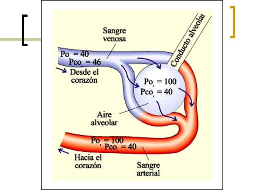
INTERCAMBIO DE GASES RESPIRATORIOS
oily surfactant collagen fibers capillary endothelial cell alveolar epithelial cell O2 air in alveolus Figure: 29-9 Title: Gas exchange between alveoli and capillaries Caption: The alveoli and capillary walls are only one cell thick and are very close to one another, with cells coated in a thin layer of fluid. This allows gases to dissolve and diffuse easily between the lungs and circulatory system. CO2 INTERCAMBIO DE GASES RESPIRATORIOS

15
(a) Transport of oxygen ( )
( ) 1) O2 diffuses through lung capillary wall. 3) O2 diffuses through tissue capillary walls. 2) O2 is carried to tissues bound to hemoglobin. lung side hemoglobin body cell side (b) Transport of carbon dioxide ( ) dissolved in plasma HCO3– as bicarbonate Figure: 29-10 Title: The chemistry and mechanism of gas exchange lung side body cell side bound to hemoglobin 2) CO2 is carried to lungs. 3) CO2 diffuses through lung capillary walls. 1) CO2 diffuses through tissue capillary wall.
 1) O2 diffuses through lung capillary wall. 3) O2 diffuses through tissue capillary walls. 2) O2 is carried to tissues bound to hemoglobin. lung side. hemoglobin. body cell side. (b) Transport of carbon dioxide. ( ) dissolved in plasma. HCO3– as bicarbonate. Figure: Title: The chemistry and mechanism of gas exchange. lung side. body cell side. bound to hemoglobin. 2) CO2 is carried to lungs. 3) CO2 diffuses through lung capillary walls. 1) CO2 diffuses through tissue capillary wall.")
18
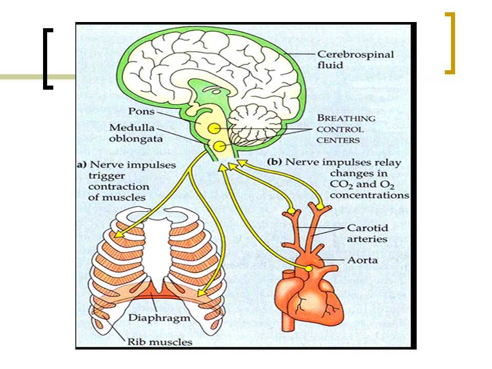
REGULACIÓN DE LA RESPIRACIÓN.
La respiración se encuentra regulada por el sistema nervioso, mediante un centro respiratorio presente en el tronco encefálico.

19
Centros Respiratorios
Constituidos por varios núcleos neuronales, ubicados en el bulbo raquídeo (grupos respiratorios dorsal y ventral). Y la protuberancia anular (centro neumotáxico y centro apnéustico). Los núcleos del bulbo se encargan de dar ritmicidad a la respiración (esto es, luego de un movimiento inspiratorio viene otro espiratorio). Los de la protuberancia anular se encargan de cambiar la frecuencia respiratoria (número de inspiraciones por minuto).
. Y la protuberancia anular (centro neumotáxico y. centro apnéustico). Los núcleos del bulbo se encargan de dar ritmicidad a la respiración (esto es, luego de un movimiento inspiratorio viene otro espiratorio). Los de la protuberancia anular se encargan de cambiar la frecuencia respiratoria (número de. inspiraciones por minuto).")
20
Regulación del ritmo respiratorio.
a) Grupo respiratorio dorsal; controla la respiración normal.
 Grupo respiratorio dorsal; controla la respiración normal.")
21
b) Grupo respiratorio ventral; controla la respiración forzada.
Al ser estimulado por el grupo respiratorio dorsal, el grupo respiratorio ventral inicia los movimientos inspiratorios y espiratorios.

22
Regulación de la frecuencia respiratoria:
Los centros neumotáxico y apnéustico actúan sobre el centro de la ritmicidad respiratoria del bulbo raquídeo (grupos respiratorios dorsal y ventral), modificando el tiempo para la inspiración respiratoria.
, modificando el tiempo para la inspiración respiratoria.")
24
Centro neumotáxico: Su función es limitar el tiempo para la etapa de inspiración, originada por el grupo neuronal dorsal del bulbo raquídeo, de modo que, al generar inspiraciones breves, aumenta la frecuencia respiratoria.

25
Centro apnéustico: Su función es aumentar el tiempo para la etapa de inspiración, originada por el grupo neuronal dorsal del bulbo raquídeo, de modo que, al generar inspiraciones más profundas, disminuye la frecuencia respiratoria.

26
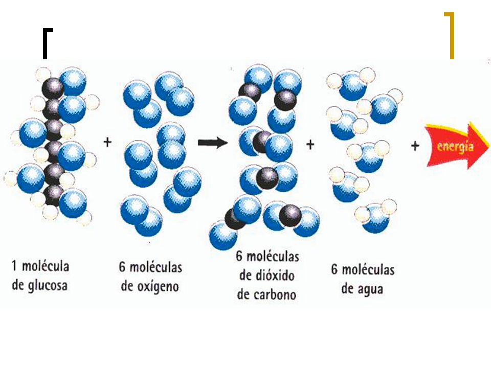
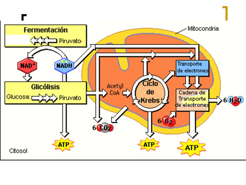
Respiración Celular

29
En presencia de OXÍGENO
CICLO DE KREBS : Matriz mitocondrial FOSFORILACIÓN OXIDATIVA : Membrana interna mitocondria

30
En ausencia de OXÍGENO FERMENTACIÓN: LÁCTICA ALCOHÓLICA

31
ALGUNAS ENFERMEDADES RESPIRATORIAS
Asma Bronquitis Edema pulmonar Embolia pulmonar (EP) Enfermedad pulmonar obstructiva crónica (EPOC) Enfisema Neumonía Rinitis Tos Cáncer
 Enfermedad pulmonar obstructiva crónica (EPOC) Enfisema. Neumonía. Rinitis. Tos. Cáncer.")
32
¡A descansar!

Presentaciones similares