Descargar la presentación
La descarga está en progreso. Por favor, espere

1
1. 0 Estructura y función de las proteínas 1
1.0 Estructura y función de las proteínas 1.1 Estructura y función de aminoácidos

2
Luciferina y luciferasa. Producción de luz.
Hemoglobina. Transporte de oxígeno. Queratina: Principal componente estructural de pelo, escamas, cuernos, lana, uñas y plumas. FIGURE 3-1 Some functions of proteins. (a) The light produced by fireflies is the result of a reaction involving the protein luciferin and ATP, catalyzed by the enzyme luciferase (see Box 13-1). (b) Erythrocytes contain large amounts of the oxygen-transporting protein hemoglobin. (c) The protein keratin, formed by all vertebrates, is the chief structural component of hair, scales, horn, wool, nails, and feathers. The black rhinoceros is nearing extinction in the wild because of the belief prevalent in some parts of the world that a powder derived from its horn has aphrodisiac properties. In reality, the chemical properties of powdered rhinoceros horn are no different from those of powdered bovine hooves or human fingernails.
 The light produced by fireflies is the result of a reaction involving the protein luciferin and ATP, catalyzed by the enzyme luciferase (see Box 13-1). (b) Erythrocytes contain large amounts of the oxygen-transporting protein hemoglobin. (c) The protein keratin, formed by all vertebrates, is the chief structural component of hair, scales, horn, wool, nails, and feathers. The black rhinoceros is nearing extinction in the wild because of the belief prevalent in some parts of the world that a powder derived from its horn has aphrodisiac properties. In reality, the chemical properties of powdered rhinoceros horn are no different from those of powdered bovine hooves or human fingernails.")
3
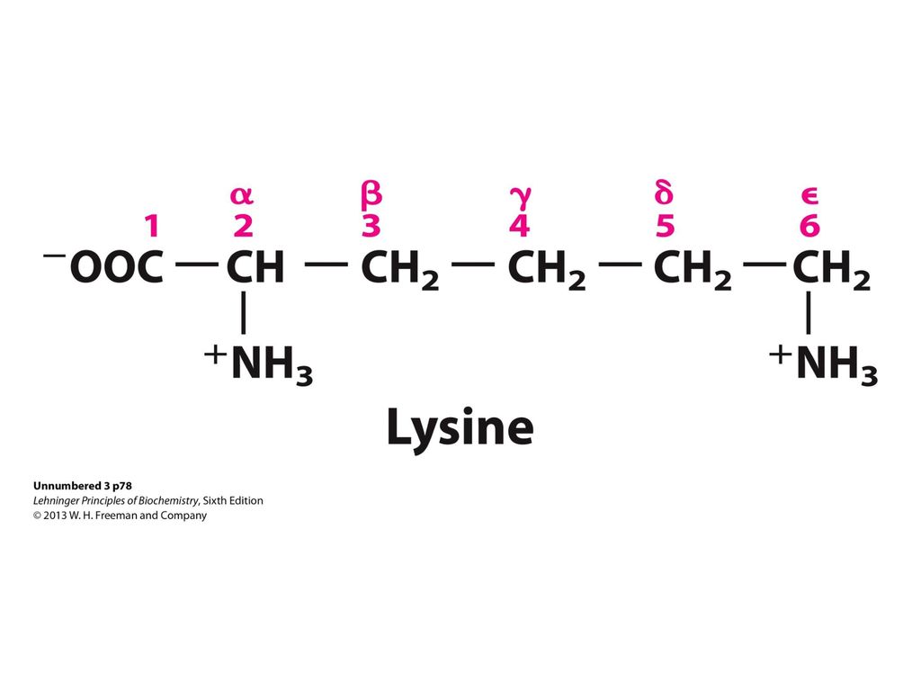
1.2.1 Estructura y función de los aminoácidos (aa)
Funciones: Formación de proteínas. Transmisión del impulso nervioso. Regulación del crecimiento celular. Biosíntesis de porfirinas, purinas, pirimidinas y urea. Participan en las vías de transmisión de señales. Señalización hormonal.

6
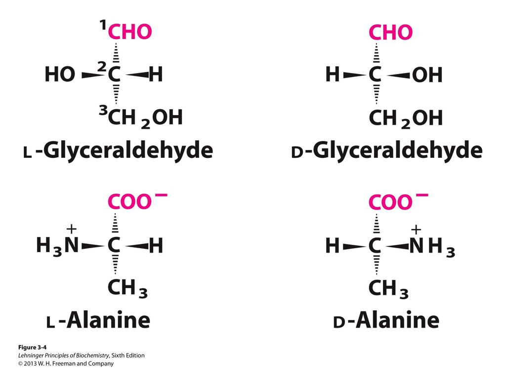
El grupo amino está a la izquierda.
El grupo amino está a la derecha.

8
Clasificación de los aminoácidos (aa)
No polares Polares Cadena lateral alifática: Gli, Ala, Pro, Val, Leu, Met, Ile. Cadena lateral con carga Cadena lateral aromática: Phe, Tir, Trp. Cadena lateral sin carga: Ser, Thr, Cis, Asn, Gln. Positiva: Lis, Arg, His (Básicos). Negativa: Asp, Glu (Ácidos).
. Negativa: Asp, Glu (Ácidos).")
9
FIGURE 3-5 (part 1) The 20 common amino acids of proteins
FIGURE 3-5 (part 1) The 20 common amino acids of proteins. The structural formulas show the state of ionization that would predominate at pH 7.0. The unshaded portions are those common to all the amino acids; the portions shaded in pink are the R groups. Although the R group of histidine is shown uncharged, its pKa (see Table 3-1) is such that a small but significant fraction of these groups are positively charged at pH 7.0. The protonated form of histidine is shown above the graph in Figure 3-12b.
 The 20 common amino acids of proteins. The structural formulas show the state of ionization that would predominate at pH 7.0. The unshaded portions are those common to all the amino acids; the portions shaded in pink are the R groups. Although the R group of histidine is shown uncharged, its pKa (see Table 3-1) is such that a small but significant fraction of these groups are positively charged at pH 7.0. The protonated form of histidine is shown above the graph in Figure 3-12b.")
10
FIGURE 3-5 (part 2) The 20 common amino acids of proteins
FIGURE 3-5 (part 2) The 20 common amino acids of proteins. The structural formulas show the state of ionization that would predominate at pH 7.0. The unshaded portions are those common to all the amino acids; the portions shaded in pink are the R groups. Although the R group of histidine is shown uncharged, its pKa (see Table 3-1) is such that a small but significant fraction of these groups are positively charged at pH 7.0. The protonated form of histidine is shown above the graph in Figure 3-12b.
 The 20 common amino acids of proteins. The structural formulas show the state of ionization that would predominate at pH 7.0. The unshaded portions are those common to all the amino acids; the portions shaded in pink are the R groups. Although the R group of histidine is shown uncharged, its pKa (see Table 3-1) is such that a small but significant fraction of these groups are positively charged at pH 7.0. The protonated form of histidine is shown above the graph in Figure 3-12b.")
11
FIGURE 3-5 (part 3) The 20 common amino acids of proteins
FIGURE 3-5 (part 3) The 20 common amino acids of proteins. The structural formulas show the state of ionization that would predominate at pH 7.0. The unshaded portions are those common to all the amino acids; the portions shaded in pink are the R groups. Although the R group of histidine is shown uncharged, its pKa (see Table 3-1) is such that a small but significant fraction of these groups are positively charged at pH 7.0. The protonated form of histidine is shown above the graph in Figure 3-12b.
 The 20 common amino acids of proteins. The structural formulas show the state of ionization that would predominate at pH 7.0. The unshaded portions are those common to all the amino acids; the portions shaded in pink are the R groups. Although the R group of histidine is shown uncharged, its pKa (see Table 3-1) is such that a small but significant fraction of these groups are positively charged at pH 7.0. The protonated form of histidine is shown above the graph in Figure 3-12b.")
12
FIGURE 3-5 (part 4) The 20 common amino acids of proteins
FIGURE 3-5 (part 4) The 20 common amino acids of proteins. The structural formulas show the state of ionization that would predominate at pH 7.0. The unshaded portions are those common to all the amino acids; the portions shaded in pink are the R groups. Although the R group of histidine is shown uncharged, its pKa (see Table 3-1) is such that a small but significant fraction of these groups are positively charged at pH 7.0. The protonated form of histidine is shown above the graph in Figure 3-12b.
 The 20 common amino acids of proteins. The structural formulas show the state of ionization that would predominate at pH 7.0. The unshaded portions are those common to all the amino acids; the portions shaded in pink are the R groups. Although the R group of histidine is shown uncharged, its pKa (see Table 3-1) is such that a small but significant fraction of these groups are positively charged at pH 7.0. The protonated form of histidine is shown above the graph in Figure 3-12b.")
13
FIGURE 3-5 (part 5) The 20 common amino acids of proteins
FIGURE 3-5 (part 5) The 20 common amino acids of proteins. The structural formulas show the state of ionization that would predominate at pH 7.0. The unshaded portions are those common to all the amino acids; the portions shaded in pink are the R groups. Although the R group of histidine is shown uncharged, its pKa (see Table 3-1) is such that a small but significant fraction of these groups are positively charged at pH 7.0. The protonated form of histidine is shown above the graph in Figure 3-12b.
 The 20 common amino acids of proteins. The structural formulas show the state of ionization that would predominate at pH 7.0. The unshaded portions are those common to all the amino acids; the portions shaded in pink are the R groups. Although the R group of histidine is shown uncharged, its pKa (see Table 3-1) is such that a small but significant fraction of these groups are positively charged at pH 7.0. The protonated form of histidine is shown above the graph in Figure 3-12b.")
15
FIGURE 3-11 Effect of the chemical environment on pKa
FIGURE 3-11 Effect of the chemical environment on pKa. The pKa values for the ionizable groups in glycine are lower than those for simple, methyl-substituted amino and carboxyl groups. These downward perturbations of pKa are due to intramolecular interactions. Similar effects can be caused by chemical groups that happen to be positioned nearby—for example, in the active site of an enzyme.

16
Figure 1-3

17
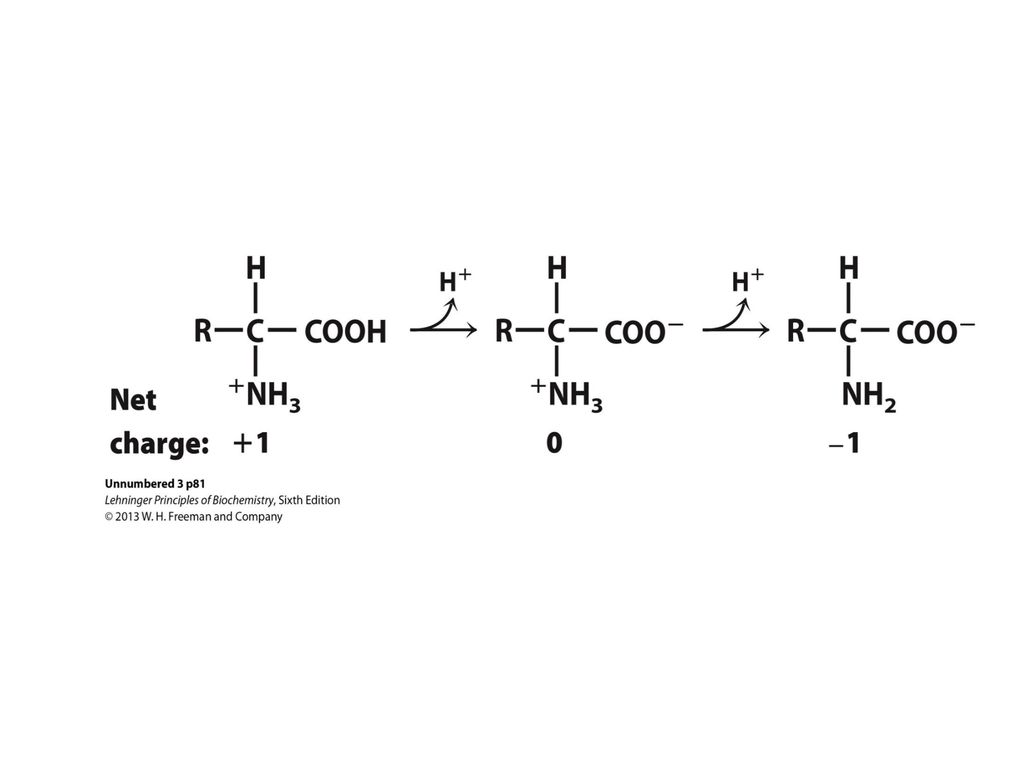
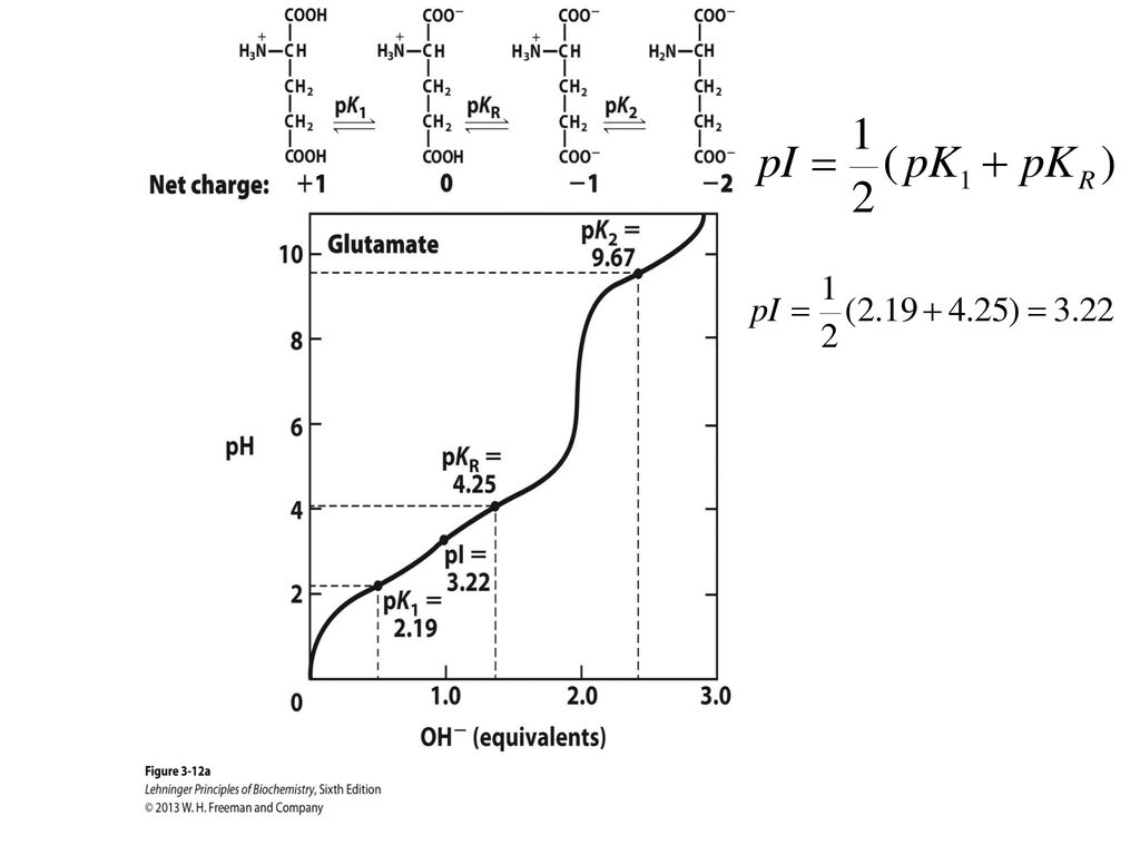
Punto isoeléctrico: Valor de pH en el que una molécula no tiene carga neta.
Si pH<pI, el aa tiene carga (+); Si pH>pI, el aa tiene carga (-).
; Si pH>pI, el aa tiene carga (-).")
19
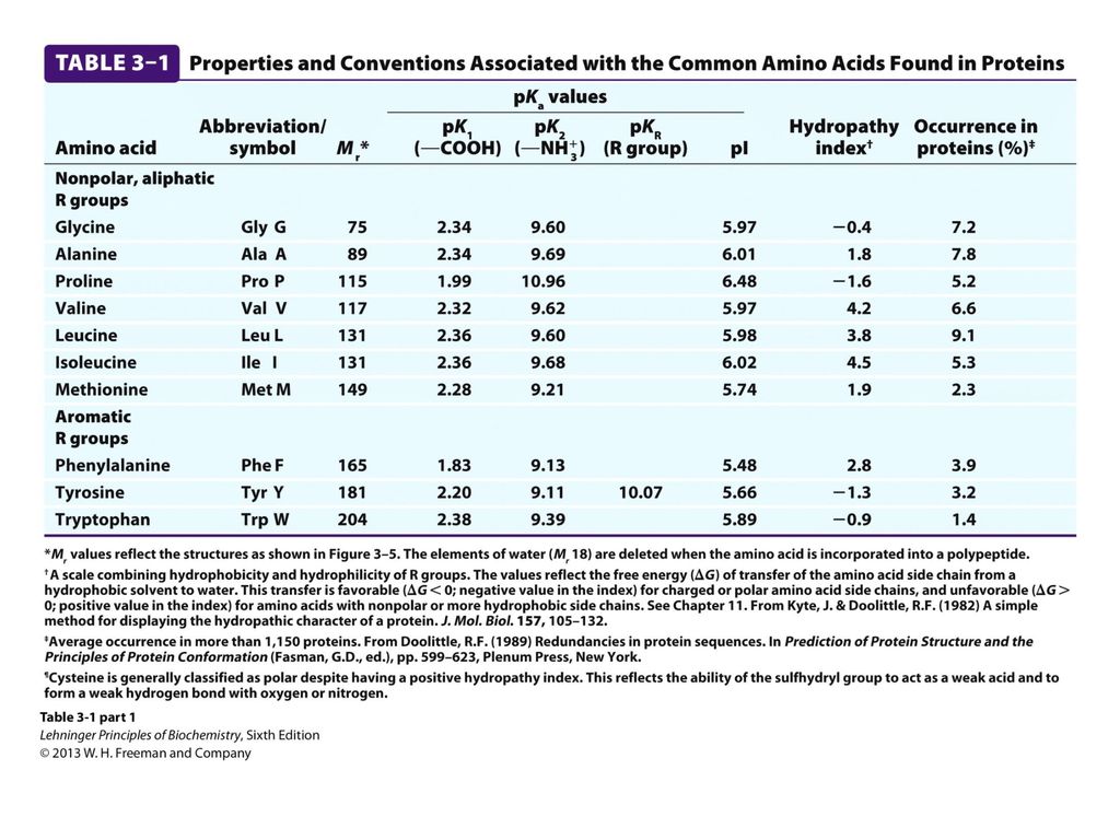
pI: Ácido aspártico: 3.02. Lisina: 9.7. Arginina: 10.8.

21
Se considera que el peso molecular promedio de un aminoácido es de 110.

22
Grupos ionizables de los aminoácidos
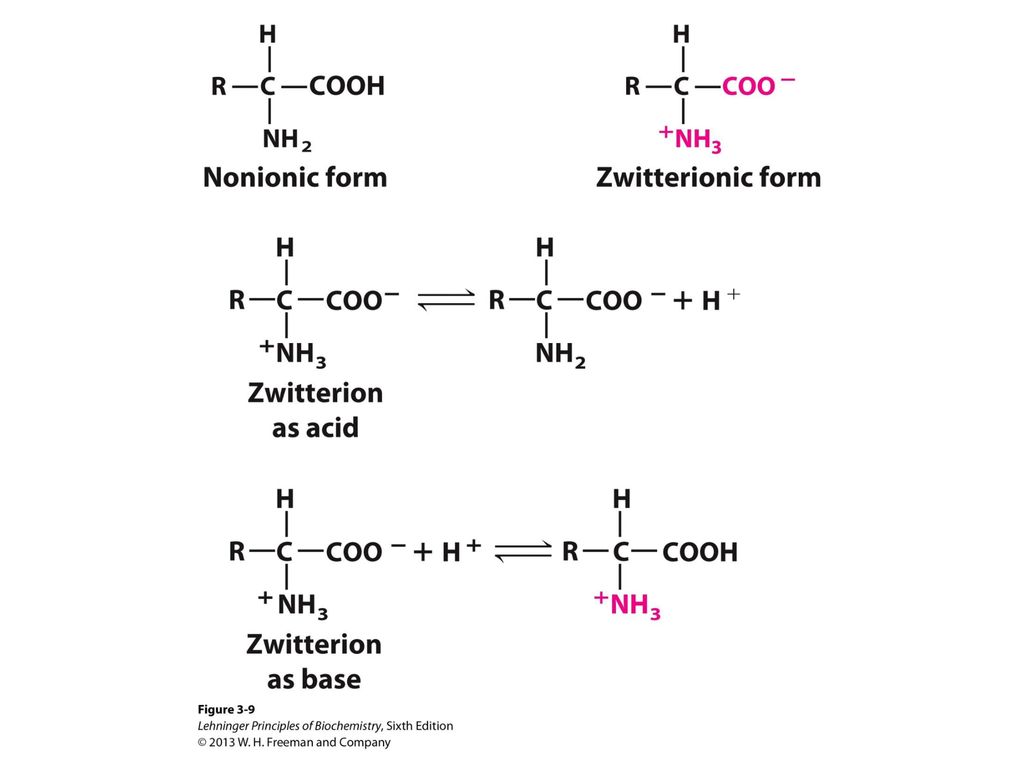
En el intervalo de pH fisiológico tanto los grupos de ácido carboxílico como los grupos amino de los a-aminoácidos se hallan completamente ionizados.

23
Ejercicio 1. Relación entre la curva de titulación y las propiedades ácido-base de la glicina
Una solución de 100 mL de glicina 0.1 M a pH 1.72 se tituló con una solución de NaOH 2 M. Se midió el pH y los resultados se graficaron. Los puntos clave en la titulación se designaron como I, II, III, IV, y V. Para cada uno de los casos, identifique el punto apropiado en la titulación y justifique la respuesta.

24
i) La glicina está presente predominantemente como +H3N-CH2-COOH.
l) La glicina está presente predominantemente en una mezcla de 50:50 de +H3N-CH2-COOH y de +H3N-CH2-COO. i) La glicina está presente predominantemente como +H3N-CH2-COOH. f) La glicina tiene su máxima capacidad amortiguadora. m) La carga neta promedio de la glicina es de +1/2. e) El pH es igual al pKa del grupo amino protonado. c) El grupo carboxilo ha sido titulado por completo. b) Este es el punto isoeléctrico. g) La carga neta de la glicina es 0. o) Peores regiones de pH para poder amortiguador. j) La especie predominante es +H3N-CH2-COO. k) La carga neta promedio de la glicina es -1. h) La mitad de los grupos amino están ionizados. d) El pH es igual al pKa del grupo carboxilo. a) La glicina está completamente titulada. n) Este es el fin de la titulación. La glicina está completamente titulada Este es el punto isoeléctrico. c) El grupo carboxilo ha sido titulado por completo. d) El pH es igual al pKa del grupo carboxilo. e) El pH es igual al pKa del grupo amino protonado. f) La glicina tiene su máxima capacidad amortiguadora. g) La carga neta de la glicina es 0. h) La mitad de los grupos amino están ionizados. i) La glicina está presente predominantemente como +H3N-CH2-COOH. j) La especie predominante es +H3N-CH2-COO. k) La carga neta promedio de la glicina es -1. l) La glicina está presente predominantemente en una mezcla de 50:50 de +H3N-CH2-COOH y de +H3N-CH2-COO. m) La carga neta promedio de la glicina es de +1/2. n) Este es el fin de la titulación. o) Peores regiones de pH para poder amortiguador.
 La glicina está presente predominantemente en una mezcla de 50:50 de +H3N-CH2-COOH y de +H3N-CH2-COO. i) La glicina está presente predominantemente como +H3N-CH2-COOH. f) La glicina tiene su máxima capacidad amortiguadora. m) La carga neta promedio de la glicina es de +1/2. e) El pH es igual al pKa del grupo amino protonado. c) El grupo carboxilo ha sido titulado por completo. b) Este es el punto isoeléctrico. g) La carga neta de la glicina es 0. o) Peores regiones de pH para poder amortiguador. j) La especie predominante es +H3N-CH2-COO. k) La carga neta promedio de la glicina es -1. h) La mitad de los grupos amino están ionizados. d) El pH es igual al pKa del grupo carboxilo. a) La glicina está completamente titulada. n) Este es el fin de la titulación. La glicina está completamente titulada. Este es el punto isoeléctrico. c) El grupo carboxilo ha sido titulado por completo. d) El pH es igual al pKa del grupo carboxilo. e) El pH es igual al pKa del grupo amino protonado. f) La glicina tiene su máxima capacidad amortiguadora. g) La carga neta de la glicina es 0. h) La mitad de los grupos amino están ionizados. i) La glicina está presente predominantemente como +H3N-CH2-COOH. j) La especie predominante es +H3N-CH2-COO. k) La carga neta promedio de la glicina es -1. l) La glicina está presente predominantemente en una mezcla de 50:50 de +H3N-CH2-COOH y de +H3N-CH2-COO. m) La carga neta promedio de la glicina es de +1/2. n) Este es el fin de la titulación. o) Peores regiones de pH para poder amortiguador.")
25
Respuesta V. I. III. II. IV. II y IV. I, III y V.

26
Ejercicio 2. Tamaño de proteínas
¿Cuál es el peso molecular aproximado para una proteína de 682 residuos de aminoácidos? 75 000

27
Ejercicio 3. Punto isoeléctrico de la pepsina
Pepsina es el nombre dado a muchas enzimas digestivas secretadas por glándulas presentes en el estómago. Estas glándulas también secretan HCl, el cual disuelve algunos componentes de la comida, permitiendo que la pepsina actúe enzimáticamente sobre las proteínas. La mezcla de comida, HCl y enzimas de digestión se conoce como Quimo y tiene un pH de 1.5. El punto isoeléctrico (pI) de la pepsina es 1.0. a) ¿Qué grupos funcionales deben de estar presentes en la pepsina para darle este punto isoeléctrico? b) ¿Qué aminoácidos de la enzima pueden contener estos grupos funcionales?
 de la pepsina es 1.0. a) ¿Qué grupos funcionales deben de estar presentes en la pepsina para darle este punto isoeléctrico b) ¿Qué aminoácidos de la enzima pueden contener estos grupos funcionales")
28
Respuesta Grupos carboxilo.
b) Ácido aspártico (Asp) y ácido glutámico (Glu).
 Ácido aspártico (Asp) y ácido glutámico (Glu).")
29
Ejercicio 4. Punto isoeléctrico de histonas
Las histonas son proteínas que se encuentran en el núcleo de células eucariotas, fuertemente ligados al ADN, el cual tiene muchos grupos fosfato. El pI de las histonas es muy alto, a) ¿Cuáles residuos de aminoácidos deben estar presentes en alta proporción en las histonas? b) ¿En qué manera contribuyen estos residuos a la unión fuerte de las histonas con el ADN?
 ¿Cuáles residuos de aminoácidos deben estar presentes en alta proporción en las histonas b) ¿En qué manera contribuyen estos residuos a la unión fuerte de las histonas con el ADN")
30
Respuesta a) Lis, His, Arg. b) Los grupos fosfato cargados negativamente en el ADN interactúan con grupos laterales de Lis (épsilon amino), His (imidazol), Arg (guanidino) cargados positivamente en las histonas.
 Lis, His, Arg. b) Los grupos fosfato cargados negativamente en el ADN interactúan con grupos laterales de Lis (épsilon amino), His (imidazol), Arg (guanidino) cargados positivamente en las histonas.")
31
Ejercicio 6. Carga de la His a diferentes pH
¿Cuál es la carga neta de la histidina a pH 1, 4, 8 y 12? Determine a cada pH si la histidina migra hacia el ánodo (+) ó hacia el cátodo (-) cuando se coloca en un campo eléctrico.
 ó hacia el cátodo (-) cuando se coloca en un campo eléctrico.")
32
Respuesta El pI de la histidina es: 7.59.
Si pH<pI el aminoácido tiene carga (+). Si pH>pI el aminoácido tiene carga (-). pH Carga Migración 1 +2 Cátodo. 4 +1 8 No migra. 12 -1 Ánodo.
. Si pH>pI el aminoácido tiene carga (-). pH. Carga. Migración Cátodo No migra Ánodo.")
33
Ejercicio 8. Estereoisómeros
La citrulina posee la siguiente estructura. ¿Es aminoácido D o L? Explique Debido a que el grupo NH3+ está a la derecha el aminoácido es D

34
FIGURE 3-6 Absorption of ultraviolet light by aromatic amino acids
FIGURE 3-6 Absorption of ultraviolet light by aromatic amino acids. Comparison of the light absorption spectra of the aromatic amino acids tryptophan and tyrosine at pH 6.0. The amino acids are present in equimolar amounts (10–3 M) under identical conditions. The measured absorbance of tryptophan is as much as four times that of tyrosine. Note that the maximum light absorption for both tryptophan and tyrosine occurs near a wavelength of 280 nm. Light absorption by the third aromatic amino acid, phenylalanine (not shown), generally contributes little to the spectroscopic properties of proteins.
 under identical conditions. The measured absorbance of tryptophan is as much as four times that of tyrosine. Note that the maximum light absorption for both tryptophan and tyrosine occurs near a wavelength of 280 nm. Light absorption by the third aromatic amino acid, phenylalanine (not shown), generally contributes little to the spectroscopic properties of proteins.")
35
aa Clasificación y funciones de los aminoácidos (aa)
Proteínicos No proteínicos Canónicos: A, C, D, E, F, G, H, I, K, L, M, N, P, Q, R, S, T, V, W, Y. Derivados: selenocisteína, hidroxiprolina, hidroxilisina. Neurotransmisores: Ácido gama amino butírico (GABA), dopamina. Hormona: tiroxina. Mediador de reacciones alérgicas: histamina. Intermediarios de reacciones metabólicas: citrulina, ornitina, homocisteína, S-adenosilmetionina. Presentes en antibióticos: D-valina (en valinomicina, gramicidina A y actinomicina D).
, dopamina. Hormona: tiroxina. Mediador de reacciones alérgicas: histamina. Intermediarios de reacciones metabólicas: citrulina, ornitina, homocisteína, S-adenosilmetionina. Presentes en antibióticos: D-valina (en valinomicina, gramicidina A y actinomicina D).")
36
1.2 Niveles de estructuración de las proteínas.
Estructura primaria

37
Niveles de estructuración en las proteínas

38
La estructura primaria de las proteínas es la descripción de todos los enlaces covalentes que unen los residuos de aminoácidos en una cadena polipeptídica: Enlace peptídico. Enlace disulfuro.

39
ENLACE PEPTÍDICO Es plano, por lo que no existe rotación alrededor del enlace. Posee un carácter de doble enlace, lo que significa que es más corto que un enlace sencillo por lo que es rígido y plano. Esta característica previene la libre rotación alrededor del enlace entre el carbono carbonílico y el nitrógeno del enlace peptídico. El resto de la molécula puede rotar libremente. Esta capacidad de rotación permite a las proteínas adoptar una inmensa gama de configuraciones.

40
ENLACE PEPTÍDICO Residuo Residuo Conocido también como enlace amida.

41
PÉPTIDOS Péptido: Cadena de residuos de aminoácidos unidos mediante enlaces peptídicos.

42
FIGURE 3-15 Alanylglutamylglycyllysine
FIGURE 3-15 Alanylglutamylglycyllysine. This tetrapeptide has one free α-amino group, one free α-carboxyl group, and two ionizable R groups. The groups ionized at pH 7.0 are in red.

43
Enlace o puente disulfuro: Enlace covalente entre los grupos tiol
(-SH2) de dos cisteínas FIGURE 3-7 Reversible formation of a disulfide bond by the oxidation of two molecules of cysteine. Disulfide bonds between Cys residues stabilize the structures of many proteins.
 de dos cisteínas. FIGURE 3-7 Reversible formation of a disulfide bond by the oxidation of two molecules of cysteine. Disulfide bonds between Cys residues stabilize the structures of many proteins.")
44
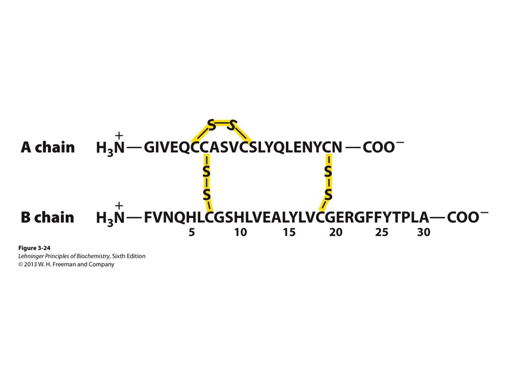
FIGURE 3-24 Amino acid sequence of bovine insulin
FIGURE 3-24 Amino acid sequence of bovine insulin. The two polypeptide chains are joined by disulfide cross-linkages. The A chain is identical in human, pig, dog, rabbit, and sperm whale insulins. The B chains of the cow, pig, dog, goat, and horse are identical.

Presentaciones similares