Descargar la presentación
La descarga está en progreso. Por favor, espere

1
DIAGNÓSTICO DE ARTRITIS PSORIÁTICA
Aparece a cualquier edad pero mas comun entre 15 a 30 años

2
CAUSAS Estudios experimentales sugieren que el dolor lumbar pueda originarse de muchas estructuras espinales: Ligamentos uniones facetarias periostio vertebral musculatura fascia paravertebral vasos sanguíneos anillo fibroso raíces nerviosas Aparece a cualquier edad pero mas comun entre 15 a 30 años

3
Aparece a cualquier edad pero mas comun entre 15 a 30 años
N Engl J Med, Vol. 344, No. 5· February 1, 2001

4
Psoriasis Enfermedad crónica de la piel
Características inflamatorias y de hiperproliferación Afecta al 1 a 2% de la población en USA Aparece a cualquier edad Distribución uniforme entre hombres y mujeres Aparece a cualquier edad pero mas comun entre 15 a 30 años Christophers E. Psoriasis – epidemiology and clinical spectrum. Clin Exp Dermatol 2001;26:314-20 Gupta A. Epidemiology of psoriasis as observed form the National Ambulatory Medical Care Survey, J Am Acad Dermatol 2005;3:184

5
Artritis Psoriática Artropatía inflamatoria crónica de las articulaciones periféricas, columna y entesis, asociada con la presencia de psoriasis y caracterizada por unos subtipos fenotípicamente distintos y un curso clínico variable Turkiewicz AM, Moreland RW. Psoriatic arthritis. Arthritis Rheum 2007;56;4:

6
Espondiloartropatías
Espondilitis anquilosante Artritis reactiva Artritis relacionada con EII Espondiloartropatía no diferenciada Artritis psoriática Braun J, Sieper J. Early diagnosis of spondyloarthritis. Nat Clin Pract Rheumatol 2006;2:536-45

7
Artritis Psoriática Prevalencia 0.04% - 0.2% de la población general
6 – 39% de los pacientes con psoriasis La prevalencia exacta de AP es desconocida y su cálculo ha resultado históricamente difícil por la falta de unos criterios diagnósticos que sean ampliamente aceptados por la comunidad médica reumatológic Gladman DD, Antoni C, Mease P, et al. Psoriatic arthritis: epidemiology, clinical features, course and outcome. Ann Rheum Dis 2005;64:14-17 Zachariae H. Prevalence of joint disease in patients with psoriasis: implications for therapy. Am J Clin Dermatol 2003;4:441-7

8
Artritis Psoriática Hombres Incidencia (por 100.000 habitantes Mujeres
La prevalencia exacta de AP es desconocida y su cálculo ha resultado históricamente difícil por la falta de unos criterios diagnósticos que sean ampliamente aceptados por la comunidad médica reumatológic Año Wilson FC, Icen M, Crowson CS, McEvoy MT, Gabriel SE, Kremers HM. Time trends in epidemiology and characteristics of psoritic arthritis over 3 decades: A population-based study. J Rheumatol 2009;36:361-67

9
Artritis Psoriática Se consideró inicialmente que el compromiso articular era de menor severidad con relación a AR 20% de los casos siguen un curso clínico severo 2 años: 47% de los pacientes tienen una erosión ósea 10 años: 50% de los pacientes tiene deformidades en por lo menos 5 articulaciones Goupille P. Psoriatic arthritis. Joint Bone Spine 2005;72:466-70

10
Compromiso Articular Usualmente en patrón asimétrico
Mono u oligoartritis Usualmente compromete las IFD La dactilitis es un punto importante para el diagnóstico(50%) Sinovitis y tendinitis Factor pronóstico La espondiloartritis ocurre en 40% de los pacientes Inflamación de sacroiliacas (usualmente unilateral) Dolor lumbar y/o glúteo inflamatorio Rigidez matutina Current Rheumatology, diagnosis and treatment, 2ª edición. New York, USA. Mc Graw-Hill Companies, 2007.capítulo 19
 Sinovitis y tendinitis. Factor pronóstico. La espondiloartritis ocurre en 40% de los pacientes. Inflamación de sacroiliacas (usualmente unilateral) Dolor lumbar y/o glúteo inflamatorio. Rigidez matutina. Current Rheumatology, diagnosis and treatment, 2ª edición. New York, USA. Mc Graw-Hill Companies, 2007.capítulo 19.")
11
Compromiso Articular Entesitis (38%) Artritis mutilante
Fascia plantar y tendón de Aquiles Artritis mutilante El compromiso en piel, precede al compromiso en 10 años (75-85%) 15-25% de los casos presentan primero el cuadro de artritis Leung YY, Li EK, Leung MH, Kun EW, Tam LS. Psoriatic arthritis in Hong Kong. Hong Kong J Dermatol Venereol 2007;15:62-7
 15-25% de los casos presentan primero el cuadro de artritis. Leung YY, Li EK, Leung MH, Kun EW, Tam LS. Psoriatic arthritis in Hong Kong. Hong Kong J Dermatol Venereol 2007;15:62-7.")
12
Kelley Figure 72-4 Patterns of peripheral joint disease. A-D, Asymmetric polyarticular disease. A, Distal interphalangeal joint involvement and forearm lymphedema; toe dactylitis with skin and nail change (B); predominant distal interphalangeal joint involvement (C); and arthritis mutilans (D). Kelly’s Testbook of Rheumatology, 8ª eidición. Philadelphia: WB Saunders Co, 2008:1102-9
; predominant distal interphalangeal joint involvement (C); and arthritis mutilans (D). Kelly’s Testbook of Rheumatology, 8ª eidición. Philadelphia: WB Saunders Co, 2008:")
13
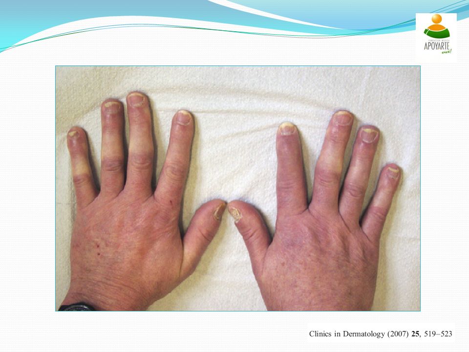
Current Dactylitis of the ring finger of a patient with psoriatic arthritis.

15
Primer on rheumatic disease
Ray distribution peripheral arthritis. Note the involvement of the second, third, and fi fth digits on the left hand, while the third right digit is totally spared.

16
Primer on rheumatic disease
Ray distribution peripheral arthritis. Note the involvement of the second, third, and fi fth digits on the left hand, while the third right digit is totally spared. Primer on the rheumatic diseases, 13º edición. New York, USA: Springer;2008.p

17
Primer on rheumatic disease
Ray distribution peripheral arthritis. Note the involvement of the second, third, and fi fth digits on the left hand, while the third right digit is totally spared.

18
Primer on rheumatic disease
Ray distribution peripheral arthritis. Note the involvement of the second, third, and fi fth digits on the left hand, while the third right digit is totally spared.

19
Nail changes in psoriasis
Primer on the rheumatic diseases, 13º edición. New York, USA: Springer;2008.p

20
Current Psoriatic nail changes with onycholysis and subungual debris

21
Kelley Nail dystrophic changes. Nail pitting (A); onycholysis (B), and severe destructive change with nail loss and pustule formation (C). Kelly’s Testbook of Rheumatology, 8ª eidición. Philadelphia: WB Saunders Co, 2008:1102-9

22
Current Psoriatic spondylitis. Extensive spinal involvement has led to exaggerated thoracic kyphosis and loss of cervical extension. As a result the patient is unable to touch the occiput to the wall when standing against the wall ("occiput-to-wall" test). There is limited chest expansion leading to a protuberant abdomen and to diaphragmatic breathing
. There is limited chest expansion leading to a protuberant abdomen and to diaphragmatic breathing.")
23
Compromiso Extra-articular
Enfermedad sistémica Ojo Pulmones Riñones Corazón Current Rheumatology, diagnosis and treatment, 2ª edición. New York, USA. Mc Graw-Hill Companies, 2007.capítulo 19

24
Hallazgos de laboratorio
No hay ningún examen específico Factor reumatoide negativo (Moll y Wright, Bennet, Fournie y CASPAR) Anti CCP: Positivo en 5 a 10% de pacientes con Aps VSG y PCR se encuentran elevados Candia L, Marquez J, Gonzalez C, Santos AM, Londoño J, Valle R et al. Low frecuency of anticyclic citrullinated peptide antibodies in psoriatic arthritis but not in cutaneous psoriasis. J Clin Rheumatol 2006;12: Korendowych E, Owen P, Ravindran J, Carmichael C, McHugh N. The clinical and genetic associations of anti-cyclic citrullinated peptide antibodies in psoriatic arthritis. Rheumatology (Oxford) 2005; 44:
 Anti CCP: Positivo en 5 a 10% de pacientes con Aps. VSG y PCR se encuentran elevados. Candia L, Marquez J, Gonzalez C, Santos AM, Londoño J, Valle R et al. Low frecuency of anticyclic citrullinated peptide antibodies. in psoriatic arthritis but not in cutaneous psoriasis. J Clin Rheumatol 2006;12: Korendowych E, Owen P, Ravindran J, Carmichael C, McHugh N. The clinical and genetic associations of anti-cyclic. citrullinated peptide antibodies in psoriatic arthritis. Rheumatology (Oxford) 2005; 44:")
25
Hallazgos radiológicos
Disminución del espacio articular Erosiones óseas y resorción: En IFD conocido como deformidad en punta de lápiz Hallazgos asimétricos Afectan IFP e IFD Se presenta con menor frecuencia osteopenia yuxta articular Usualmente respeta las MCF y muñecas Zayas VM, Monu JU. Imaging of psoriatic arthritis. Contemp Diagn Radiol 2008;31:1-6

26
Current Radiographic changes from psoriatic arthritis of the distal interphalangeal joint. A: Subtle periosteal erosions at the margins of the joint space are an initial appearance. B: Progressive erosive and proliferative changes occur over time and can be greatly destructive. C: Distinctive "pencil-in-cup" appearance with severe disease.

27
pencil-in-cup deformities

28
The autoimmune disease
Radiological changes in psoriatic arthritis. Both destructive and proliferative changes are characteristic.

29
Conventional radiography of the right hand of a patient with psoriatic arthritis (PsA) demonstrating
periostitis or new bone formation in the extracapsular region (arrow heads). These radiographic changes can be used to help establish a diagnosis of PsA but are often absent in early disease.
. These radiographic changes. can be used to help establish a diagnosis of PsA but are often absent in early disease.")
30
Frontal radiograph of the hand of
a patient with a long history of psoriasis shows fusion of the radiocarpal and intercarpal joints. Extensive sclerosis of the first metacarpal simulates an “ivory digit,” a classic finding in psoriatic arthritis.

31
Plain oblique radiograph of the sternum
in this patient with sternal pain and history of psoriasis shows irregularity of the manubriosternal junction (arrowheads) that is consistent with erosions in the sternum. Psoriatic arthritis affects synovial, cartilaginous, and fibrous joints.
 that is. consistent with erosions in the sternum. Psoriatic arthritis affects synovial, cartilaginous, and fibrous joints.")
32
Criterios diagnósticos

33
Criterios diagnósticos
Sensibilidad: 91% Especificidad: 98% Moll JM, Wright V. Psoriatic arthritis. Semin Arthritis Rheum 1973;3:55-78

34
Criterios diagnósticos
5 patrones de compromiso clínico 1. Poliarticular, con artritis simétrica, semejante a la AR (15%) 2. Oligoarticular (4 ó menos articulaciones) con artritis asimétrica (70%) 3. Compromiso predominante de AID (5%) 4. Predominio de espondiloartritis (5%) 5. Artritis mutilante (5%) Moll JM, Wright V. Psoriatic arthritis. Semin Arthritis Rheum 1973;3:55-78
 2. Oligoarticular (4 ó menos articulaciones) con artritis asimétrica (70%) 3. Compromiso predominante de AID (5%) 4. Predominio de espondiloartritis (5%) 5. Artritis mutilante (5%) Moll JM, Wright V. Psoriatic arthritis. Semin Arthritis Rheum 1973;3:")
35
S: 44% E: 100% Bennett RM. Psoriatic arthritis. En: McCarty DJ, editor. Arthritis and Related Conditions. Philadelphia: Lea and Febyger, 1979:645

36
S: 97% E: 96% Vasey FB, Espinoza LR. Psoriatic arthritis. En: Calin A, editor. Spondyloarthropathies. Orlando: Grune and Stratton, 1984:151-85

37
Criterios diagnósticos
Sensibilidad: 74% Especificidad: 91% Dougados M, Van der Linden S, Juhlin R, Huitfeldt B, Amor B, Calin A, et al. The European Spondyloarthropathy Study Group preliminary criteria for the classification of spondyloarthropathy. Arthritis Rheum 1991;34:

38
Criterios diagnósticos
Sensibilidad: 98% Especificidad: 91% McGonagle D, Conaghan PG, Emery P. Psoriatic arthritis: a unified concept twenty years on. Arthritis Rheum 1999;42:1080-6

39
S: 94% E: 95% Fournie B, Crognier L, Arnaud C, Zabraniecki L, Lascaux-Lefebvre V, Marc V, et al. Proposed classification criteria of psoriatic arthritis. A preliminary study in 260 patients. Rev Rheum Engl Ed 1999;66:446-56

40
S: 91.4% E: 98.7% Taylor W, Gladman D, Helliwell P, Marchesoni A, Mease P, Mielants H. Classification criteria for psoriatic arthritis, development of new criteria from a large international study. Arthritis Rheum 2006;54:

41
Criterios diagnósticos

42
Criterios diagnósticos

43
Conclusiones La artritis psoriática es una entidad clínica que debe tenerse en cuenta en la práctica clínica diaria Su diagnóstico debe basarse en los criterios de CASPAR Es fundamental la detección temprana para un manejo oportuno y de esta forma evitar secuelas y discapacidad, ya que hoy en día se dispone de mejores herramientas terapéuticas que detienen el proceso inflamatorio, como los medicamentos de terapia biológica Las clínicas de psoriasis, integradas por médicos dermatólogos y reumatólogos son una estrategia adecuada para llegar en forma mas temprana al diagnóstico

Presentaciones similares


, fue un dramaturgo, poeta y actor inglés. Conocido en ocasiones como el Bardo de Avon (o.>")