Descargar la presentación
La descarga está en progreso. Por favor, espere

1
Fracturas Acetabulares
Dr Pérez Ortopedia HCG

2
Tipicamente ocurren de gente joven, son secundarias a traumas de alta energia.
Hay màs de 50 %de probabilidades de tener lesiones asociadas: fx de columna, trauma abdominal y toràcico, lesiones genito uninarias, fx de extremidades, lesiones de los ligamentos de las rodillas,luxaciones, trauma craneoencefalico. El tratamiento quirùrgico es frecuente para restaurar la anstomìa articular.

3
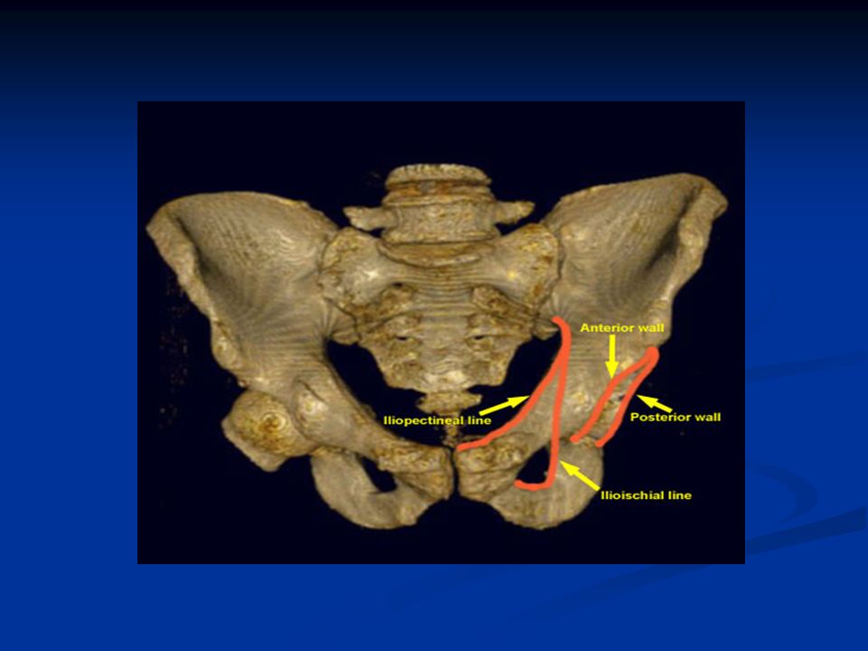
Anatomía Está formado por el hueso innominado.
La unión de 3 huesos: ilium, ischium, and pubis joined by the tri-radiate cartilage El acetábulo está dividido en 2 columnas: anterior y posterior Las 2 columnas se describen tiene la forma de unaY invertida, o la letra Griega lambda (l). Columna anterior: ant border of the iliac wing, the entire pelvic brim, the ant wall, and the superior pubic ramus Columna posterior: the ischial portion of bone ( lesser and greater sciatic notches), post wall, and the ischial tunerocity
. Columna anterior: ant border of the iliac wing, the entire pelvic brim, the ant wall, and the superior pubic ramus. Columna posterior: the ischial portion of bone ( lesser and greater sciatic notches), post wall, and the ischial tunerocity.")
6
Radiología Five (5) Pelvic XRs Proyección anteroposterior (AP)
Oblicuas Bilateral 45 grados, o proyecciones de Judet de la pelvis. Inlet y Outlet Tomografía computarizada, TAC, provee información adicional de la configuracion delas fracturas.

7
Pelvis XR: Inlet: Pt supino con XR paralelo al plano del sacro.
AP de pelvis con inclinación grados caudalmente. Outlet: Pt en supine con XR perpendicular al plane del sacro AP de pelvis con inclinación grados cefálico.

8
Judet hip XR Iliac oblique:
Pt is supine with involved side of pelvis rotated anteriorly 45 deg, beam directed vertically toward affected hip shows iliopectineal line, AC and PW Obturator oblique: Pt is supine with uninvolved side of pelvis rotated ant. 45 degrees, beam directed vertically toward the affected hip shows ilioischial line, PC and AW

9
AP Pelvis XR

10
Teardrop Internal limb = outer wall of obturator canal
External limb = middle 1/3 of cotyloid fossa Inferior border = ischiopubic notch

11
Inlet Pelvis XR

12
Outlet Pelvis XR

13
Iliac oblique

14
Obturator oblique

15
Classificación Inicialmete publicado por Judet en 1964,y despues modificado por Letournel Judet and Letournel sistema de clasificación: tipos simples y complejoss Simples: posterior wall (PW), posterior column (PC), anterior wall (AW), anterior column (AC), transverse Complejos: T-shaped, anterior column and posterior hemitransverse (AC-PHT) , both-column (BC), posterior column and wall (PC-PW), transverse posterior wall (T-PW)
, posterior column (PC), anterior wall (AW), anterior column (AC), transverse. Complejos: T-shaped, anterior column and posterior hemitransverse (AC-PHT) , both-column (BC), posterior column and wall (PC-PW), transverse posterior wall (T-PW)")
16
Simple types

17
Complex types

18
PW

19
PC

20
AW

21
Transverse

22
T-PW

23
AC-PHT

24
BC

25
PC-PW

27
Nonoperative tx Nondisplaced fx, <5mm, or articular step-off of <2mm Operative contraindications: local or systemic infection, severe osteoporosis Operative relative contraindications: advanced age, associated medical conditions (ESRD on dialysis, ESLD, Seizure Disorder, uncontrolled DM, CHF, Neurological Disorder), associated soft tissue and visceral injuries, or a multiply injured pt not stable for a big acetabular sx Displaced fx: large portion of acetabulum remains intact with a congruous femoral head, or secondary congruence with a both-column fx
, associated soft tissue and visceral injuries, or a multiply injured pt not stable for a big acetabular sx. Displaced fx: large portion of acetabulum remains intact with a congruous femoral head, or secondary congruence with a both-column fx.")
28
PW: if less than 50% of the width of the articular cartilage is displaced (ST), some authors say less than 25% Many low AW fx A minority of low T-shaped fx Infratectal transverse fx In assesing the intact portion of acetabulum, it is useful to obtain roof arc measurements Matta first described these angles in 1986 Stable fx=all roof arc angles >45 degrees CT subchondral arc technique of Olsen: no involvement of the upper 10mm of the acetabulum by CT corresponds to an intact 45 degrees roof arc on all 3 plain XRs

29
Roof Arc Angles A vertical line is drawn from roof of acetabulum to geometric center of the femoral head, and second line is drawn from fracture to the geometric center 1. Medial Roof Arc (AP pelvis) 2. Anterior Roof Arc (Obturator oblique) 3. Posterior Roof Arc (Iliac oblique)
 2. Anterior Roof Arc (Obturator oblique) 3. Posterior Roof Arc (Iliac oblique)")
30
Roof arc measurement

31
Operative tx Any displaced fx, > 5mm, or articular step-off of >2mm Allows early ambulation and decreases chance of post-traumatic arthritis Usually undertaken 2-3 days after injury, when initial fx and intrapelvic vessel bleeding has subsided Ideally performed before 10 days, so fx fragments remain mobile Three weeks after injury, a bony callus has formed, making reduction more difficult (typically not done)
")
32
Surgical approaches Kocher-Langenbeck: best access to posterior column (prone) Ilioinguinal: best access to anterior column and inner aspect of innominate bone (supine) Extended iliofemoral: best simultaneous access to the two columns (lateral) Combined approaches performed concurrently or successively is less desirable Extended iliofemoral approach has the highest incidence of ectopic bone formation (HO) and longest postoperative recovery
 Extended iliofemoral: best simultaneous access to the two columns (lateral) Combined approaches performed concurrently or successively is less desirable. Extended iliofemoral approach has the highest incidence of ectopic bone formation (HO) and longest postoperative recovery.")
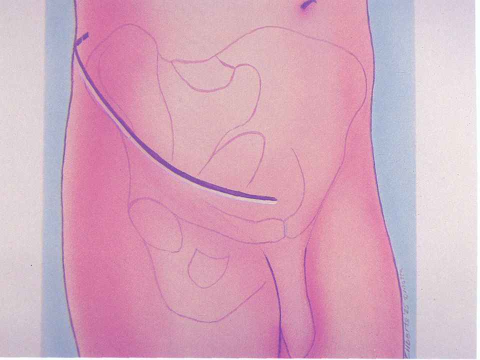
33
Kocher-Langenbeck approach
Posterior wall fractures Posterior column fractures Posterior column-posterior wall fractures Juxta-tectal/Infra-tectal transverse or transverse-posterior wall fractures Some T-shaped fractures

35
Ilioinguinal approach
Anterior column fractures Anterior wall fractures Some anterior column-posterior hemitransverse fractures May also be used for both column fractures with large single posterior fragment, with reduction being achieved indirectly through reduction of the quadrilateral plate Fractures with associated superior ramus and symphysis pubis fractures

37
Extended Iliofemoral approach
T-shaped fractures Transverse fractures with extended posterior wall T-shaped fractures with wide separations of the vertical stem of the "T" or those with associated pubic symphysis dislocations Certain associated both column fractures Associated fracture patterns or transverse fractures which are operated greater than 21 days following injury

39
Other approaches Stoppa approach (supine): Cole and Bolhofner
Allows access to the medial wall of the acetabulum, quadrilateral surface, and sacroiliac joint Triradiate approach (prone): Alternate exposure to the external aspect of the innominate bone, with almost same exposure as iliofemoral but visualization of the posterior part of the ilium is not as good
: Alternate exposure to the external aspect of the innominate bone, with almost same exposure as iliofemoral but visualization of the posterior part of the ilium is not as good.")
40
Postoperative care If the fx has been reduced accurately, 90% of normal ROM will be obtained without difficulty by the pt Pt is placed on bedrest initially, allowing ambulation when symptoms allow Iliofemoral approach= 5 days of absolute bedrest, to allow for edema to subside and initial wound healing PROM of the hip can be instituted by PT or by a CPM Gait training can usually begun on POD#2 15kg WB is allowed

41
The pt is encouraged to ambulate with a step-through gait and a heel-toe walking motion, using crutches or walker Pt is instructed on active flexion, abduction, and extension exercises to be performed at the hip while standing AP Pelvis XR should be obtained after gait training and before discharge to confirm that loss of reduction has not occurred Iliofemoral approach: active abduction and passive adduction are not allowed for the first 3 weeks Limited weight bearing is continued for 8 weeks, then WBAT with external support is begun PT is directed at regaining muscle strength at the hip, particularly the abductors Note: NWB for 12 weeks is typically performed at LSU

42
Complications Operative wound infection: decreased with the liberal use of drains, and intraoperative hemostasis Iatrogenic nerve palsy: Peroneal branch of Sciatic N (Kocher-Langenbeck), Sciatic N (Iliofemoral), Femoral N (Ilioingiunal) Periarticular ectopic bone formation: greatest with lateral exposure of the innominate bone, highest with iliofemoral approach, followed by Kocher-Langenbeck, and almost nonexistent with ilioingiunal or Stoppa approaches Indomethacin 25mg POTID or a localized single-dose of XRT significantly decreases risk (both equally effective- Burd et.al JBJS 2001) Thromboembolic complications (DVT, PE): Coumadin started 48 hours postop and cont for 6 wks, or LMW Heparin started POD#1 and cont for 3 wks
, Sciatic N (Iliofemoral), Femoral N (Ilioingiunal) Periarticular ectopic bone formation: greatest with lateral exposure of the innominate bone, highest with iliofemoral approach, followed by Kocher-Langenbeck, and almost nonexistent with ilioingiunal or Stoppa approaches. Indomethacin 25mg POTID or a localized single-dose of XRT significantly decreases risk (both equally effective- Burd et.al JBJS 2001) Thromboembolic complications (DVT, PE): Coumadin started 48 hours postop and cont for 6 wks, or LMW Heparin started POD#1 and cont for 3 wks.")
43
Morel-Lavale lesion A closed degloving injury over the greater trochanter Results from the blunt trauma that caused the fx The subcutaneous tissue is torn away from the underlying fascia, and a significant cavity results Cavity contains hematoma and liquified fat These areas must be drained and debrided before or during surgery to decrease the chance of infection Advisable to leave this area open through the surgical incision or a separate incision Dressing changes and wound packing are sometimes needed for a prolonged period of time Primary excision of the necrotic fat and closure over a drain has not been routinely successful

Presentaciones similares


.>")


 Usted tiene – You have (Formal) El tiene – He has Ella.>")


