Descargar la presentación
La descarga está en progreso. Por favor, espere

1
Seminario 3: Sistema Muscular
CATEDRA DE FISIOLOGIA ANIMAL Facultad de Ciencias Naturales y Museo Universidad Nacional de La Plata Seminario 3: Sistema Muscular

2
Temario SISTEMA MUSCULAR
Características y funciones de las proteínas contráctiles y regulatorias. Mecanismo de contracción el músculo estriado esquelético. Placa motora. Músculo estriado cardíaco: características funcionales. Músculo liso : estructura y mecanismo de contracción-relajación. Papel del Calcio y del ATP en los distintos tipos musculares. Contracciones isométricas e isotónicas, ejemplos. Curvas tensión-longitud. Características funcionales de las fibras fásicas y tónicas.

3
Propiedades del musculo
Contractibilidad Excitabilidad Extensibilidad Elasticidad

6
Características y funciones de las proteínas contráctiles y regulatorias.
Mecanismo de contracción el músculo estriado esquelético. Placa motora.

7
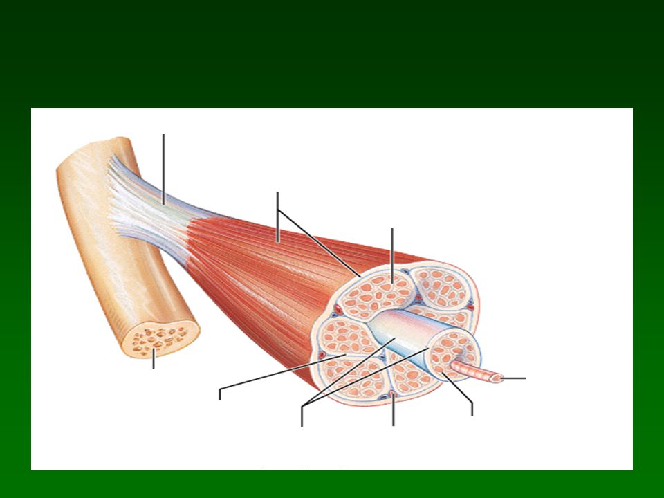
Músculo esquelético Ubicación Núcleos Control

16
Resumiendo Troponina Tropomiosina Miosina

18
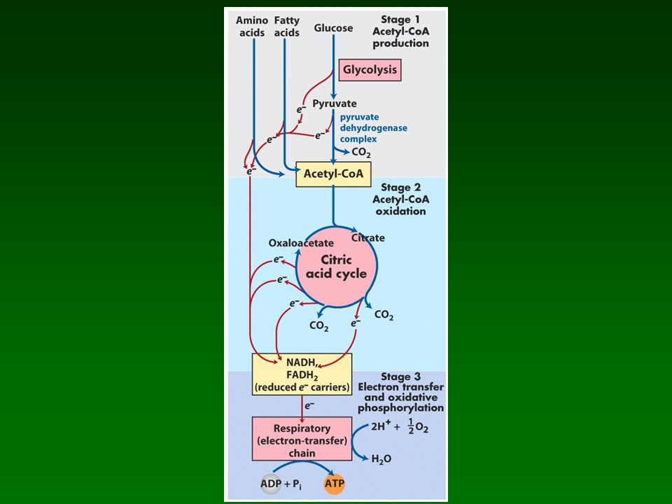
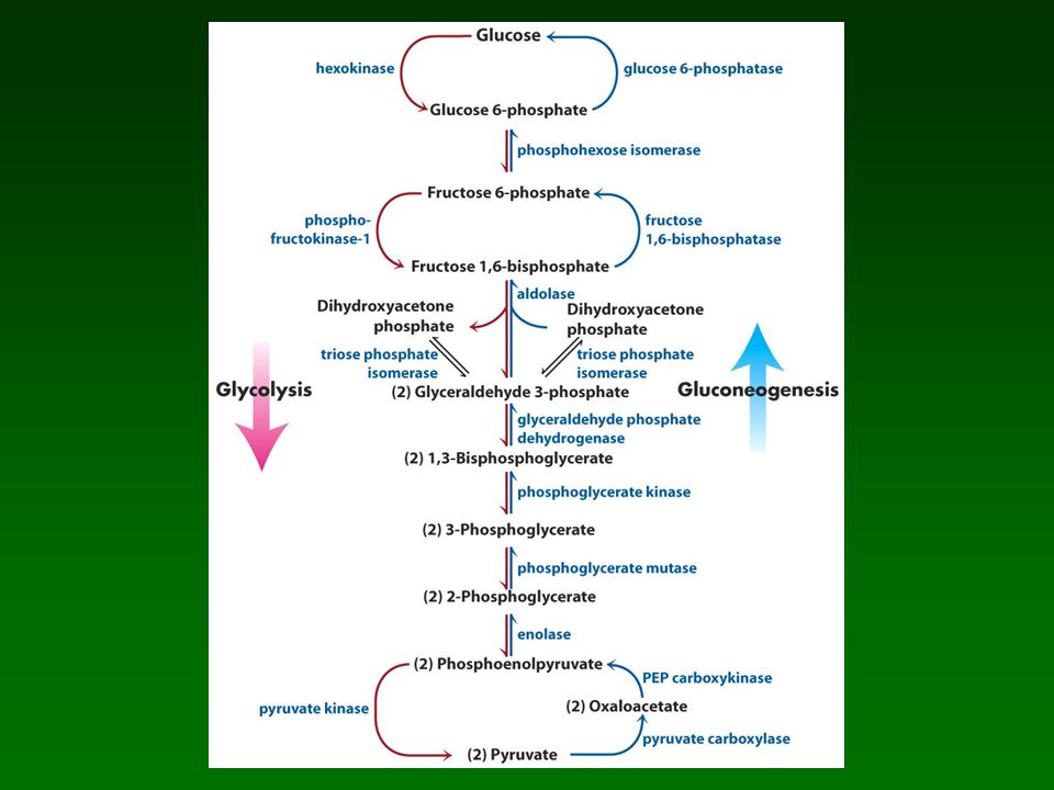
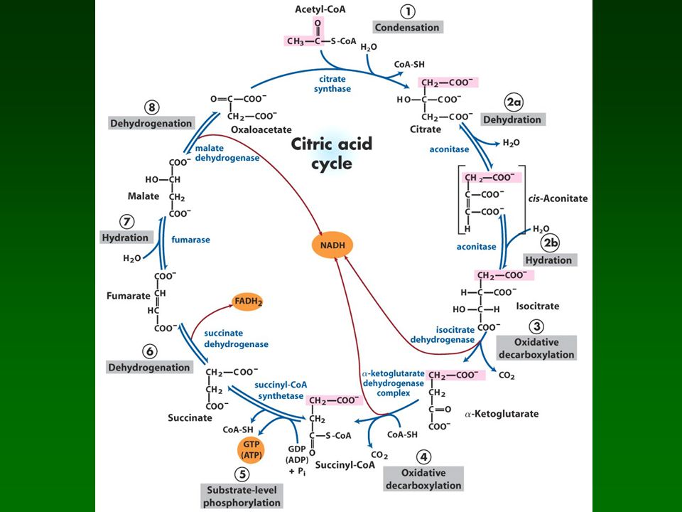
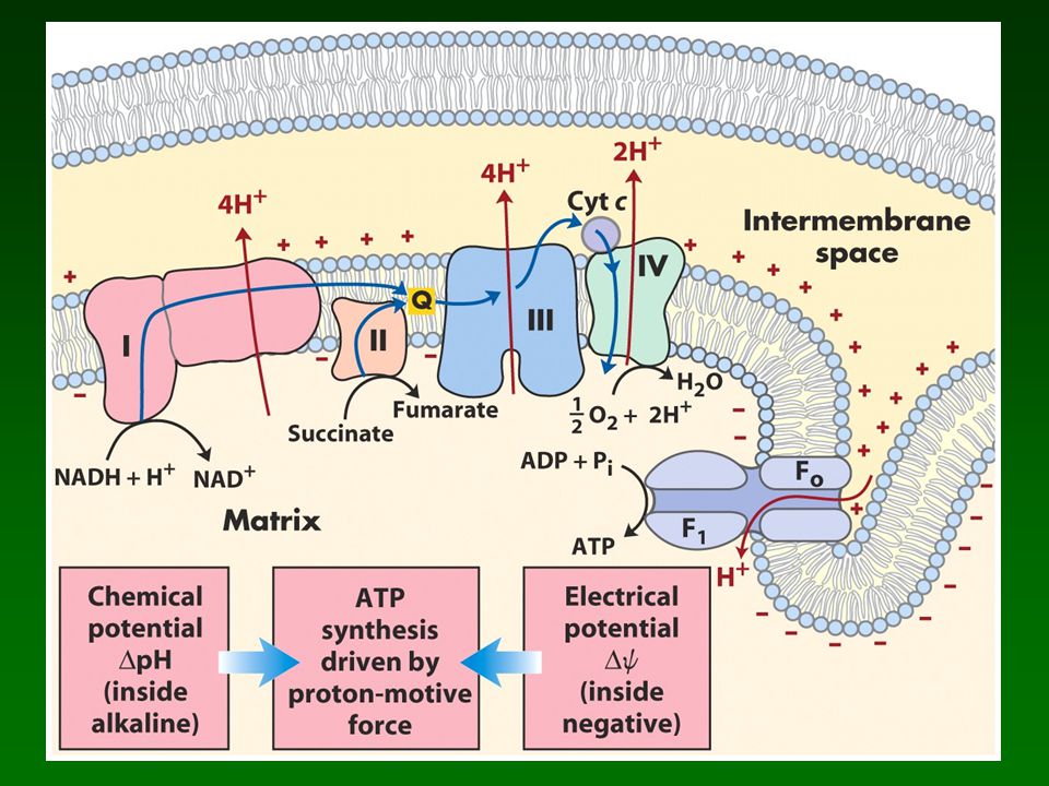
Fuentes de ATP Glucolisis CP TCA-OP Duracion Uso Requerimientos

22
Velocidad de contraccion

23
Músculo estriado cardíaco: características funcionales.
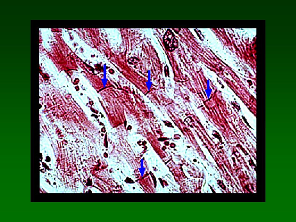
Ubicación Núcleos Control Uniones

27
Actividad eléctrica en el corazón

28
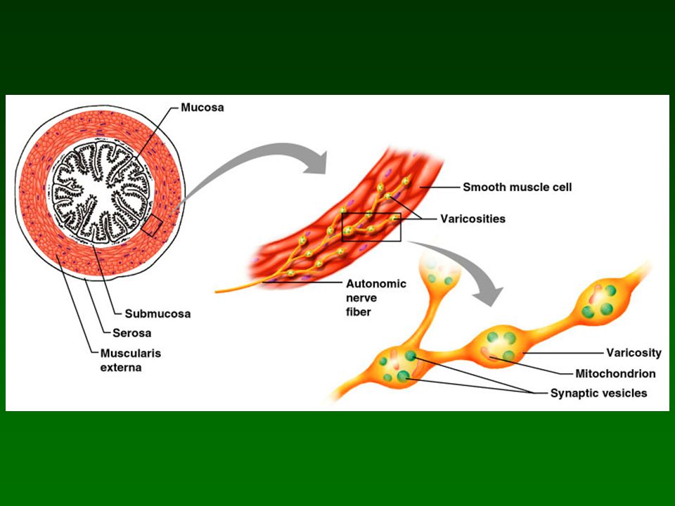
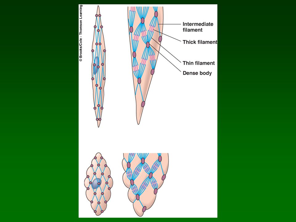
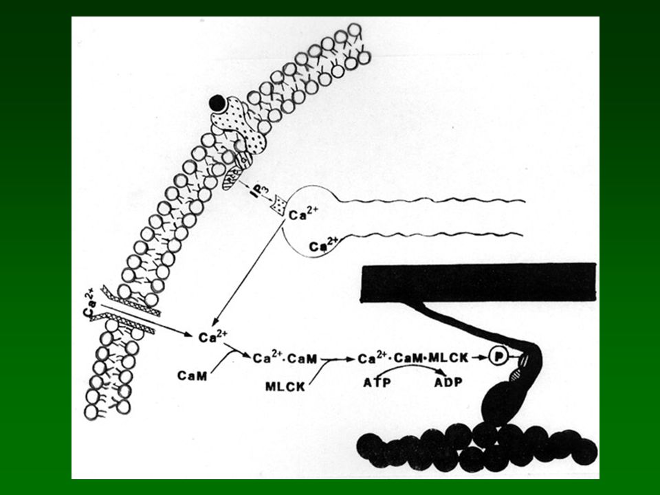
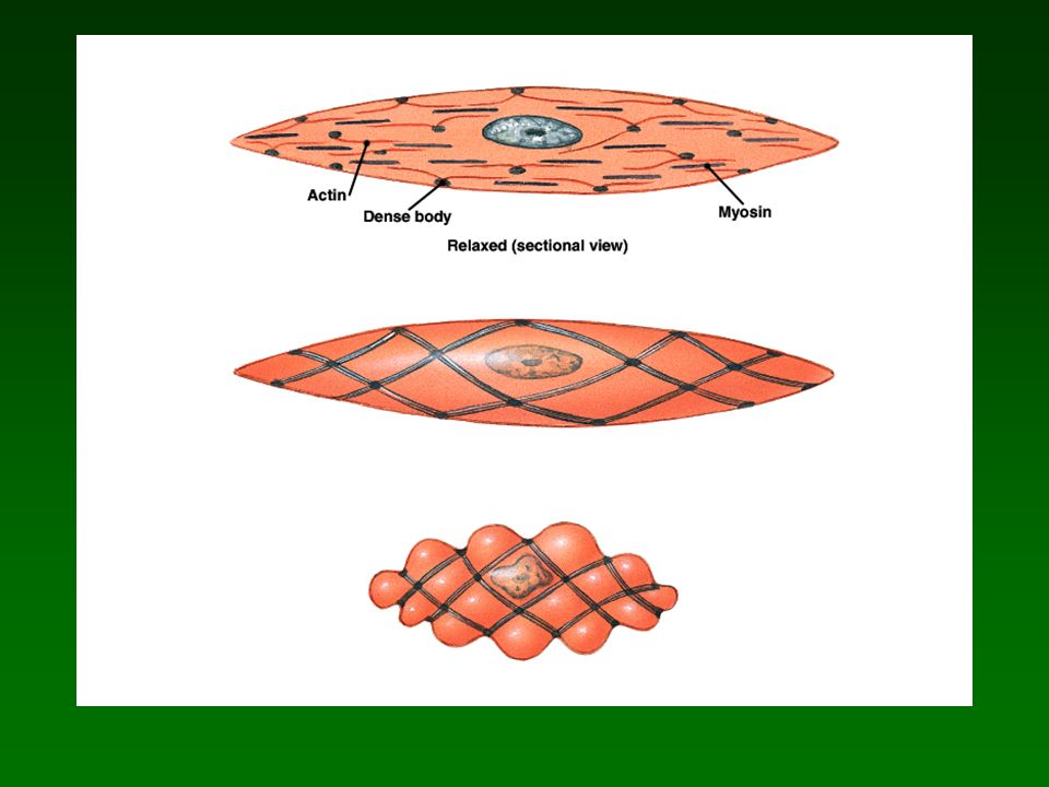
Músculo liso : estructura y mecanismo de contracción-relajación.
Ubicación Núcleos Control Uniones

34
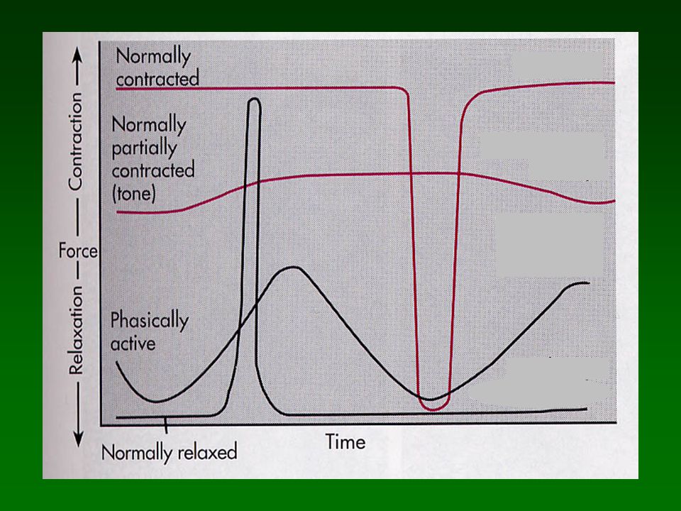
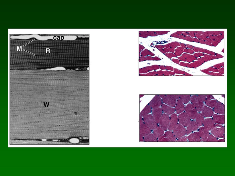
Características funcionales de las fibras fásicas y tónicas.
Contracción Diámetro Irrigación Color Mitocondrias Depositos de CH2O

35
En resumen, ¿Qué determina las características de las fibras?
Graduada o todo o nada? Velocidad de contracción Resistencia a la fatiga

41
Después del esfuerzo: Recuperación

42
Contracciones isométricas e isotónicas, ejemplos
Contracciones isométricas e isotónicas, ejemplos. Curvas tensión-longitud.

43
Longitud (% largo relativo)
60 0.5 1.0 1.25m 1.65m 2.0m 2.25m 3.65m Longitud (% largo relativo) Tensión relativa
 Tensión relativa.")
44
Tension vs. tiempo

45
Temas especiales Organización neuromuscular en vertebrados e invertebrados Esqueletos rígidos e hidráulicos Músculos del vuelo en insectos

49
Musculo estriado Ubicación Nucleos Control

51
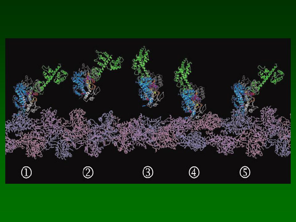
SLIDING FILAMENT THEORY

53
Muscle Tissue Types Skeletal Smooth Cardiac Attached to bones
Nuclei multiple and peripherally located Striated, Voluntary and involuntary (reflexes) Smooth Walls of hollow organs, blood vessels, eye, glands, skin Single nucleus centrally located Not striated, involuntary, gap junctions in visceral smooth Cardiac Heart Striations, involuntary, intercalated disks
 Smooth. Walls of hollow organs, blood vessels, eye, glands, skin. Single nucleus centrally located. Not striated, involuntary, gap junctions in visceral smooth. Cardiac. Heart. Striations, involuntary, intercalated disks.")
54
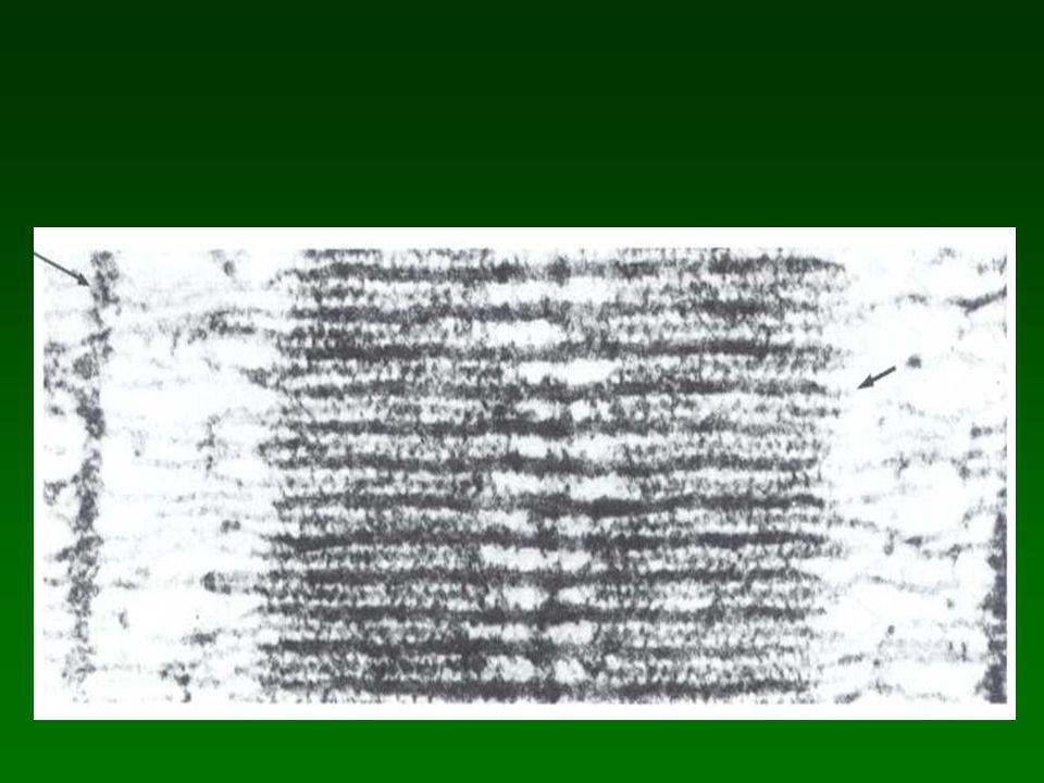
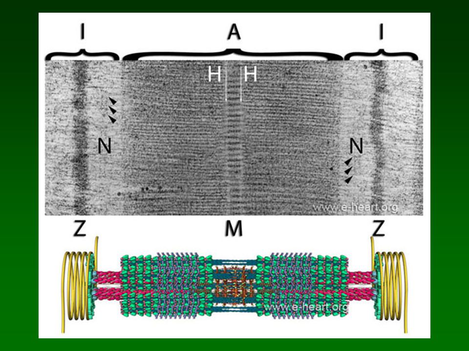
The sarcomere. The upper panel shows a transmission electron micrograph of a human cardiac sarcomere. The boundaries of the sarcomere are the 2 Z-discs (Zwischencheibe) which are dark and electron dense. On each side of the Z-discs, there is an area of electron-lucent material with fine filaments that run perpendicular to the Z-disc. These represent actin filaments which insert into the Z-disc. The area that encompasses a Z-disc and adjacent electron-lucent area of actin filaments from two sarcomeres is designated the I band (Isotropic band). In the middle of this electron-lucent actin rich area, there are some subtle electron-dense streaks that run parallel to the Z-disc and are named the N-disc (Nebenscheibe) . In this area, there are some proteins that interact with titin, actin and α-actinin. The mid portion of the sarcomere is comprised of the A band (Anisotropic band). Within the A band, there are several areas with specific patterns of electron density. There is a middle line or M band (Mittellinie) which bisects the A band. Adjacent to the M band, there are two lighter electron-lucent areas designated the H band (Helle). The M band corresponds to the insertion site of the tails of the thick filaments into a matrix of proteins that keep them organized and in register. Within the M band, there are subtle electron-dense core of proteins which correspond to myomesin and other proteins that serve to anchor the thick filaments. The larger dark areas that flank the M and H bands are the actual rows of thick filaments. In the middle third of these areas, there are some vertical striations which correspond to electron-dense myosin binding protein C. The lower portion of the figure shows a simplified illustration of the sarcomere. The M band shows the tails of the thick filaments interacting with a light brown lattice of myomesin molecules. The thick filaments extend away from the M band with the heads (green) protruding away from the axis of the filament. Myosin binding protein C is visible as small blue subunits that cover about one third of the length of the thick filament. The thin filaments (red) are made of several proteins (see The Thick and Thin Filaments). The yellow coils shown at the level of the Z-disc represent intermediate filaments of desmin which connect adjacent myofibrils to each other and to the sarcolemma. An animation of the sarcomere contraction is shown here and here
 which are dark and electron dense. On each side of the Z-discs, there is an area of electron-lucent material with fine filaments that run perpendicular to the Z-disc. These represent actin filaments which insert into the Z-disc. The area that encompasses a Z-disc and adjacent electron-lucent area of actin filaments from two sarcomeres is designated the I band (Isotropic band). In the middle of this electron-lucent actin rich area, there are some subtle electron-dense streaks that run parallel to the Z-disc and are named the N-disc (Nebenscheibe) . In this area, there are some proteins that interact with titin, actin and α-actinin. The mid portion of the sarcomere is comprised of the A band (Anisotropic band). Within the A band, there are several areas with specific patterns of electron density. There is a middle line or M band (Mittellinie) which bisects the A band. Adjacent to the M band, there are two lighter electron-lucent areas designated the H band (Helle). The M band corresponds to the insertion site of the tails of the thick filaments into a matrix of proteins that keep them organized and in register. Within the M band, there are subtle electron-dense core of proteins which correspond to myomesin and other proteins that serve to anchor the thick filaments. The larger dark areas that flank the M and H bands are the actual rows of thick filaments. In the middle third of these areas, there are some vertical striations which correspond to electron-dense myosin binding protein C. The lower portion of the figure shows a simplified illustration of the sarcomere. The M band shows the tails of the thick filaments interacting with a light brown lattice of myomesin molecules. The thick filaments extend away from the M band with the heads (green) protruding away from the axis of the filament. Myosin binding protein C is visible as small blue subunits that cover about one third of the length of the thick filament. The thin filaments (red) are made of several proteins (see The Thick and Thin Filaments). The yellow coils shown at the level of the Z-disc represent intermediate filaments of desmin which connect adjacent myofibrils to each other and to the sarcolemma. An animation of the sarcomere contraction is shown here and here v=-pg09F5V63U..")
Presentaciones similares


 Usted tiene – You have (Formal) El tiene – He has Ella.>")


 of verbs: those that end in –AR, those that end in –ER, and those that end in –IR. This.>")

. It features two verb changes that we will see very soon.>")


To learn about where people speak Spanish 2) to find out what Spanish is really called 3) to revise the colours 4) to do some brain exercises.>")


nos (us) te(you) le (him/her/you.>")
