Descargar la presentación
La descarga está en progreso. Por favor, espere

1
Por: Arlenne Méndez XII Semestre
EL PERITONEO Por: Arlenne Méndez XII Semestre

2
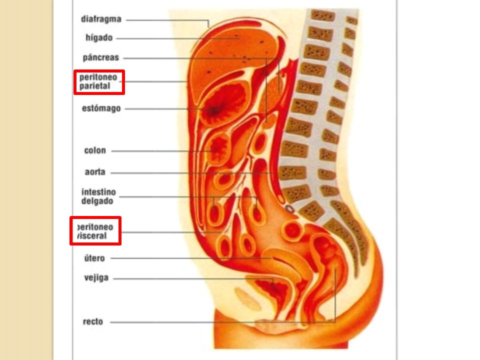
GENERALIDADES Membrana serosa, lisa, brillante, casi transparente y delgada. Constituida por dos hojas: Parietal y Visceral Peritoneo Parietal Peritoneo Visceral

4
CAVIDAD PERITONEAL Cavidad virtual delimitada por las hojas parietal y visceral del peritoneo Completamente cerrada en el hombre y abierta en la mujer Se divide en cavidad peritoneal abdominal y cavidad peritoneal pélvica

5
CAVIDAD PERITONEAL ABDOMINAL CAVIDAD PERITONEAL PÉLVICA

6
FUNCIONES DEL PERITONEO
Sostén Movilidad (mesos) Movimientos peristálticos y desplazamiento de órganos (líquido peritoneal) Reabsorción (líquido peritoneal) Almacena grasa Pedículo Defensa frente a las infecciones intraperitoneales (tabica y aisla/tabica y reabsorbe/reconstrucción)
 Movimientos peristálticos y desplazamiento de órganos (líquido peritoneal) Reabsorción (líquido peritoneal) Almacena grasa. Pedículo. Defensa frente a las infecciones intraperitoneales (tabica y aisla/tabica y reabsorbe/reconstrucción)")
7
TERMINOLOGÍA ASOCIADA AL PERITONEO

8
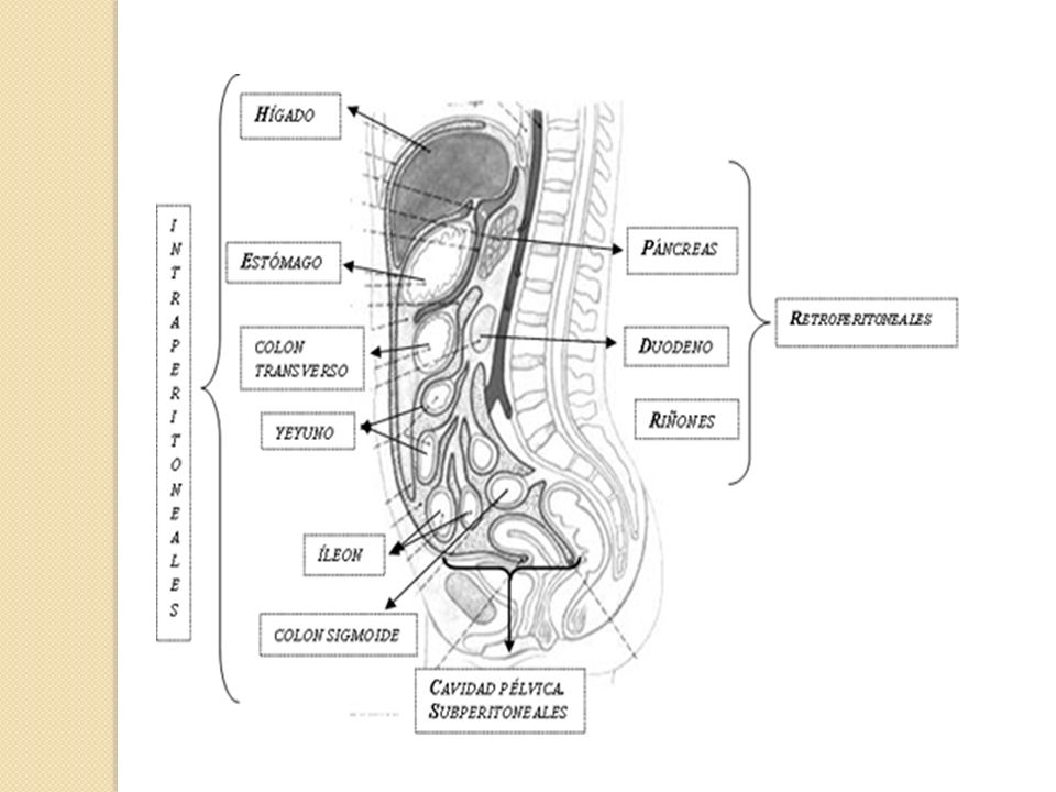
Órgano Intraperitoneal completamente recubiertos por peritoneo
(estómago, bazo, hígado, yeyuno, íleon) Órganos Extraperitoneales Órganos Retroperitoneales: recubiertos por peritoneo sólo en su cara anterior. (páncreas, duodeno, riñones, glándulas suprarrenales, colon asc, colon desc) Órganos Subperitoneales o Infraperitoneales: órganos de la cavidad abdominal pélvica recubiertos por peritoneo en su cara superior. (vejiga urinaria, porción inferior del recto)
 Órganos Extraperitoneales. Órganos Retroperitoneales: recubiertos por peritoneo sólo en su cara anterior. (páncreas, duodeno, riñones, glándulas suprarrenales, colon asc, colon desc) Órganos Subperitoneales o Infraperitoneales: órganos de la cavidad abdominal pélvica recubiertos por peritoneo en su cara superior. (vejiga urinaria, porción inferior del recto)")
10
Órgano Toracoabdominal
se localiza total o parcialmente en la cavidad torácica (hígado, vesícula biliar, bazo, esófago abdominal, fondo y cuerpo gástrico, ángulos hepático y esplénico del colon, parte del colon transverso) Órgano supramesocólico (estómago, hígado y bazo) Órgano inframesocólico (ID y colon asc y desc)
 Órgano supramesocólico. (estómago, hígado y bazo) Órgano inframesocólico. (ID y colon asc y desc)")
13
TÉRMINOS PERITONEALES

14
Meso Doble hoja de peritoneo que une la víscera de la cavidad abdominal a la pared abdominal posterior. Mesoesófago Mesenterio Mesocolon Mesosigmoides Mesoapéndice Epiplón Doble hoja de peritoneo que se refleja desde el estómago para cubrir órganos vecinos. Epiplón Menor Epiplón Mayor

15
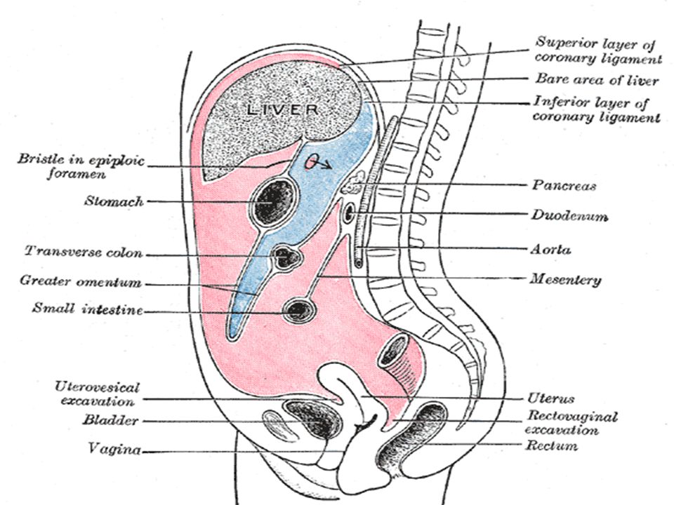
Figure 2. 19. Principal formations of peritoneum. A
Figure Principal formations of peritoneum. A. In this anterior view of the opened peritoneal cavity, parts of the greater omentum, transverse colon, and the small intestine and its mesentery have been cut away to reveal deep structures and the layers of the mesenteric structures. The mesentery of the jejunum and ileum (small intestine) and sigmoid mesocolon have been cut close to their parietal attachments. B. The median section of the abdominopelvic cavity of a male shows the relationships of the peritoneal attachments. C. The greater omentum is shown in its “normal†position, covering most of the abdominal viscera. D. The lesser omentum, attaching the liver to the lesser curvature of the stomach, is shown by reflecting the liver and gallbladder superiorly. The greater omentum has been removed from the greater curvature of the stomach and transverse colon to reveal the intestines. E. The greater omentum has been reflected superiorly and the small intestine has been retracted to the right side to reveal the mesentery of the small intestine and the mesentery of the transverse colon (transverse mesocolon).
 and sigmoid mesocolon have been cut close to their parietal attachments. B. The median section of the abdominopelvic cavity of a male shows the relationships of the peritoneal attachments. C. The greater omentum is shown in its “normal†position, covering most of the abdominal viscera. D. The lesser omentum, attaching the liver to the lesser curvature of the stomach, is shown by reflecting the liver and gallbladder superiorly. The greater omentum has been removed from the greater curvature of the stomach and transverse colon to reveal the intestines. E. The greater omentum has been reflected superiorly and the small intestine has been retracted to the right side to reveal the mesentery of the small intestine and the mesentery of the transverse colon (transverse mesocolon).")
16
Se subdivide en 3 ligamentos:
Epiplón Mayor Pliegue peritoneal prominente que cuelga a modo de “delantal” de la curvatura mayor del estómago y de la porción proximal del duodeno. Después de descender se repliega para insertarse en la cara anterior del colon transverso y en su mesenterio Se subdivide en 3 ligamentos: Ligamento gastrofrénico Ligamento gastroesplénico Ligamento gastrocólico

17
Ligamentos del epiplón menor:
Doble hoja de peritoneo que une la curvatura menor del estómago y la parte proximal de la primera porción del duodeno con el hígado. Ligamentos del epiplón menor: Ligamento gastrohepático (pars flácida) Ligamento hepatoduodenal (pars densa):. Contiene el pedículo hepático
 Ligamento hepatoduodenal (pars densa):. Contiene el pedículo hepático.")
18
Figure 2. 20. Parts of greater and lesser omenta
Figure Parts of greater and lesser omenta. The blue arrows indicate that the liver and gallbladder have been reflected superiorly. The central part of the greater omentum has been cut out to show its relation to the transverse colon and mesocolon. The term greater omentum is often used as a synonym for the gastrocolic ligament, but it actually also includes the gastrosplenic and gastrophrenic ligaments, all of which have a continuous attachment to the greater curvature of the stomach. The hepatoduodenal ligament (free edge of the lesser omentum) conveys the portal triad: hepatic artery, bile duct, and portal vein.
 conveys the portal triad: hepatic artery, bile duct, and portal vein.")
19
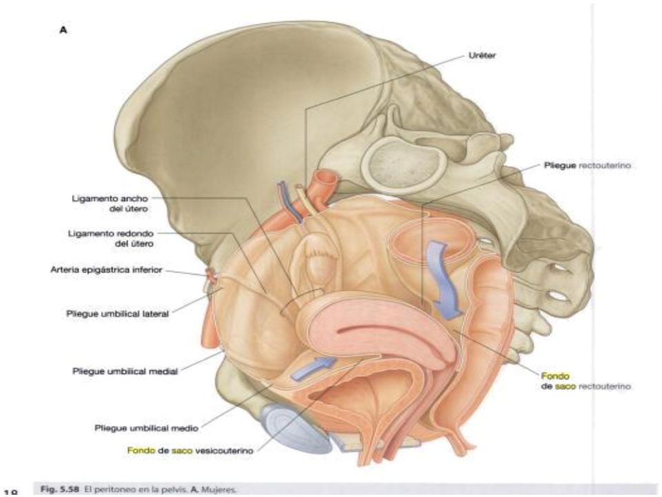
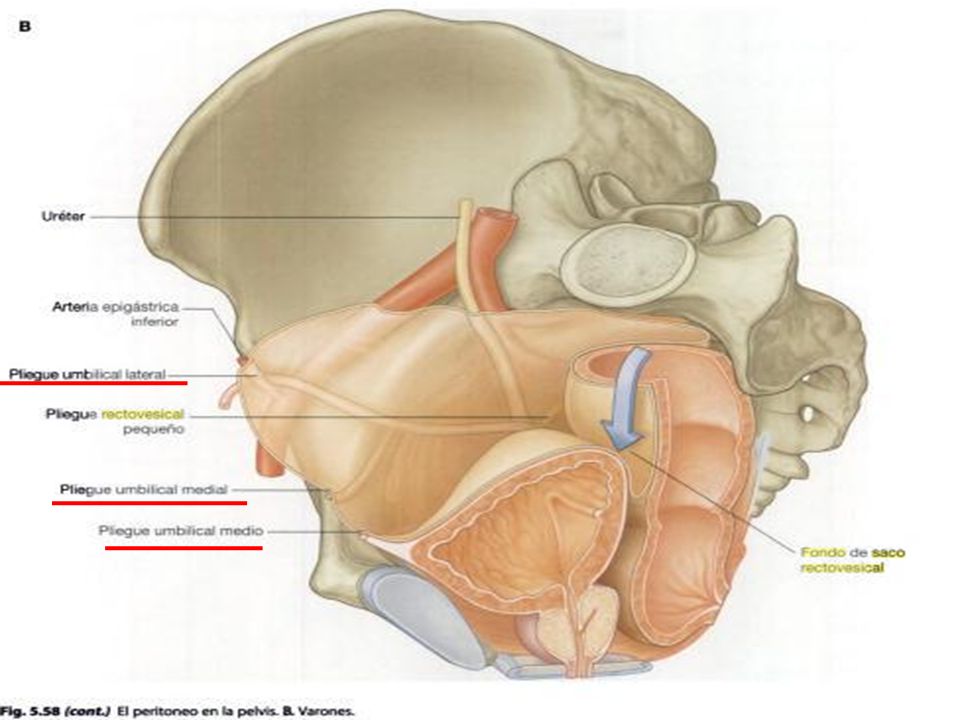
Ligamentos Doble hoja de peritoneo pueden contener o no elementos vasculonerviosos. Fondo de saco Sitio donde el peritoneo se pliega para formar una bolsa que está cerrada en un extremo y por el otro se comunica con la cavidad peritoneal Fondo de saco rectouterino o de Douglas Fondo de saco vesicorectal Fondo de saco pararectal

22
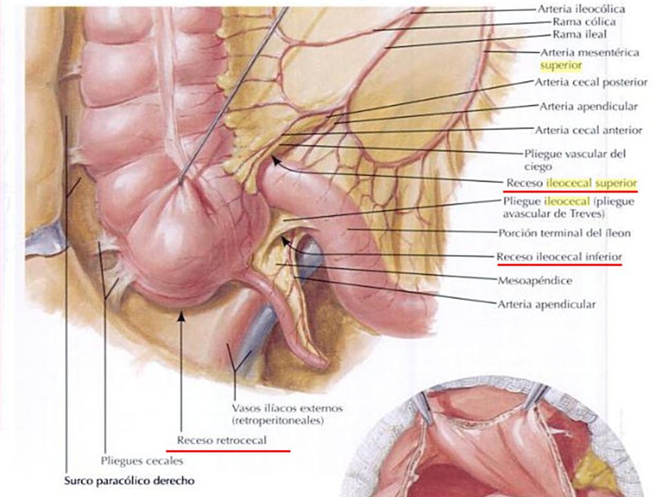
Fosas: Fondo de sacos pequeños
Fosa retrocecal Fosa ileocecal superior Fosa ileocecal inferior Fosa intersigmoidea Pliegues Reflexión de peritoneo que se forma del peritoneo que cubre vasos sanguíneos, conductos o vasos obliterados Espacios subfrénicos Espacios entre la cara inferior del diafragma y cara superior del hígado

25
BOLSA MENOR BOLSA OMENTAL TRANSCAVIDAD DE LOS EPIPLONES

26
La bolsa omental tiene:
Cavidad extensa, con forma de saco, que se encuentra posterior al estómago , estructuras adyacentes y omento menor La bolsa omental tiene: Receso superior: limitado por arriba por el diafragma y capas posteriores del ligamento coronario del hígado Receso inferior: detrás del estómago y entre las capas dobles del ligamento gastrocólico del epiplón mayor Receso esplénico: se extiende hacia la izq. quedando cerrada por los ligamentos pancreatoesplénico y gastroesplénico

28
Límites del Hiato de Winslow:
Anterior: pedículo hepático Posterior: VCI y pilar derecho del diafragma Superior: lóbulo caudado del hígado Inferior: porción superior o primera porción del duodenoy conducto biliar

29
Figure 2. 17. Transverse section of abdomen at level of omental bursa
Figure Transverse section of abdomen at level of omental bursa. The orientation figure indicates the level of the section superficially. The dark arrow passes from the greater sac of the peritoneal cavity through the omental (epiploic) foramen and across the full extent of the omental bursa (lesser sac).
 foramen and across the full extent of the omental bursa (lesser sac).")
30
Figure 2. 22. Omental (epiploic) foramen and omental bursa. A
Figure Omental (epiploic) foramen and omental bursa. A. The index finger is passing from the greater sac through the omental foramen into the omental bursa (lesser sac). B. The hepatic artery is being compressed between the index finger (passing through the omental foramen into the omental bursa) and the thumb (on the anterior wall of the foramen).
 foramen and omental bursa. A. The index finger is passing from the greater sac through the omental foramen into the omental bursa (lesser sac). B. The hepatic artery is being compressed between the index finger (passing through the omental foramen into the omental bursa) and the thumb (on the anterior wall of the foramen).")
31
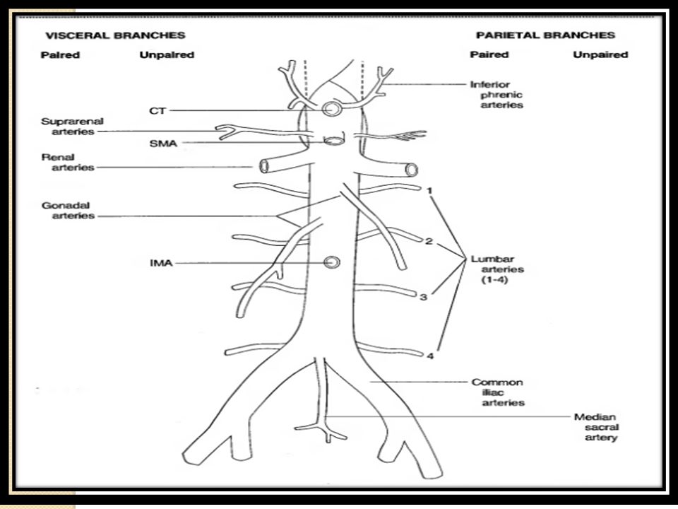
AORTA ABDOMINAL

34
GRACIAS!!!!!

Presentaciones similares


 1. Hígado 2>")


