Descargar la presentación
La descarga está en progreso. Por favor, espere

2
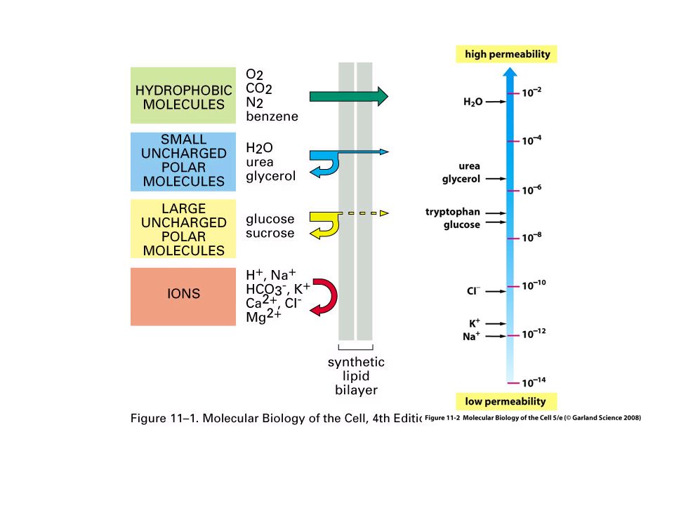
Cabeza hidrofilica Cola hidrofobica AGUA

7
LE 7-5b Viscous Fluid Unsaturated hydrocarbon tails with kinks Membrane fluidity Saturated hydro- carbon tails

8
LE 7-5a Lateral movement (~10 7 times per second) Flip-flop (~ once per month) Movement of phospholipids
 Flip-flop (~ once per month) Movement of phospholipids")
9
LE 7-5c Cholesterol Cholesterol within the animal cell membrane

10
Region hidrofilica de la proteina Region hidrofobica de la proteina Bicapa fosfolipido

11
EXTRACELLULAR SIDE N-terminus C-terminus CYTOPLASMIC SIDE Helix

13
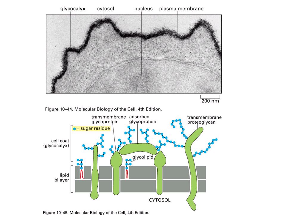
Glico- proteina reconocimiento celula-celula Intercellular joining Attachment a el Citoesqueleto y matriz extra- Celular (ECM)
")
14
Enzimas Signal Receptor ATP Transporte Actividad enzimatica transduccion señal

15
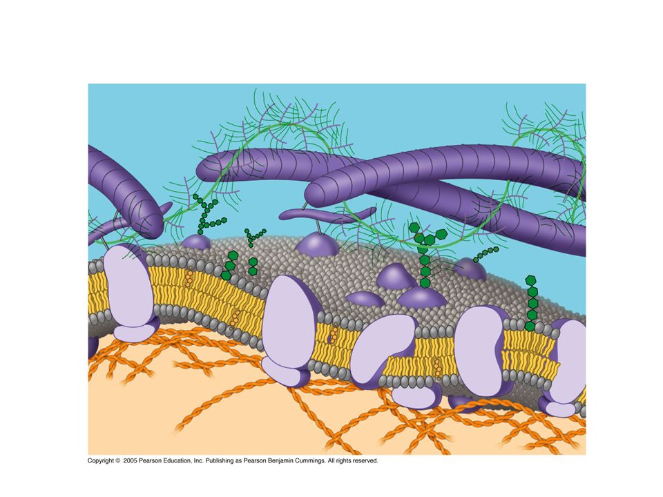
Fibras de matriz extracellular (ECM) Glicoproteina Carbohidrato Microfilamentos De citoesqueleto Colesterol Proteina Integral proteinas Perifericas LADO CITOPLASMATICO DE MEMBRANA LADO EXTRACELLULAR DE MEMBRANA Glicolipido
 Glicoproteina Carbohidrato Microfilamentos De citoesqueleto Colesterol Proteina Integral proteinas Perifericas LADO CITOPLASMATICO DE MEMBRANA LADO EXTRACELLULAR DE MEMBRANA Glicolipido")
18
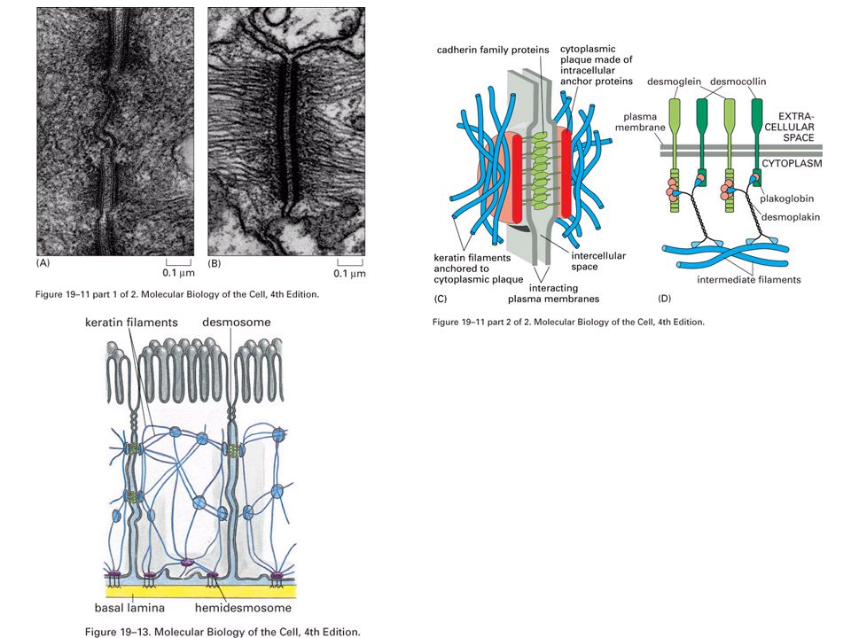
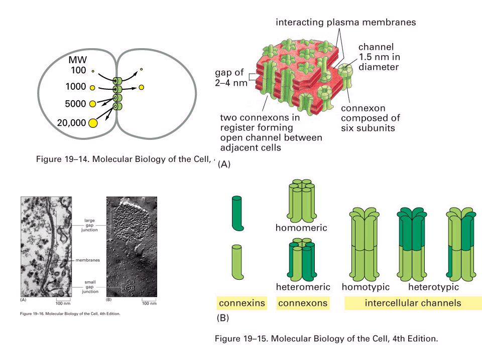
Hemi- desmosoma Lamina basal Superficie Basal Desmosoma Matriz extracelular (ECM) Superficie apical Conexon Unión impermeable Unión adherencia Unión comunicante
 Superficie apical Conexon Unión impermeable Unión adherencia Unión comunicante")
19
Microvellosidad Hemidesmosoma Lamina Basal Tejido conectivo Superficie basal Superficie lateral Superficie apical Filamentos intermedios Desmosoma Unión comunicante Filamentos actina y miosina Unión adherencia Unión impermeable

26
Moleculas de dyeMembrana (sección a traves) AGUA difusión neta Equilibrio Difusión de un soluto
 AGUA difusión neta Equilibrio Difusión de un soluto")
27
difusión neto Equilibrio Difusión de dos solutos difusión neto Equilibrio

28
Baja concentración de soluto (azucar) Alta concentración de azucar Igual concentración de azucar Selectiva Permeabilidad de Membrana: moléculas de azucar no pueden pasar a traves de los Poros, pero el agua si puede H2OH2O Osmosis
 Alta concentración de azucar Igual concentración de azucar Selectiva Permeabilidad de Membrana: moléculas de azucar no pueden pasar a traves de los Poros, pero el agua si puede H2OH2O Osmosis")
29
“Celula” 0.03 M sucrosa 0.02 M glucosa 0.01 M sucrosa 0.01 M glucosa 0.01 M fructosa ambiente

30
Medio isotónico Medio hipertónico Medio hipotónico

31
Célula Animal Lisis H2OH2O H2OH2O H2OH2O Normal Solución hipotónica Solucion Isotónica Solución Hipertónica H2OH2O Crenación H2OH2O H2OH2O H2OH2O H2OH2O Célula Planta Turgencia (normal) FlacidezPlasmolisis
 FlacidezPlasmolisis")
32
Filling vacuole 50 µm Contracting vacuole

37
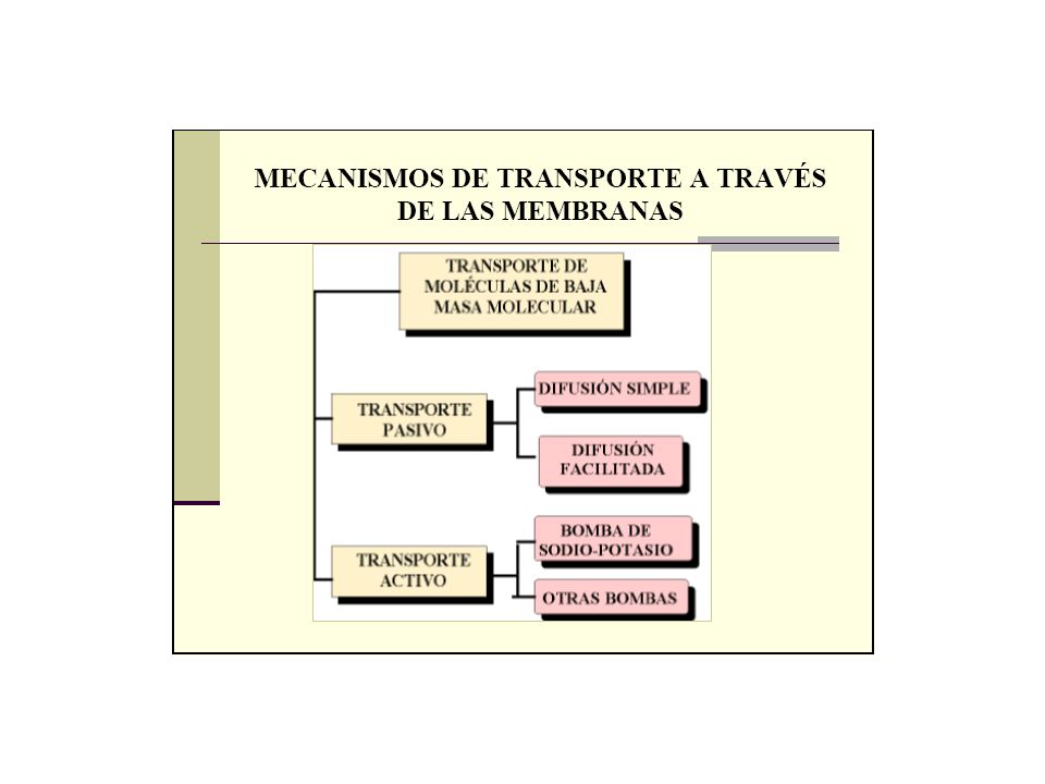
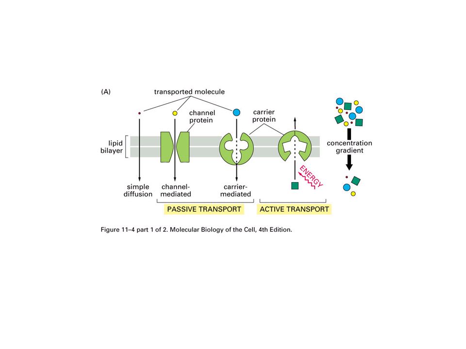
Difusion Difusion facilitada Transporte pasivo ATP Transporte activo

38
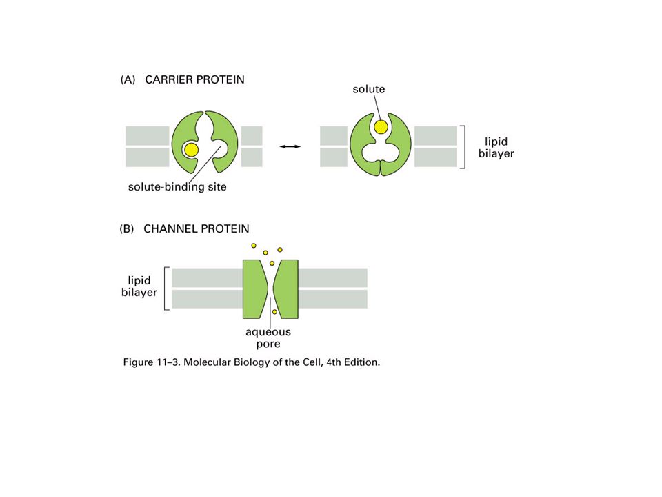
FLUIDO EXTRACELULAR Canal proteico Soluto CITOPLASMA

39
proteina carrier Soluto

40
LE 7-17b ATP Active transport

42
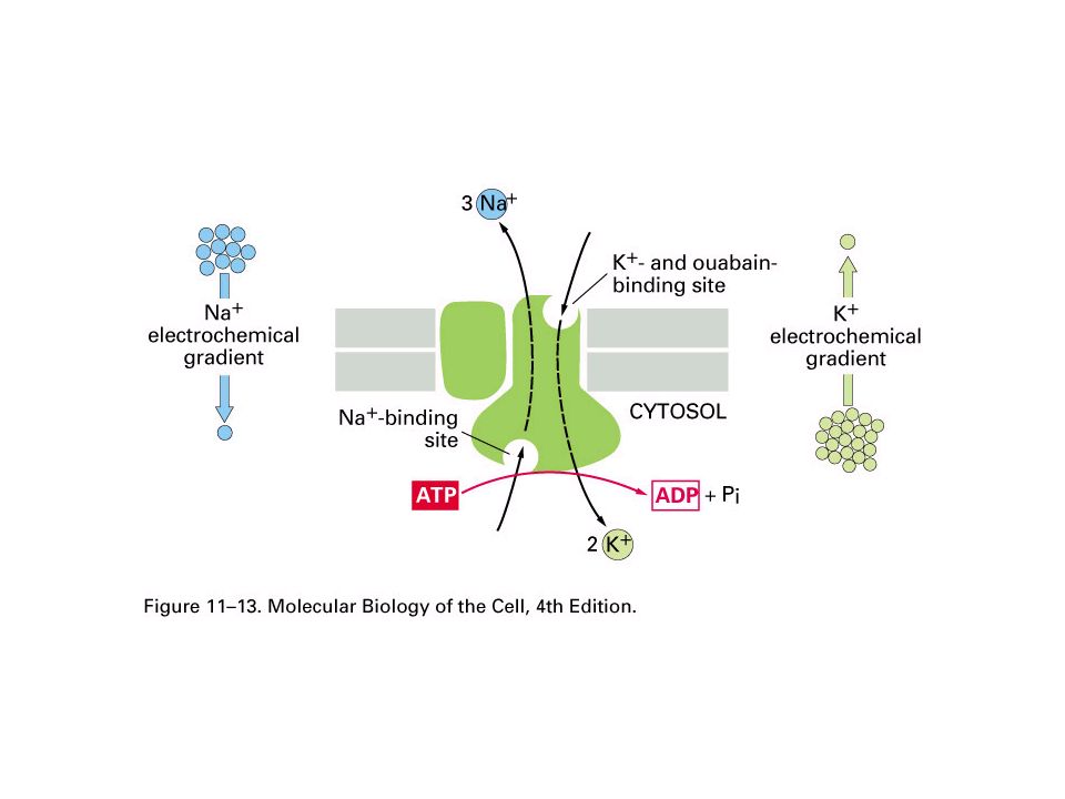
LE 7-16 Cytoplasmic Na + bonds to the sodium-potassium pump CYTOPLASM Na + [Na + ] low [K + ] high Na + EXTRACELLULAR FLUID [Na + ] high [K + ] low Na + ATP ADP P Na + binding stimulates phosphorylation by ATP. Na + K+K+ Phosphorylation causes the protein to change its conformation, expelling Na + to the outside. P Extracellular K + binds to the protein, triggering release of the phosphate group. P P Loss of the phosphate restores the protein’s original conformation. K + is released and Na + sites are receptive again; the cycle repeats. K+K+ K+K+ K+K+ K+K+ K+K+
![LE 7-16 Cytoplasmic Na + bonds to the sodium-potassium pump CYTOPLASM Na + [Na + ] low [K + ] high Na + EXTRACELLULAR FLUID [Na + ] high [K + ] low Na + ATP ADP P Na + binding stimulates phosphorylation by ATP.](http://images.slideplayer.es/33/10360743/slides/slide_42.jpg "Na + K+K+ Phosphorylation causes the protein to change its conformation, expelling Na + to the outside. P Extracellular K + binds to the protein, triggering release of the phosphate group. P P Loss of the phosphate restores the protein’s original conformation. K + is released and Na + sites are receptive again; the cycle repeats. K+K+ K+K+ K+K+ K+K+ K+K+.")
43
LE 7-18 H+H+ ATP CYTOPLASM EXTRACELLULAR FLUID Proton pump H+H+ H+H+ H+H+ H+H+ H+H+ + + + + + – – – – –

44
LE 7-19 H+H+ ATP Proton pump Sucrose-H + cotransporter Diffusion of H + Sucrose H+H+ H+H+ H+H+ H+H+ H+H+ H+H+ + + + + + + – – – – – –

47
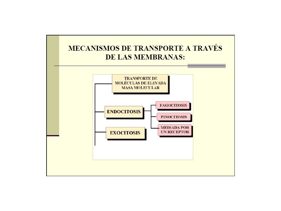
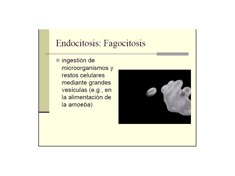
CITOPLASMA Pseudopodo “Alimento” u otra particula FLUIDO EXTRACELULAR Bacterium Food vacuole An amoeba engulfing a bacterium via phagocytosis (TEM) Pseudopodium of amoeba 1 µm Vacuola alimenticia FAGOCITOSIS
 Pseudopodium of amoeba 1 µm Vacuola alimenticia FAGOCITOSIS")
49
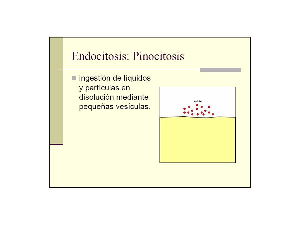
Membrana Plasmatica Pinocytosis vesicles forming (arrows) in a cell lining a small blood vessel (TEM). 0.5 µm Vesicula PINOCITOSIS

51
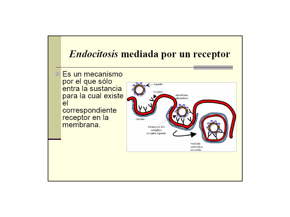
Receptor RECEPTOR-MEDIATED ENDOCYTOSIS Ligand Coated pit Coated vesicle Coat protein Coat protein Plasma membrane 0.25 µm A coated pit and a coated vesicle formed during receptor- mediated endocytosis (TEMs).
.")
Presentaciones similares