Descargar la presentación
La descarga está en progreso. Por favor, espere

1
UNIVERSIDAD VERACRUZANA NUEVO MODELO EDUCATIVO
PROGRAMA DE ESTUDIOS LICENCIATURA DE ENFERMERIA FACULTAD DE ENFERMERIA FEBRERO DE 2006

2
INICIACIÓN A LA DISCIPLINA
EXPERIENCIA EDUCATIVA: ANATOMIA Y FISIOLOGIA AREA DE CONOCIMIENTO: INICIACIÓN A LA DISCIPLINA ACADEMIA A LA QUE PERTENECE: AREA I

3
Dr. Carlos Blázquez Domínguez.
ACADEMICO: Dr. Carlos Blázquez Domínguez.

4
ANATOMIA Y FISIOLOGIA DEL TEJIDO
UNIDAD I TEJIDOS CORPORALES ANATOMIA Y FISIOLOGIA DEL TEJIDO

5
ANATOMIA Y FISIOLOGIA DEL TEJIDO
La histología humana tiene su origen en las tres capas del embrión (gástrula), a las que denominamos capas germinales porque de su crecimiento y diferenciación se forman todos los tejidos que integran a un ser completo y adulto.
, a las que denominamos capas germinales porque de su crecimiento y diferenciación se forman todos los tejidos que integran a un ser completo y adulto.")
6
ANATOMIA Y FISIOLOGIA DEL TEJIDO
Los tejidos se diferencias por su estructura y su función, atendiendo además a la naturaleza y cantidad de la sustancia que se interpone entre las células especificas o sustancia intercelular

7
ANATOMIA Y FISIOLOGIA DEL TEJIDO
La sustancia intercelular puede presentarse en mucha o poca cantidad y su estado físico varia del liquido y semiliquido al sólido.

10
The Simple Epithelial Tissue Types
Simple Squamous Epithelium:

11
Simple Cuboidal Epithelium:

12
Simple Columnar Epithelium:

13
The Pseudostratified Epithelial Tissue Type
Pseudostratified Columnar Epithelium:

14
A Schematic of Pseudostratified Columnar Epithelium

15
The Stratified Epithelial Tissue Type
Stratified Squamous Epithelium:

18
Embryonic Connective Tissue Types
Mesenchyme:

19
Mucoid:

20
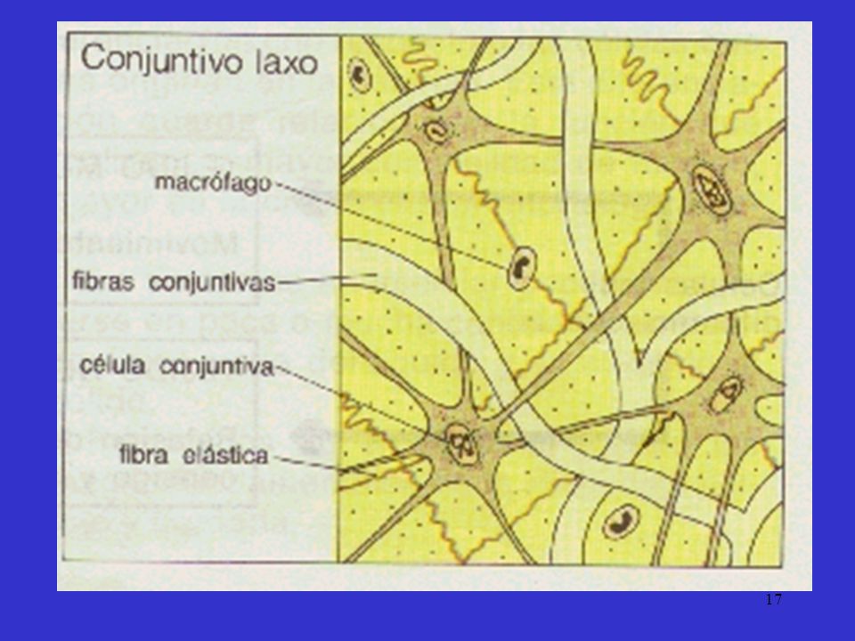
Ordinary Connective Tissue Types
Loose:

21
Dense:

22
Tissue Types: Cartilage, Bone and Blood
Special Connective Tissue Types: Cartilage, Bone and Blood

23
Cartilage and Adipose Cartilage:

24
Adipose:

26
Bone: 1 Haversian Canal 1 Haversian Canal 2 Canaliculi
3 Lamellae 4 Lacunae 1 Haversian Canal 2 Haversian System

27
A Schematic of a Long Bone

28
Blood

29
Sickle cell

30
Sickle cell A computer-coloured scanning electron microscope image of a sickled red blood cell surrounded by normal cells. Sickle cell anaemia results from a genetic defect, and is particularly common in people of Afro-Caribbean origin. Affected red blood cells contain an abnormal type of haemoglobin, haemoglobin S, which crystallizes in regions of low oxygen concentration, such as the blood capillaries, causing a sickle-like distortion of the red blood cells. Sickled cells are fragile and easily disrupted, reducing the oxygen-carrying capacity of the blood and resulting in severe anaemia (actual size of sickle cell: about 19 thousandths of a millimetre from end to end). Dr Jackie Lewin, E M Unit, Royal Free Hospital School of Medicine, London
. Dr Jackie Lewin, E M Unit, Royal Free Hospital School of Medicine, London.")
31
Muscle Tissue Smooth Involuntary (Smooth) Muscle Tissue
 Muscle Tissue")
32
Striated Voluntary (Skeletal) Muscle Tissue
 Muscle Tissue")
33
A Schematic of Skeletal Muscle

34
A Schematic of Skeletal Muscle

35
Cardiac Muscle Tissue 1 Cardiac Muscle Cell 2 Nuclei
3 Intercalated Discs

37
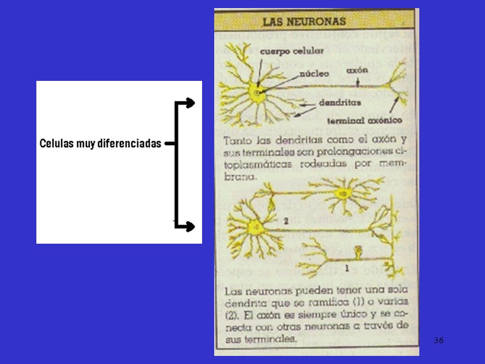
Nervous Tissue

38
Rod cells in retina

39
Rod cells in retina A colour-enhanced scanning electron micrograph of the retina in a guinea-pig eye. To the left of the picture is the photoreceptor layer which contains the light-sensitive rod cells that help convert light impulses into an image in the brain. To the right is the choroid layer of the retina which contains a network of blood vessels like the one shown emerging here. Dr David Furness, Department of Communication and Neuroscience, Keele University

40
BIBLIOGRAFÍA RECOMENDADA
1. Boya Vegue J. Atlas de Histología y Organografía Microscópica. Madrid. Ed Médica Panamericana, 1996.Burkitt HG, 2. Young s, Heath JW. Wheater Histología Funcional. Texto y Atlas en color (3a ed). Madrid. Ed. Churchill Livingstone, 1993 3. Fawcett DW. Tratado de Histología (128 ed.) Madrid: Interamericana. McGraw-Hill 4. Geneser E. Histología (28 ed.). Madrid. Ed: Médica Panamericana, 1993. 5. Junqueira LC, Carneiro J. Histoloda Básica. Texto y Atlas. (4é ed). Barcelona. ed: Masson 6. Ross MH, Reith EJ, Romrell Lj. Histología. Texto y aflas en color. (28 ed.) Buenos Aires: Panamericana, 1992. 7. Stevens A, Lowe J. Histology (1. ed.) London: Gower med. Publish.Co., 1992.
. Madrid. Ed. Churchill Livingstone, Fawcett DW. Tratado de Histología (128 ed.) Madrid: Interamericana. McGraw-Hill Geneser E. Histología (28 ed.). Madrid. Ed: Médica Panamericana, Junqueira LC, Carneiro J. Histoloda Básica. Texto y Atlas. (4é ed). Barcelona. ed: Masson Ross MH, Reith EJ, Romrell Lj. Histología. Texto y aflas en color. (28 ed.) Buenos Aires: Panamericana, Stevens A, Lowe J. Histology (1. ed.) London: Gower med. Publish.Co.,")
Presentaciones similares


—to melt rock. This.>")


. Zones 1)Conducting -Warm -Filter -Moisten 2) Respiratory - Gas Exchange.>")


