Descargar la presentación
La descarga está en progreso. Por favor, espere

1
BLOQUEOS ATRIOVENTRICULARES
Dr. José Antonio Espejel Santana Urgencias Médico Quirúrgicas Unidad Medica de Alta de Especialidad de Centro Médico “La Raza” México

2
MIOCARDIO Filamentos de actina y miocina Sarcolema Discos Intercalares
Función de “sincitio” Sincitio atrial y ventricular

3
SISTEMA DE CONDUCCIÓN

4
ELECTROCARDIOGRAMA Registro de la actividad eléctrica del corazón
Onda P: Despolarización de las aurículas. Intervalo PR: Retardo del impulso en el nodo auriculoventricular. Segmento QRS: Despolarización de los ventrículos. Onda T: Repolarización ventricular. QRS waveform nomenclature The ECG consists of a small deflection called the P wave, arising from the atria, a more complicated deflection called the QRS complex due to ventricular depolarisation and a final T wave resulting from repolarisation of the ventricles. The QRS complex of waves is the largest deflection of the ECG and is always spiky in shape. All sharp deflections resulting from electrical activation of the ventricles are called QRS complexes. However, these waves can vary immensely in size, and arrangement. The QRS complex is very important when diagnosing myocardial infarction. In order to be able to describe these complexes, a nomenclature for the waves is needed. This can be done using combinations of the letters q, r, s, Q, R, S, lower case letters denoting small waves and upper case larger waves. The first positive wave is labelled with r or R Any second positive wave is labelled r´ or R´ A negative wave which follows an R wave or r wave is labelled S or s A negative wave that precedes an R or r wave, is labelled a q or Q wave Any wave that is entirely negative is labelled qs or QS. Using these rules and nomenclature all QRS complexes can be described, enabling more accurate diagnosis.

5
Intervalo PR mide normalmente: 0.12 a 0.20 seg.
ELECTROCARDIOGRAMA Intervalo PR mide normalmente: 0.12 a 0.20 seg. Location of infarction and its relation to the ECG: inferior infarction ST elevation in leads II, III and aVF, and often ST depression in I, aVL, and precordial leads are signs of an inferior (lower) infarction. Inferior infarctions may occur due to occlusion of the right circumflex coronary arteries resulting in infarction of the inferior surface of the left ventricle, although damage can be made to the right ventricle and interventricular septum. This type of infarction often results in bradycardia due to damage to the atrioventricular node.
 infarction. Inferior infarctions may occur due to occlusion of the right circumflex coronary arteries resulting in infarction of the inferior surface of the left ventricle, although damage can be made to the right ventricle and interventricular septum. This type of infarction often results in bradycardia due to damage to the atrioventricular node.")
6
BLOQUEOS ATRIOVENTRICULAR
Retrazo ó interrupción de la conducción entre aurículas y los ventrículos Characteristic changes in AMI The 12-lead ECG is the most useful investigation for confirming the diagnosis of acute myocardial infarction, locating the site of the infarct and monitoring the progress. It is therefore very important to know the changes that occur in this situation. The only diagnostic evidence of a completed myocardial infarction seen on the ECG are those in the QRS complexes. In the early stages changes are also seen in the ST segment and the T wave, and these can be used to assist diagnosis of myocardial infarctions. Shortly after infarction there is an elevation of the ST segment seen over the area of damage, and opposite changes are seen in the opposite leads. Several hours later pathological Q waves begin to form, and tend to persist. Later the R wave becomes reduced in size, or completely lost. Later still, the ST segment returns to normal, and at this point the T wave also decreases, eventually becoming deeply and symmetrically inverted. Although these changes occur sequentially, it is very unlikely they will all be clearly observed by the paramedic or GP. A patient can present at any stage and a progression through the ECG changes will not be seen. It is important to recognise these features as they occur rather than in association with each other. All these changes imply myocardial infarction, and will be discussed in more detail over the next few slides.

7
ETIOLOGIA DE LOS BLOQUEOS AV
Cardiopatías: Isquémica Valvulopatías Miocardiopatías Enfermedad degenerativa Medicamentos: Metoprolol Digoxina Alteraciones en el K Hiperkalemia Rule 10 In leads I, II, and V2 to V6 the T wave must be upright.

8
CLASIFICACIÓN DE LOS BLOQUEOS AV
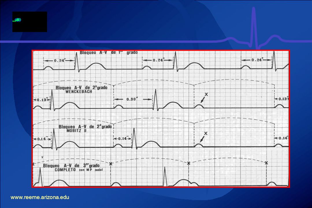
Bloqueo AV de primer grado Bloqueo AV de segundo grado Bloqueo AV de segundo grado – Mobitz I (Fenómeno de Wenckebach) Bloqueo AV de segundo grado – Mobitz II Bloqueo AV completo
 Bloqueo AV de segundo grado – Mobitz II. Bloqueo AV completo.")
10
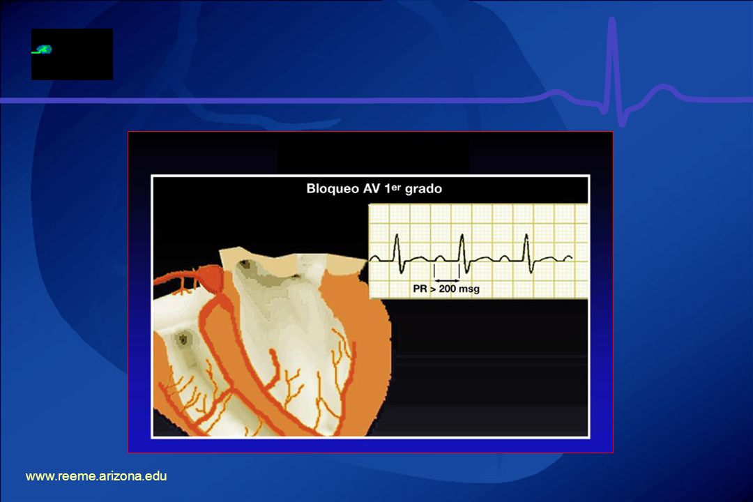
BLOQUEO AV DE PRIMER GRADO
Intervalo PR > 0.20 seg. ó 200 mseg. Intervalo PR prolongado es fijo. Siempre a la onda P le sigue un QRS. Ritmo es regular. The 10 rules for a normal ECG For an ECG to be determined as normal, Chamberlain has described 10 rules which must be met.1 The next ten slides will outline these rules. Chamberlain DAC. Personal communications.

11
BLOQUEO AV DE PRIMER GRADO
Rule 1 As described in Module 3, the PR interval is the time from initiation of depolarisation of the atria to initiation of the depolarisation of the ventricles. The PR interval should be 120 to 200 milliseconds, or 3 to 5 little squares. A longer PR may imply a block in conduction and a shorter interval indicates a vulnerability to arrhythmias.

13
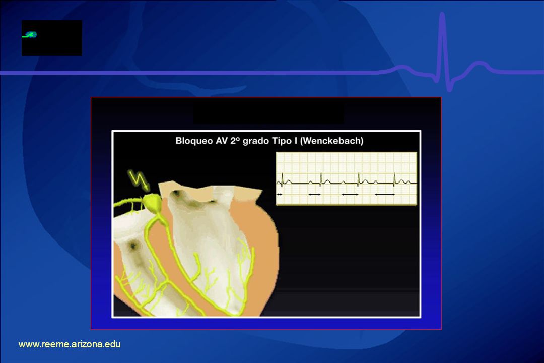
BLOQUEO AV DE SEGUNDO GRADO MOBITZ I (FENOMENO DE WENCKEBACH)
Prolongación del intervalo PR hasta que una P no se conduce. Complejo QRS de morfología normal. Ritmo es irregular. Rule 3 The QRS complex should be dominantly upright in leads I and II. Slight disparities are likely to be acceptable.

14
BLOQUEO AV DE SEGUNDO GRADO MOBITZ I (FENOMENO DE WENCKEBACH)
Rule 4 The QRS and T waves tend to have the same direction in the standard leads.

16
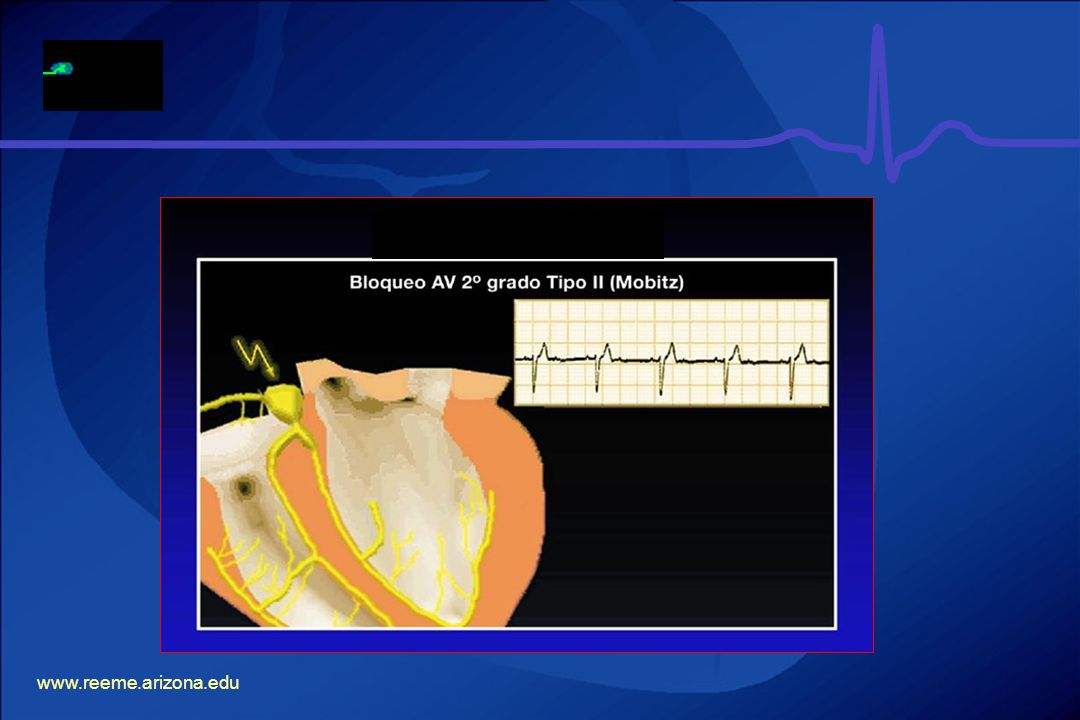
BLOQUEO AV DE SEGUNDO GRADO MOBITZ II
Intervalo PR es constante hasta que una P no conduce. Fijo: 2:1, 3:1, 4:1 Variable: 2:1, 4:1, 3:1 Avanzado: Dos ó mas P son bloqueadas. Rule 5 All waves are negative in lead aVR. This has to be so: aVR represents electrical activity as “seen” from the right shoulder. The sinus node is placed top right in the heart nearest the right shoulder, and the electrical activity is moving downwards and leftwards towards the left ventricle.

17
BLOQUEO AV DE SEGUNDO GRADO MOBITZ II (3:1)
Rule 6 The normality of QRS complexes recorded from the precordial leads is dependent on both morphological and dimensional criteria.

18
BLOQUEO AV DE SEGUNDO GRADO MOBITZ II (AVANZADO)
Location of infarction and its relation to the ECG: anterior infarction As was discussed in the previous module, the different leads look at different aspects of the heart, and so infarctions can be located by noting the changes that occur in different leads. The precordial leads (V1–6) each lie over part of the ventricular myocardium and can therefore give detailed information about this local area. aVL, I, V5 and V6 all reflect the anterolateral part of the heart and will therefore often show similar appearances to each other. II, aVF and III record the inferior part of the heart, and so will also show similar appearances to each other. Using these we can define where the changes will be seen for infarctions in different locations. Anterior infarctions usually occur due to occlusion of the left anterior descending coronary artery resulting in infarction of the anterior wall of the left ventricle and the intraventricular septum. It may result in pump failure due to loss of myocardium, ventricular septal defect, aneurysm or rupture and arrhythmias. ST elevation in I, aVL, and V2–6, with ST depression in II, III and aVF are indicative of an anterior (front) infarction. Extensive anterior infarctions show changes in V1–6 , I, and aVL.
 each lie over part of the ventricular myocardium and can therefore give detailed information about this local area. aVL, I, V5 and V6 all reflect the anterolateral part of the heart and will therefore often show similar appearances to each other. II, aVF and III record the inferior part of the heart, and so will also show similar appearances to each other. Using these we can define where the changes will be seen for infarctions in different locations. Anterior infarctions usually occur due to occlusion of the left anterior descending coronary artery resulting in infarction of the anterior wall of the left ventricle and the intraventricular septum. It may result in pump failure due to loss of myocardium, ventricular septal defect, aneurysm or rupture and arrhythmias. ST elevation in I, aVL, and V2–6, with ST depression in II, III and aVF are indicative of an anterior (front) infarction. Extensive anterior infarctions show changes in V1–6 , I, and aVL.")
20
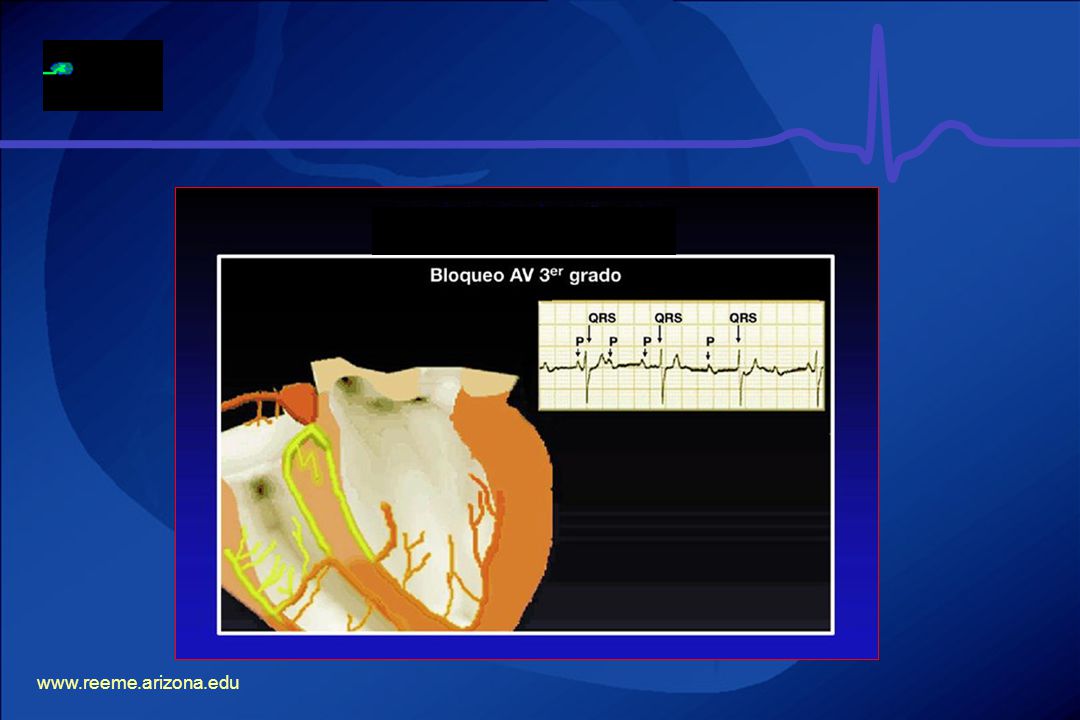
BLOQUEO AV COMPLETO Ó DE TERCER GRADO
Disociación entre aurículas y ventrículos PP es constante RR es constante Frecuencia es mayor en las P que en los complejos QRS. La morfología del QRS depende del marcapaso subsidiario. Rule 7 The ST segment should start isoelectric except in V1 and V2 where it may be elevated.

21
BLOQUEO AV COMPLETO Ó DE TERCER GRADO
Rule 8 In leads I, II, and V2 to V6 the P waves should be upright.

22
BLOQUEO AV COMPLETO Ó DE TERCER GRADO
Sequence of changes in evolving AMI The ECG changes that occur due to myocardial infarction do not all occur at the same time. There is a progression of changes correlating to the progression of infarction. Within minutes of the clinical onset of infarction, there are no changes in the QRS complexes and therefore no definitive evidence of infarction. However, there is ST elevation providing evidence of myocardial damage. The next stage is the development of a new pathological Q wave and loss of the r wave. These changes occur at variable times and so can occur within minutes or can be delayed. Development of a pathological Q wave is the only proof of infarction. As the Q wave forms the ST elevation is reduced and after 1 week the ST changes tend to revert to normal, but the reduction in R wave voltage and the abnormal Q waves usually persist. The late change is the inversion of the T wave and in a non-Q wave myocardial infarct, when there is no pathological Q wave, this T wave change may be the only sign of infarction. Months after an MI the T waves may gradually revert to normal, but the abnormal Q waves and reduced voltage R waves persist. In terms of diagnosing AMI in time to make thrombolysis a life-saving possibility, the main change to look for on the ECG is ST segment elevation.

24
¿ CUAL ES EL MANEJO ? ST elevation
ST segment elevation usually occurs in the early stages of infarction, and may exhibit quite a dramatic change. ST elevation is often upward and concave, although it can appear convex or horizontal. These changes occur in leads facing the infarction. ST elevation is not unique to MIs and therefore is not confirming evidence. Basic requirements of ST changes for diagnosis are: elevation of at least 1 mm in two or more adjoining leads for inferior infarctions (II, III, and aVF), and at least 2 mm in two or more precordial leads for anterior infarction. You should be aware that ST elevation can be seen in leads V1 and V2 normally. However, if there is also elevation in V3 the cause is unlikely to be physiological.
, and at least 2 mm in two or more precordial leads for anterior infarction. You should be aware that ST elevation can be seen in leads V1 and V2 normally. However, if there is also elevation in V3 the cause is unlikely to be physiological.")
25
Bradicardia sintomática Atropina Marcapaso transcutáneo ó transvenoso
TRATAMIENTO Bradicardia sintomática Atropina Marcapaso transcutáneo ó transvenoso Dopamina Epinefrina Isoproterenol Deep Q wave The only diagnostic changes of acute myocardial infarction are changes in the QRS complexes and the development of abnormal Q waves. However, this may be a late change and so is not useful for the diagnosis of AMI in the pre-hospital situation. Remember that Q waves of more than 0.04 seconds , or 1 little square, are not generally seen in leads I, II or the precordial leads.

26
T wave inversion The T wave is the most unstable feature of the ECG tracing and changes occur very frequently under normal circumstances, limiting their diagnostic value. Subtle changes in T waves are often the earliest signs of myocardial infarction. However, their value is limited for the reason above, but for approximately 20 to 30% of patients presenting with MI, a T wave abnormality is the only ECG sign. The T wave can be lengthened or heightened by coronary insufficiency. T wave inversion is a late change in the ECG and tends to appear as the ST elevation is returning to normal. As the ST segment returns towards the isoelectric line, the T wave also decreases in amplitude and eventually inverts.

28
GRACIAS Bundle branch block
Bundle branch block is the pattern produced when either the right bundle or the entire left bundle fails to conduct an impulse normally. The ventricle on the side of the failed bundle branch must be depolarised by the spread of a wave of depolarisation through ventricular muscle from the unaffected side. This is obviously a much slower process and usually the QRS duration is prolonged to at least 0.12 seconds (for right bundle branch block) and 0.14 seconds (for left bundle branch block). The ECG pattern of left bundle branch block (LBBB) resembles that of anterior infarction, but the distinction can readily be made in nearly all cases. Most importantly, in LBBB the QRS is widened to 140 ms or more. With rare exceptions there is a small narrow r wave (less than 0.04 seconds) in V1 to V3 which is not usually seen in anteroseptal infarction. There is also notching of the QRS best seen in the anterolateral leads, and the T wave goes in the opposite direction to the QRS in all the precordial leads. This combination of features is diagnostic. In the rare cases where there may be doubt assume the correct interpretation is LBBB. This will make up no difference to the administration of a thrombolytic on medical direction but for the present will be accepted as a contraindication for paramedics acting autonomously (see later slide). Right bundle branch block is characterised by QRS of 0.12 seconds or wider, an s wave in lead I, and a secondary R wave (R’) in V1. As abnormal Q waves do not occur with right bundle branch block, this remains a useful sign of infarction.
 and 0.14 seconds (for left bundle branch block). The ECG pattern of left bundle branch block (LBBB) resembles that of anterior infarction, but the distinction can readily be made in nearly all cases. Most importantly, in LBBB the QRS is widened to 140 ms or more. With rare exceptions there is a small narrow r wave (less than 0.04 seconds) in V1 to V3 which is not usually seen in anteroseptal infarction. There is also notching of the QRS best seen in the anterolateral leads, and the T wave goes in the opposite direction to the QRS in all the precordial leads. This combination of features is diagnostic. In the rare cases where there may be doubt assume the correct interpretation is LBBB. This will make up no difference to the administration of a thrombolytic on medical direction but for the present will be accepted as a contraindication for paramedics acting autonomously (see later slide). Right bundle branch block is characterised by QRS of 0.12 seconds or wider, an s wave in lead I, and a secondary R wave (R’) in V1. As abnormal Q waves do not occur with right bundle branch block, this remains a useful sign of infarction.")
Presentaciones similares
 es el registro gráfico, en función del tiempo, de las variaciones de potencial eléctrico.>")


