Descargar la presentación
La descarga está en progreso. Por favor, espere

1
Hipertensión Arterial Secundaria
Presenta: Dra. Ma. Del Carmen Ojeda López R2MI Revisó: Dr. César Vega López R4MI Profesor Titular: Dr. Enrique J. Díaz Greene Profesor Adjunto: Dr. Federico Rodríguez Weber

2
Caso clínico Mujer de 32 años de edad, diagnosticada hace cuatro años de hipertensión arterial. Desde entonces seguía tratamiento de forma discontinua con medidas higiénico- dietéticas, IECA y beta- bloqueadores sin control de cifras tensionales. AHF : madre y tía materna hipertensas, no enfermedad cardiovascular prematura. APNP: Tabaquismo de 20 c/día desde los 18 años. APP: Migraña que trataba con paracetamol e hipertensión gestacional (sin preeclampsia) en su único embarazo. Exploración física normal con auscultación cardiopulmonar y examen neurológico normales, pulsos periféricos conservados y simétricos, sin apreciarse soplos carotídeos ni abdominal. Talla: 1,60 m, peso: 55,3 Kg, IMC 21,5. TA decúbito: 170/102 mmHg, TA sedestación: 164/106 mmHg, FC: 82 lpm. Fondo de ojo: retinopatía hipertensiva grado II de Keith Wagener
 en su único embarazo. Exploración física normal con auscultación cardiopulmonar y examen neurológico normales, pulsos periféricos conservados y simétricos, sin apreciarse soplos carotídeos ni abdominal. Talla: 1,60 m, peso: 55,3 Kg, IMC 21,5. TA decúbito: 170/102 mmHg, TA sedestación: 164/106 mmHg, FC: 82 lpm. Fondo de ojo: retinopatía hipertensiva grado II de Keith Wagener.")
3
BH, QS normales Creatinina en sangre: 0,9 mg/dl, aclaramiento de creatinina: 102ml/min EKG y RxTx: normales Derivados urinarios de catecolaminas, renina y aldosterona plasmática normales Gamagrama renal: disminución de tamaño del riñón derecho con retraso del aporte vascular al mismo e intensa afectación funcional

4
Definición Presencia de una condición específica que se conoce produce hipertensión presence of a specific condition known to cause hypertension.

5
Hipertensión Arterial Secundaria
Puede afectar de 5 a 10% de los hipertensos. La investigación de todos los pacientes para su detección sería costosa e impráctica. Base de detección – datos clínicos. it may affect 5% to 10% of hypertensive patients. Investigation of all to detect these few would be costly and impractical and may exaggerate the risk to some patients without benefit, particularly because some secondary causes may not be correctable and others may be potentially treatable but at substantial risk. Nevertheless, for the few with a treatable secondary cause, detection and correction may be highly rewarding, even life prolonging. Knowledge of key clinical clues to secondary hypertension is essential to select which patients should be evaluated further and to what extent.

6
Causas Renal Enfermedad parenquimatosa renal Obstrucción ureteral o vesical Renovascular Displasia Fibromuscular Enfermedad ateroesclerótica Coartación aórtica Endocrina Hiperaldosteronismo primario Feocromocitoma Enfermedad de Cushing Hipo o hipertiroidismo Hiperparatiroidismo Otra Apnea obstructiva del sueño Renal parenchymal disease, commonly termed chronic kidney disease (CKD), is the most common secondary cause, but urinary outlet obstruction should be considered. Renovascular disease occurs in young women as fibromuscular dysplasia and in older individuals because of atherosclerotic renal artery stenosis. Endocrine causes include primary aldosteronism, pheochromocytoma, cortisol excess, and thyroid or parathyroid abnormalities. In the current obesity epidemic, obstructive sleep apnea (OSA) is an increasingly common problem and may comprise sympathetic nervous system activation [1] and a relative aldosterone excess state
, is the most common secondary cause, but urinary outlet obstruction should be considered. Renovascular disease occurs in young women as fibromuscular dysplasia and in older individuals because of atherosclerotic renal artery stenosis. Endocrine causes include primary aldosteronism, pheochromocytoma, cortisol excess, and thyroid or parathyroid abnormalities. In the current obesity epidemic, obstructive sleep apnea (OSA) is an increasingly common problem and may comprise sympathetic nervous system activation [1] and a relative aldosterone excess state.")
7
Datos clínicos clave Edad de inicio temprano
Historia Edad de inicio temprano Cualquier causa secundaria Curso grave o acelerado Ausencia de historia familiar de HAS Hipertensión resistente

8
Hipokalemia al tomar diurético
Intolerancia a fármacos Hipokalemia al tomar diurético Hiperaldosteronismo primario o secundario, exceso de corticoesteroide Emperoamiento después de beta-bloqueo Feocromocitoma Insuficiencia renal aguda después del inicio o incremento de dosis de IECA, ARA Hipertensión renovascular HistoricalPossible causesEarly age of onsetAny secondary causeSevere or accelerated courseAny secondary causeAbsent family history of hypertensionAny secondary causeResistant hypertensionAny secondary causeSpecific drug intolerancesMarked hypokalemia while taking a diuretic medicationPrimary or secondary hyperaldosteronism, corticosteroid excessWorsening hypertension after beta-blockadePheochromocytomaAcute renal failure after initiation or dose increase of ACEI, ARBRenovascular hypertension

9
Crisis, labilidad, ortostatismo Feocromocitoma
Síntomas Crisis, labilidad, ortostatismo Feocromocitoma Obstrucción urinaria, ronquidos, hipersomnolencia diurna SAOS Edema pulmonar Hipertensión renovascular SymptomsSpells, lability, orthostatismPheochromocytomaProstatismUrinary obstructionSnoring, daytime hypersomnolenceOSAFlash pulmonary edemaRenovascular hypertensionPhysical examination signsCafé au lait spots, neurofibromasPheochromocytomaCervical fat pad, moon facies, pigmented striaeCushing's diseaseThigh BP lower than brachial BP, continuous murmur over backAortic coarctationGoiter or thyroid noduleThyroid diseaseLarge neck, narrow pharynx with soft tissue crowdingOSAAbdominal systolic-diastolic bruit, multiple arterial bruitsRenovascular hypertensionLarge palpable kidneysPolycystic kidney disease

10
Manchas café con leche, neurofibromas Feocromocitoma
Signos Manchas café con leche, neurofibromas Feocromocitoma Jiba cervical, fascies en luna llena, estrías pigmenteadas Enfermedad de Cushing Soplo abdominal sistolo-diastólico, soplos arteriales múltiples Hipertensión renovascular TA en muslo mayor a la braquial, soplo continuo en espalda Coartación de la aorta

11
Hallazgos de laboratorio
Hiperkalemia Enfermedad del parénquima renal, obstrucción urinaria Hipokalemia Hipertensión renovascular, hipoaldosteronismo primario o secundario Elevación de creatinina sérica EGO anormal Enfermedad del parénquima renal Daño a órgano blanco desproporcionado (inf lacunares, retinopatía hipertensiva, HVI, IR) Cualquier causa secundaria Caída nocturna de la TA Laboratory findingsHyperkalemiaRenal parenchymal disease, urinary obstructionHypokalemiaRenovascular hypertension, primary or secondary hyperaldosteronismElevated serum creatinineRenal parenchymal disease, urinary obstruction, renovascular hypertensionAbnormal urinalysisRenal parenchymal diseaseLoss or blunting of nocturnal BP decreaseAny secondary causeDisproportionate target organ damage (cerebral lacunar infarcts, hypertensive retinopathy, left ventricular hypertrophy, renal failure)Any secondary cause
 Cualquier causa secundaria. Caída nocturna de la TA. Laboratory findingsHyperkalemiaRenal parenchymal disease, urinary obstructionHypokalemiaRenovascular hypertension, primary or secondary hyperaldosteronismElevated serum creatinineRenal parenchymal disease, urinary obstruction, renovascular hypertensionAbnormal urinalysisRenal parenchymal diseaseLoss or blunting of nocturnal BP decreaseAny secondary causeDisproportionate target organ damage (cerebral lacunar infarcts, hypertensive retinopathy, left ventricular hypertrophy, renal failure)Any secondary cause.")
13
Causas Renal Enfermedad parenquimatosa renal Obstrucción ureteral o vesical Renal parenchymal disease, commonly termed chronic kidney disease (CKD), is the most common secondary cause, but urinary outlet obstruction should be considered. Renovascular disease occurs in young women as fibromuscular dysplasia and in older individuals because of atherosclerotic renal artery stenosis. Endocrine causes include primary aldosteronism, pheochromocytoma, cortisol excess, and thyroid or parathyroid abnormalities. In the current obesity epidemic, obstructive sleep apnea (OSA) is an increasingly common problem and may comprise sympathetic nervous system activation [1] and a relative aldosterone excess state
, is the most common secondary cause, but urinary outlet obstruction should be considered. Renovascular disease occurs in young women as fibromuscular dysplasia and in older individuals because of atherosclerotic renal artery stenosis. Endocrine causes include primary aldosteronism, pheochromocytoma, cortisol excess, and thyroid or parathyroid abnormalities. In the current obesity epidemic, obstructive sleep apnea (OSA) is an increasingly common problem and may comprise sympathetic nervous system activation [1] and a relative aldosterone excess state.")
14
Hipertensión Renal Causa más común de HAS secundaria.
Renal parenchymal disease is the most common cause of secondary hypertension. A basic classification of renal diseases is included in Table 3. Although full coverage of renal diseases and their treatment is beyond the scope of this review, it is essential that the clinician recognize major types of renal disease in order to initiate early treatment and appropriate referral. A recent movement to have clinical laboratories report an estimated glomerular filtration rate (eGFR) concurrent with serum creatinine measurements should assist practitioners in recognizing renal function impairment (CKD) earlier, initiating renal protective measures, and making earlier specialist referrals [10]. Hypertension may be a presenting sign of renal disease and may be severe, even before a decline in renal function is evident. Further, aggressive treatment of hypertension in this setting may delay progressive renal function decline.
 concurrent with serum creatinine measurements should assist practitioners in recognizing renal function impairment (CKD) earlier, initiating renal protective measures, and making earlier specialist referrals [10]. Hypertension may be a presenting sign of renal disease and may be severe, even before a decline in renal function is evident. Further, aggressive treatment of hypertension in this setting may delay progressive renal function decline.")
15
Hipertensión Renal En 2.5% de hipertensos. Hipertensión – causa común de enfermedad renal crónica. - segunda causa más comun de IRCT en la población. Hipertensión + enfermedad renal pérdida de función renal acelerada. Renal parenchymal disease is the most common secondary cause of hypertension and is present in 2% to 5% of persons with elevated blood pressure. Also, hypertension is a common cause of chronic kidney disease and is the second most common cause of end-stage renal failure in the population. Persons with chronic kidney disease and hypertension are at high risk of cardiovascular disease. Contributing to this increased risk is a loss of the normal nocturnal decline in blood pressure (“non-dipper”) in persons with renal disease. Regardless of the cause of chronic kidney disease, hypertension is associated with a more rapid loss of renal function. This may be due to transmission of higher pressures into the glomerulus as afferent renal artery resistance fails to limit transmission of higher systemic pressures into the nephron. Higher glomerular transcapillary pressures and flows injure glomerular cells by several mechanisms, ultimately leading to glomerulosclerosis. In addition, the degree of proteinuria is an independent predictor of progressive loss of renal function.
 in persons with renal disease. Regardless of the cause of chronic kidney disease, hypertension. is associated with a more rapid loss of renal function. This may be. due to transmission of higher pressures into the glomerulus as afferent. renal artery resistance fails to limit transmission of higher systemic. pressures into the nephron. Higher glomerular transcapillary. pressures and flows injure glomerular cells by several mechanisms, ultimately. leading to glomerulosclerosis. In addition, the degree of proteinuria. is an independent predictor of progressive loss of renal. function.")
16
Hipertensión Renal - Mecanismos
Eliminación de Na y H2O impedida Expansión de volumen Hipersecreción de renina Disminución de la producción de vasodilatadores renales Prostaglandinas, kalikreina, kinina Acumulación de mediadores de estrés oxidativo Disminuye disponibilidad de ON Incrementa niveles de endotelina At least three major mechanisms are involved in the hypertension of renal disease: volume expansion from impaired renal elimination of salt and water, oversecretion of renin, and decreased production of renal vasodilators (i.e., prostaglandins, kallikrein, or kinin). In addition, accumulation of mediators of oxidative stress, reducing the availability of the vasodilator nitric oxide, and increased levels of the vasoconstrictor endothelin may also contribute.
. In addition, accumulation of mediators of oxidative stress, reducing the availability of the vasodilator nitric oxide, and increased. levels of the vasoconstrictor endothelin may also contribute.")
17
Hipertensión Renal TA objetivo 130/80 Restricción de sodio Diurético
Disminuye riesgo de deterioro progresivo de función renal Hasta 3 o más antihipertensivos Restricción de sodio Diurético Si Cr > 2.0mg/dL / GFR <30mL/min Diuréticos de asa Combinación de asa + tiazida Hipersecreción de renina en pequeño % IECAs reducen proteinuria y presión transcapilar glomerular The blood pressure goal in chronic kidney disease is less than 130/80 mm Hg. Achieving this goal lessens the risk of progressive loss of renal function and often requires the use of three or more antihypertensive drugs. Treatment should begin with dietary sodium restriction. In addition, diuretic therapy is often essential for blood pressure control. If serum creatinine is more than 2.0 mg/dL (GFR <30 mL/min), the more potent loop agents or metolazone is necessary. In diuretic-resistant persons, the combination of a loop agent with a thiazide may be required. Oversecretion of renin occurs in only a small proportion of persons with chronic kidney disease; however, ACEIs reduce proteinuria and high glomerular transcapillary pressures by decreasing resistance in the efferent arteriole of the nephron.
, the more potent loop agents or metolazone is necessary. In diuretic-resistant persons, the combination of a loop agent. with a thiazide may be required. Oversecretion of renin occurs in. only a small proportion of persons with chronic kidney disease; however, ACEIs reduce proteinuria and high glomerular transcapillary pressures. by decreasing resistance in the efferent arteriole of the nephron.")
18
Causas Renovascular Displasia Fibromuscular Enfermedad ateroesclerótica Coartación aórtica Renal parenchymal disease, commonly termed chronic kidney disease (CKD), is the most common secondary cause, but urinary outlet obstruction should be considered. Renovascular disease occurs in young women as fibromuscular dysplasia and in older individuals because of atherosclerotic renal artery stenosis. Endocrine causes include primary aldosteronism, pheochromocytoma, cortisol excess, and thyroid or parathyroid abnormalities. In the current obesity epidemic, obstructive sleep apnea (OSA) is an increasingly common problem and may comprise sympathetic nervous system activation [1] and a relative aldosterone excess state
, is the most common secondary cause, but urinary outlet obstruction should be considered. Renovascular disease occurs in young women as fibromuscular dysplasia and in older individuals because of atherosclerotic renal artery stenosis. Endocrine causes include primary aldosteronism, pheochromocytoma, cortisol excess, and thyroid or parathyroid abnormalities. In the current obesity epidemic, obstructive sleep apnea (OSA) is an increasingly common problem and may comprise sympathetic nervous system activation [1] and a relative aldosterone excess state.")
19
Hipertensión Renovascular
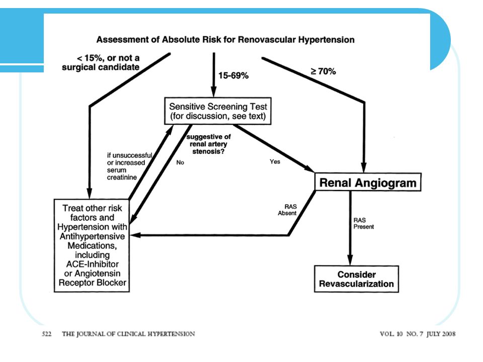
(dx clínico retrospectivo) VS Estenosis de Arteria Renal (dx anatómico) Renovascular Hypertension vs Renal Artery Stenosis Unlike most other cardiovascular and renal conditions, renovascular hypertension can be diagnosed only retrospectively. Classically, renovascular hypertension can be correctly and properly diagnosed 6 to 12 weeks after an intervention (see below), only if the BP is lower than it was before the intervention, with the patient taking the same or fewer antihypertensive medications. In contrast to renovascular hypertension (which has a physiologic basis for its diagnosis), renal artery stenosis is an anatomic diagnosis. Classically, renal artery stenosis was diagnosed when there was a >75% narrowing of the diameter of a main renal artery or >50% luminal narrowing with a poststenotic dilatation. These criteria were based on planar images derived from renal angiograms done in the mid-1970s; typically, a 50% luminal narrowing is the minimum in the current literature.
 VS. Estenosis de Arteria Renal. (dx anatómico) Renovascular Hypertension vs Renal Artery Stenosis. Unlike most other cardiovascular and renal conditions, renovascular hypertension can be diagnosed. only retrospectively. Classically, renovascular. hypertension can be correctly and properly. diagnosed 6 to 12 weeks after an intervention (see. below), only if the BP is lower than it was before. the intervention, with the patient taking the same. or fewer antihypertensive medications. In contrast. to renovascular hypertension (which has a physiologic. basis for its diagnosis), renal artery stenosis. is an anatomic diagnosis. Classically, renal artery. stenosis was diagnosed when there was a >75% narrowing of the diameter of a main renal artery. or >50% luminal narrowing with a poststenotic. dilatation. These criteria were based on planar. images derived from renal angiograms done in the. mid-1970s; typically, a 50% luminal narrowing is. the minimum in the current literature.")
20
Hipertensión Renovascular
Subtipos Displasia fibromuscular Enfermedad ateroesclerótica Otras causas de enfermedad renovascular First, many older persons have relatively advanced renal arterial stenoses on angiography, but few have resistant hypertension.10 Second, surgical removal of a small kidney due to presumed ischemic nephropathy (from renovascular hypertension) has been followed by normal BP values in only about 25% of the patients in whom it was attempted in the mid-1950s. Third, diagnostic performance of screening tests for renovascular hypertension are different from those for renal artery stenosis, because the latter analysis usually includes 2 arteries per person, whereas the former is done based on responses in individual patients
 has been followed by normal BP. values in only about 25% of the patients in whom it was attempted in the mid-1950s. Third, diagnostic performance of screening tests for renovascular. hypertension are different from those for renal artery stenosis, because the latter analysis usually includes 2 arteries per person, whereas the former. is done based on responses in individual patients.")
21
Subtipos

22
Displasia Fibromuscular
Enfermedad no ateroesclerosa, no inflamatoria de arterias pequeñas y medianas. intima (1%–5%) media (60%–85%) “hilo de cuentas” periadventicia (10%–25%) Fibromuscular dysplasia (FMD) is a nonatherosclerotic and noninflammatory disease of small and medium arteries. The location on the vessel wall and frequency of FMD is intimal (1%–5%), medial (60%–85%), or periadventitial (10%–25%).3 Medial FMD is further divided into subtypes, but medial fibroplasia, described as a “string of beads” (Figure 1), is the most common. The classic arteriographic appearance results from proliferation of the extracellular matrix, with disruption of the internal elastic lamina causing multiple stenoses and saccular aneurysms.
 media (60%–85%) hilo de cuentas periadventicia (10%–25%) Fibromuscular dysplasia (FMD) is a nonatherosclerotic. and noninflammatory disease of small and. medium arteries. The location on the vessel wall. and frequency of FMD is intimal (1%–5%), medial. (60%–85%), or periadventitial (10%–25%).3. Medial FMD is further divided into subtypes, but. medial fibroplasia, described as a string of beads (Figure 1), is the most common. The classic arteriographic. appearance results from proliferation. of the extracellular matrix, with disruption of the. internal elastic lamina causing multiple stenoses. and saccular aneurysms.")
23
Displasia Fibromuscular media
The classic arteriographic appearance results from proliferation of the extracellular matrix, with disruption of the internal elastic lamina causing multiple stenoses and saccular aneurysms Figure 1. Fibromuscular dysplasia of the renal artery showing alternating constrictions and saccular aneurysms,described as a “string of beads.” The top panel is the renal arteriogram and the bottom panel is the digital subtraction image.

24
Displasia Fibromuscular
60-75% afecta arterias renales 1) Arteria renal derecha 2) Bilateral 39-66% DFM íntima o adventicia mayor trombosis o disección DFM media se encuentra confinada a los 2/3 distales de la arteria renal Involucra a las ramas en 39% Aneurismas Progresión hasta 38%, la oclusión total es rara Pérdida de parénquima FMD affects the renal arteries with a frequency of 60%–75%.4 Medial FMD is confined to the distal two thirds of the renal artery and involves the branch vessels in about 39% of patients.5 The right renal artery is the dominant site of FMD, but it occurs bilaterally in 39%–66% of cases Other vascular beds may be affected, including carotid (the most common), cerebral, vertebral, coronary, aorta, subclavian, axillary, iliac, popliteal, hepatic, splenic, celiac, and mesenteric arteries.
 Arteria renal derecha. 2) Bilateral 39-66% DFM íntima o adventicia mayor trombosis o disección. DFM media se encuentra confinada a los 2/3 distales de la arteria renal. Involucra a las ramas en 39% Aneurismas. Progresión hasta 38%, la oclusión total es rara. Pérdida de parénquima. FMD affects the renal arteries with a frequency of 60%–75%.4 Medial FMD is confined to the distal two thirds of the renal artery and involves. the branch vessels in about 39% of patients.5 The right renal artery is the dominant site of FMD, but it occurs bilaterally in 39%–66% of cases. Other vascular beds may be affected, including carotid (the most common), cerebral, vertebral, coronary, aorta, subclavian, axillary, iliac, popliteal, hepatic, splenic, celiac, and mesenteric arteries.")
25
DFM - Fisiopatología Estenosis con repercusión hemodinámica Disminución en perfusión renal Disminución en GFR, excreción de Na y H2O Incremento en actividad de renina y ATII Vasoconstricción Hipertensión Hemodynamically significant stenosis of the renal artery reduces renal perfusion pressure and blood flow causing a decline in the glomerular filtration rate and sodium and water excretion.11 To compensate for this, increased activity of renin and angiotensin II produce vasoconstriction and blood pressure elevation.

26
DFM – Factores predisponentes
HLA-DRw6 AD? Tabaquismo ECA (alelo I) A genetic predisposition, HLA-DRw6 antigen, and cigarette smoking are thought to be of etiologic importance in FMD, but the cause is unknown.12 In a pedigree analysis of 20 index cases of FMD, 60% had at least one family member with suspected disease.13 The investigators speculated that FMD was an autosomal dominant trait with variable penetrance, but there was no documentation of FMD. A better study reported a familial occurrence of 11% in first-degree relatives of 33 women and was more common in patients with bilateral renal FMD.6 There was no vertical transmission to offspring.
 A genetic predisposition, HLA-DRw6 antigen, and cigarette smoking are thought to be of etiologic importance in FMD, but the cause is unknown.12. In a pedigree analysis of 20 index cases of FMD, 60% had at least one family member with suspected disease.13 The investigators speculated that. FMD was an autosomal dominant trait with variable penetrance, but there was no documentation of FMD. A better study reported a familial occurrence. of 11% in first-degree relatives of 33 women and was more common in patients with bilateral renal FMD.6 There was no vertical transmission. to offspring.")
27
Displasia fibromuscular
Mujeres : Hombres 81% - 40% Menos Africano-americanos Menor historia familiar de HAS que en la hipertensión esencial Menor duración de hipertensión TA similar Soplos abdominal (55%), flanco (20%) Nivel de K más bajo In the Cooperative Study of Renovascular Hypertension, cases of essential hypertension were compared with 84 cases of renovascular hypertension due to FMD. Among patients with FMD, there were more women (81% vs 40%; P<.01), fewer African Americans (10% vs 29%, P<.01), a lower rate of a family history of hypertension (41% vs 67%; P<.01), and a thinner body habitus (30% vs 6%; P<.05).18 Duration of hypertension was shorter with FMD than essential hypertension (2.0 vs 3.1 years; P<.01). Blood pressure was similar, but abdominal (55% vs 6%; P<.05) and flank (20% vs 1%; P<.05) bruits were more common. A potassium level <3.4 mEq/L was present in 17% of fibromuscular patients compared with 7% of patients with essential hypertension (P<.05) If other vessels are involved with FMD, other symptoms (eg, angina pectoris, intestinal angina, claudication, headache, transient ischemic attack) and signs may be present. Several reports have documented the association of a pheochromocytoma with fibromuscular dyspasia.20,21
, flanco (20%) Nivel de K más bajo. In the Cooperative Study of Renovascular Hypertension, cases of essential hypertension were compared with 84 cases of renovascular hypertension due to FMD. Among patients with FMD, there were more women (81% vs 40%; P<.01), fewer African Americans (10% vs 29%, P<.01), a lower rate of a family history of hypertension (41% vs 67%; P<.01), and a thinner body habitus (30% vs 6%; P<.05).18 Duration of hypertension was shorter with FMD than essential hypertension (2.0 vs 3.1 years; P<.01). Blood pressure was similar, but abdominal (55% vs 6%; P<.05) and flank (20% vs 1%; P<.05) bruits were more common. A potassium level <3.4 mEq/L was present in 17% of fibromuscular patients compared with 7% of patients with essential hypertension (P<.05) If other vessels are involved with FMD, other symptoms (eg, angina pectoris, intestinal angina, claudication, headache, transient ischemic attack) and signs may be present. Several reports have documented the association of a pheochromocytoma. with fibromuscular dyspasia.20,21.")
28
Enfermedad renovascular ateroesclerótica
Causa más común de HAS renovascular en personas de edad media o más. Enfermedad progresiva, oclusiva, estrecha el ostium y del tercio proximal de la arteria renal principal y la aorta cercana. Factores de riesgo: Edad avanzada, diabetes, dislipidemia, tabaquismo, historia de eventos cardiovasculares. Atheromatous disease is the most common cause of renovascular hypertension in middle-aged or older persons. It accounts for 85% of renovascular hypertension in the population and is more common in men than in women. Probably about 90% of current patients with renovascularhypertension have atherosclerotic disease as the underlying pathologic reason for the arterial stenosis. This progressive, occlusive process typically narrows the ostium and proximal third of the main renal artery, as well as the nearby aorta. As with all other atherosclerotic vascular diseases, it is found with increasing frequency with advancing age and has the usual associated risk factors (diabetes, dyslipidemia, tobacco use, and history of cardiovascular events).
.")
29
Enfermedad renovascular ateroesclerótica
Bilateral 30% Progresivo 35% A pesar de control de TA The lesions usually are in the proximal third of the renal artery, often near or at the orifice (Fig. 13-2). In many instances, renal artery obstruction is due to extension of aortic atheromatous disease across the orifice of the artery. The disease is bilateral in 30% of cases, and in 35% of cases it progresses even if blood pressure is controlled. Atherosclerosis of the renal artery can progress to occlusion of the vessel. Progressive disease can cause end-stage renal disease.
. In. many instances, renal artery obstruction is due to extension of aortic. atheromatous disease across the orifice of the artery. The disease is bilateral in 30% of cases, and in. 35% of cases it progresses even if blood pressure is controlled. Atherosclerosis of the renal artery can progress to occlusion of the. vessel. Progressive disease can cause end-stage renal disease.")
30
Diagnóstico

31
Diagnóstico MEDICINA GENERAL 2001; 30: 55-58

32
Diagnóstico Figure 3. Stenotic accessory artery in a 55-year-old patient with hypertension. (a) Baseline Tc-99m DTPA scintigrams (sequential posterior views obtained at 2-minute intervals from top left to bottom right) show slight retention of the radiopharmaceutical in the left renal pelvis. (b) Captopril scintigrams show markedly decreased uptake in the upper half of the right kidney, a finding consistent with renovascular disease of a polar artery. RadioGraphics 2000; 20:1355–1368
 show slight retention. of the radiopharmaceutical in the left renal pelvis. (b) Captopril scintigrams show markedly decreased uptake in the upper half of the right kidney, a finding consistent with renovascular disease of a polar artery. RadioGraphics 2000; 20:1355–1368.")
33
Diagnóstico Figure 5. Severe stenosis in a patient with a solitary left kidney who experienced recurring hypertension and renal failure 3 months after stent placement in the left renal artery. (a) Doppler spectrum from the proximal left renal artery shows flow acceleration of close to 300 cm/sec inside the stent. (b) Intrarenal Doppler spectrum shows a waveform with a pulsus tardus configuration, which indicates severe hemodynamic repercussions. (c) Arteriogram shows severe stenosis inside the stent (arrow). Thrombosis of the infrarenal aorta is also noted. RadioGraphics 2000; 20:1355–1368
 Doppler spectrum from the proximal left renal. artery shows flow acceleration of close to 300 cm/sec inside. the stent. (b) Intrarenal Doppler spectrum shows a. waveform with a pulsus tardus configuration, which indicates. severe hemodynamic repercussions. (c) Arteriogram. shows severe stenosis inside the stent (arrow). Thrombosis. of the infrarenal aorta is also noted. RadioGraphics 2000; 20:1355–1368.")
34
Diagnóstico Figure 11. Renovascular disease in a patient with hypertension and two right renal arteries. (a) Coronal maximum- intensity projection image from MR angiography shows an eccentric atheromatous lesion of the abdominal aorta adjacent to the upper right renal artery (black arrow); however, this lesion does not cause stenosis. The lower right renal artery has a proximal stenosis (arrowhead). A stenosis of the left renal artery is also demonstrated (white arrow). (b) Conventional aortogram shows the findings seen on the MR angiogram (a) with good correlation. RadioGraphics 2000; 20:1355–1368
 Coronal maximum- intensity projection image from MR angiography shows an eccentric atheromatous lesion of the abdominal aorta adjacent to the upper right renal artery (black arrow); however, this lesion does not cause stenosis. The lower right renal artery has a proximal stenosis (arrowhead). A stenosis of the left renal artery is also demonstrated (white arrow). (b) Conventional aortogram shows the findings seen on the MR angiogram (a) with good correlation. RadioGraphics 2000; 20:1355–1368.")
36
Diagnóstico

38
Tratamiento

40
Causas Endocrina Hiperaldosteronismo primario Feocromocitoma Enfermedad de Cushing Hipo o hipertiroidismo Hiperparatiroidismo Renal parenchymal disease, commonly termed chronic kidney disease (CKD), is the most common secondary cause, but urinary outlet obstruction should be considered. Renovascular disease occurs in young women as fibromuscular dysplasia and in older individuals because of atherosclerotic renal artery stenosis. Endocrine causes include primary aldosteronism, pheochromocytoma, cortisol excess, and thyroid or parathyroid abnormalities. In the current obesity epidemic, obstructive sleep apnea (OSA) is an increasingly common problem and may comprise sympathetic nervous system activation [1] and a relative aldosterone excess state
, is the most common secondary cause, but urinary outlet obstruction should be considered. Renovascular disease occurs in young women as fibromuscular dysplasia and in older individuals because of atherosclerotic renal artery stenosis. Endocrine causes include primary aldosteronism, pheochromocytoma, cortisol excess, and thyroid or parathyroid abnormalities. In the current obesity epidemic, obstructive sleep apnea (OSA) is an increasingly common problem and may comprise sympathetic nervous system activation [1] and a relative aldosterone excess state.")
41
Feocromocitoma Tumor de origen neuroectodérmico productor de catecolaminas. 40% se descubre durante cirugía, imagen o autopsia. <1% de HAS (pb más alto) Neurofibromatosis Pheochromocytomas occur in no more than 1% or 2% of all patients with neurofibromatosis, but 5% to 25% of patients with pheochromocytoma will have neurofibromatosis; thus, patients with neurofibromatosis, symptomatic or not, should be regularly screened for pheochromocytoma A pheochromocytoma is a tumor of neuroectodermal origin that produces excess quantities of catecholamines as well as numerous other physiologically active peptides. This overabundance of catecholamines causes blood pressure (BP) to increase, accompanied by a constellation of signs and symptoms that can imitate those seen with a diverse grouping of medical and surgical disorders. Earlyrecognition, precise localization and attentive management of a benign pheochromocytoma in most instances leads to a complete cure. In up to 40% of patients with a pheochromocytoma, it is discovered during surgery, found in the course of abdominal imaging, or uncovered at the time of autopsy. These tumors can prove life-threatening, particularly during surgical and obstetric procedures.3
 Neurofibromatosis. Pheochromocytomas occur in no more than 1% or 2% of all patients with neurofibromatosis, but 5% to 25% of patients with pheochromocytoma will have neurofibromatosis; thus, patients with neurofibromatosis, symptomatic or not, should be regularly screened for pheochromocytoma. A pheochromocytoma is a tumor of neuroectodermal origin that produces excess quantities of catecholamines as well as numerous other physiologically active peptides. This overabundance of catecholamines causes blood pressure (BP) to increase, accompanied by a constellation of signs and symptoms that can imitate those seen with a diverse grouping of medical and surgical disorders. Earlyrecognition, precise localization and attentive management of a benign pheochromocytoma in most instances leads to a complete cure. In up to 40% of patients with a pheochromocytoma, it is discovered during surgery, found in the course of abdominal imaging, or uncovered at the time of autopsy. These. tumors can prove life-threatening, particularly during surgical and obstetric procedures.3.")
42
Feocromocitoma Patrón de TA: Hipotensión ortostática
Crisis hipertensivas Cefalea Diaforesis Palpitaciones The triad of symptoms that typifies pheochromocytoma is headache, excessive/generalized sweating, and palpitations; however, the symptom pattern of a pheochromocytoma can be quite varied, with other common symptoms including pallor, weight loss, and feelings of panic and anxiety. There is not a specific BP pattern that could be held as the sine qua non for a pheochromocytoma. The BP patterns that can develop with a pheochromocytoma include 1) a sustained hypertensive state without BP spikes; 2) a persistent hypertensive state with intermittent hypertensive spikes potentially reaching crisis levels; and 3) a normotensive state with brief, sudden, and striking BP elevations. The symptoms that accompany a pheochromocytoma-related hypertensive crisis include dizziness, flushing, visual disturbances, panic/anxiety, nausea, vomiting, or an epileptic aura.
 a sustained hypertensive state without BP spikes; 2) a persistent hypertensive state with intermittent hypertensive spikes potentially reaching crisis levels; and 3) a normotensive state with brief, sudden, and striking BP elevations. The symptoms that accompany a pheochromocytoma-related hypertensive crisis include dizziness, flushing, visual disturbances, panic/anxiety, nausea, vomiting, or an epileptic aura.")
43
Patients with signs and symptoms consistent with a pheochromocytoma should be evaluated on a priority basis. Biochemical evidence of excessive catecholamine production is a necessary step for the diagnosis of pheochromocytoma (Figure). Traditional biochemical tests include measurements of urinary and plasma catecholamines, urinary metanephrines (normetanephrine and metanephrine), and urinary vanillylmandelic acid; each of these tests has its advantages and disadvantages.8–11 The assay of catecholamines and their metabolites in timed urinary samples has heretofore been considered the best methodologic approach for pheochromocytoma screening despite the difficulties inherent to accurately timed urine collections. Accumulating evidence, however, suggests that measurements of plasma free metanephrines or urinary fractionated metanephrines (normetanephrine and metanephrine separately) are the most sensitive tests for diagnosis and are the most suitable for reliable exclusion of pheochromocytoma.11 In particular, plasma and urinary catecholamines can be normal when testing is carried out between spells when normotensive and asymptomatic patients with pheochromocytoma are screened for the tumor because of a hereditary predisposition or the incidental finding of an adrenal mass in the course of imaging studies. Increased sensitivity of metanephrines compared with plasma or urinary catecholamines is due to the continuous production of O-methylated metabolites from catecholamines seeping from chromaffin stores in tumors. The generation of O-methylated metabolites is self-regulating and therefore independent of the highly variable release of catecholamines in tumor tissue.12 Measurements of fractionated (and not total) metanephrines are superior to measurements of total metanephrines in that they allow tumors that produce predominantly or only one of the 3 O-methylated metabolites to be better detected. High suspicion with a 2-fold or greater elevation in urinary metanephrines should prompt a localizing imaging study and based on findings a 123I-MIBG scan. High suspicion with normal urinary catecholamines warrants rechecking in conjunction with a spell. Low suspicion with elevated urinary catecholamines dictates an imaging study and, if urinary catecholamines are normal, investigation for other causes of spells.
 metanephrines are superior to measurements of total metanephrines in that they allow tumors that produce predominantly or only one of the 3 O-methylated metabolites to be better detected. High suspicion with a. 2-fold or greater elevation in urinary metanephrines should prompt a localizing imaging study and based on findings a 123I-MIBG. scan. High suspicion with normal urinary catecholamines warrants rechecking in conjunction with a spell. Low suspicion with elevated. urinary catecholamines dictates an imaging study and, if urinary catecholamines are normal, investigation for other causes of. spells.")
44
Feocromocitoma Tratamiento quirúrgico.
Manejo prequirúrgico – reposición de volumen, bloqueo alfa y beta. Bloqueo B puede producir respuestas paradójicas Seguimiento obligatorio (un % significativo recurre) Surgical treatment is the only effective therapeutic approach for pheochromocytoma, be it benign or malignant. Adequate preoperative management includes careful attention to volume replacement (many of these patients are subclinically volume-contracted). Drug treatment that offers combined a- and b-blockade is essential for successful surgery, which is performed in most cases by laparoscopy.14 b-Blockade alone can result in paradoxical pressor responses.15 If the tumor is removed, biochemical evidence of the disease disappears within 1 week; however, although prognosis after tumor resection is excellent, a significant proportion of pheochromocytomas recur, some as metastases; thus, routine follow-up is mandatory.
 Surgical treatment is the only effective therapeutic approach for pheochromocytoma, be it benign or malignant. Adequate preoperative management includes careful attention to volume replacement (many of these patients are subclinically volume-contracted). Drug treatment that offers combined a- and b-blockade is essential for successful surgery, which is performed in most cases by laparoscopy.14 b-Blockade alone can result in paradoxical pressor responses.15 If the tumor is removed, biochemical evidence of the disease disappears within 1 week; however, although prognosis after tumor resection is excellent, a significant proportion of pheochromocytomas recur, some. as metastases; thus, routine follow-up is mandatory.")
45
Hiperaldosteronismo primario
Cualquier edad. 30-50 años Sx asociados a severidad de hipokalemia y/o complicaciones de hipertensión. Dx a considerar en cualquier px con: Hipokalemia espontánea Hipokalemia severa inducida por dosis usuales de diurético Hipertensión refractaria. Primary aldosteronism can occur at all ages, although in most reported series, patients fall in the 30-to-50–year age range. Primary aldosteronism is a common cause of resistant hypertension in black and white patients.16 The symptoms associated with primary aldosteronism are not directly linked to excess aldosterone per se, rather they relate to the severity of the accompanying hypokalemia and/or the complications of hypertension. Primary aldosteronism should be considered as a diagnostic possibility in any patient with spontaneous hypokalemia, moderately severe hypokalemia induced by usual doses of diuretics, or refractory hypertension.

46
Hiperaldosteronismo primario
Alcalosis hipokalemica Debilidad, poliuria/polidipsia, nicturia Hipertensión No edema HVI, insuficiencia cardiaca c/s fx sistólica conservada Patients with primary aldosteronism can present with little more than a mild hypokalemic alkalosis and hypertension; however, if hypokalemia is severe enough, weakness, polyuria/polydipsia, and nocturia may be present.21,22 Primary aldosteronism,however, does not cause edema, because of aldosterone escape in which an increase in Na+-wasting forces offsets the Na+-retaining effect of excess aldosterone. Although early thinking held that aldosterone-producing adenomas caused token endorgan damage, recent evidence has proven this to be an incorrect assumption. Patients with primary aldosteronism frequently develop left ventricular hypertrophy as well as heart failure with and without preserved systolic function.23 The myocardial susceptibility to damage in primary hyperaldosteronism may relate to aldosterone being both profibrotic and a neurohumoral substance capable of increasing oxidative stress. Treatment of primary aldosteronism is also characterized by partial reversibility of renal dysfunction and frequent return to normoalbuminuria from microalbuminuria.24

47
Hiperaldosteronismo primario
Hipokalemia Alcalosis metabólica Supresión de renina y aldosterona Sx de exceso aparente de mineralocorticoides, Sx de Liddle, Ingesta de Regaliz These hypertensive syndromes are typified by hypokalemia, metabolic alkalosis, and suppression of both plasma renin and aldosterone levels. These entities include the syndrome of apparent mineralocorticoid excess, Liddle’s syndrome, and licorice ingestion.21 Regarding the latter, the active component of licorice, glycyrrhizic acid, is hydrolyzed to glycyrrhetinic acid, which inhibits renal 11b-hydroxysteroid dehydrogenase type 2 (a steroid metabolizing enzyme) and by that mechanism increases access of cortisol to its receptors with resultant renal Na+ retention and potassium loss.25 Cushing’s syndrome may also present with hypokalemia and hypertension, which occurs most frequently in cases of ectopic adrenocorticotropic hormone (ACTH) or adrenal carcinoma.
 and by that mechanism increases access of cortisol to its receptors. with resultant renal Na+ retention and potassium loss.25 Cushing’s syndrome may also present with hypokalemia and hypertension, which occurs. most frequently in cases of ectopic adrenocorticotropic hormone (ACTH) or adrenal carcinoma.")
48
Hiperaldosteronismo primario
Until relatively recently, the diagnosis of primary aldosteronism was mainly considered when hypertension and hypokalemia coexisted. It is now obvious that this approach missed many surgically correctable cases of primary aldosteronism in that hypokalemia may only occur in as few as 30% of all cases. The diagnosis of primary aldosteronism can be established in the patient with a serum potassium value <3.0 mEq/L, inappropriate kaliuresis (>30 mmol of potassium excretion) in the low potassium state. a reduced plasma renin activity (PRA) value (<1.0 ng/mL/h, but typically much lower), and elevated plasma or urinary aldosterone values. Unfortunately, many cases of primary aldosteronism do not meet all such criteria. In these equivocal cases, measurement of urinary aldosterone excretion during salt loading (>250 mmol excretion of Na+) can help strengthen the diagnosis. An aldosterone excretion rate >14 mg/d following 3 days of salt loading sets apart 95% of patients with primary aldosteronism from those with essential hypertension. a reduced plasma renin activity (PRA) value (<1.0 patients with primary aldosteronism from those with essential hypertension. In contrast to the diagnostic usefulness of elevated urinary aldosterone excretion under conditions of salt loading, plasma aldosterone values are of limited usefulness in that 60% of patients with primary aldosteronism have plasma aldosterone values that fall within the range for essential hypertension. Interpretation of plasma aldosterone values can prove difficult in that they are influenced by a diurnal rhythm (highest in the morning), the presence of hypokalemia (low potassium suppresses production), and concurrent medications (angiotensin-converting enzyme inhibitors and b-blockers tend to reduce values).1 Finally, PRA values are increasingly used as a way to index the appropriateness of a plasma aldosterone value (plasma aldosterone-PRA ratio). This ratio assumes some diagnostic significance when it is >30 and the plasma aldosterone value is elevated. Although this computation is viewed as the screening test of choice for primary aldosteronism, there are drawbacks with its use. First, there is inherent variability in PRA and plasma aldosterone values, even in the presence of a tumor; second, PRA values remain suppressed or stimulated for some time after medication discontinuation, which can complicate interpretation of this calculation; and finally, extremely low PRA values can drive this ratio to >30 with a small change in the absolute PRA value (eg, with a plasma aldosterone of 15 ng/dL and a PRA value of 0.5 ng/ mL/h, the ratio is 30; however, a plasma aldosterone value of 15 ng/dL and a PRA value of 0.3 ng/mL/h computes to a ratio of 45) (Table).1
 in the low potassium state. a reduced plasma renin activity (PRA) value (<1.0. ng/mL/h, but typically much lower), and elevated plasma or urinary aldosterone values. Unfortunately, many cases of primary aldosteronism do not meet all such criteria. In these equivocal cases, measurement of urinary aldosterone excretion during salt loading (>250 mmol excretion of Na+) can help strengthen the diagnosis. An aldosterone excretion rate >14 mg/d following 3 days of salt loading sets apart 95% of. patients with primary aldosteronism from those with essential hypertension. a reduced plasma renin activity (PRA) value (<1.0. patients with primary aldosteronism from those with essential hypertension. In contrast to the diagnostic usefulness of elevated. urinary aldosterone excretion under conditions of salt loading, plasma aldosterone values are of limited usefulness in that 60% of patients with primary aldosteronism have plasma aldosterone values that fall within the range for essential hypertension. Interpretation of plasma aldosterone values can prove difficult in that they are influenced by a diurnal rhythm (highest in the morning), the presence of hypokalemia (low. potassium suppresses production), and concurrent medications (angiotensin-converting enzyme inhibitors and b-blockers tend to reduce values).1. Finally, PRA values are increasingly used as a way to index the appropriateness of a plasma aldosterone value (plasma aldosterone-PRA ratio). This ratio assumes some diagnostic significance when it is >30 and the plasma aldosterone value is elevated. Although this computation is viewed as the screening test of choice for primary aldosteronism, there are drawbacks with its use. First, there is inherent variability. in PRA and plasma aldosterone values, even in the presence of a tumor; second, PRA values remain suppressed or stimulated for some time after medication discontinuation, which can complicate interpretation of this calculation; and finally, extremely low PRA values can drive this ratio to >30 with a small change in the absolute PRA value (eg, with a plasma aldosterone of 15 ng/dL and a PRA value of 0.5 ng/ mL/h, the ratio is 30; however, a plasma aldosterone value of 15 ng/dL and a PRA value of 0.3 ng/mL/h computes to a ratio of 45) (Table).1.")
49
Hiperaldosteronismo primario
Adenomas unilaterales – Qx En casi todos los casos mejora HAS Mayor edad, mayor duración de hipertensión y otras condicones asociadas (obesidad, alteraciones del sueño, etc) predicen respuesta menos favorable Tratamiento médico indicado en hiperplasia suprarrenal bilateral o adenomas o en alto riesgo Qx Espironolactona Eplerenona Diurético Bloqueadores Ca++ Unilateral adrenal adenomas are best treated surgically, either by an open or preferably a laparoscopic procedure. In most but not all instances, adenoma removal alleviates or substantially improves the hypertension. Older age, longer duration of hypertension, and the presence of other conditions associated with hypertension (obesity, sleep abnormalities, etc) predict a less favorable response to surgical intervention. Medical therapy is indicated in patients with bilateral adrenal hyperplasia or adenomas and in those patients with adenomas in whom there exists a high operative risk. Medical therapy with the aldosterone receptor antagonist spironolactone is generally effective in reversing the biochemical abnormalities of primary aldosteronism, but additional antihypertensive medication may be required for full BP control. In particular, sustained salt and water depletion with aggressive diuretictherapy noticeably improves the BP-reducing effect of spironolactone.26 Calcium channel blockers do not suppress aldosterone production, as is commonly believed; therefore, they fall short in correcting the primary metabolic abnormalities of this disease. The dosage of spironolactone may be limited by symptoms of gynecomastia and impotence.27 Eplerenone is a newer aldosterone receptor antagonist with fewer endocrine side effects than spironolactone and can be used in its place; however, it is less potent than spironolactone on a milligram-for-milligram basis
 predicen respuesta menos favorable. Tratamiento médico indicado en hiperplasia suprarrenal bilateral o adenomas o en alto riesgo Qx. Espironolactona. Eplerenona. Diurético. Bloqueadores Ca++ Unilateral adrenal adenomas are best treated surgically, either by an open or preferably a laparoscopic procedure. In most but. not all instances, adenoma removal alleviates or substantially improves the hypertension. Older age, longer duration of hypertension, and the presence of other conditions associated with hypertension (obesity, sleep abnormalities, etc) predict a less favorable response to surgical intervention. Medical therapy is indicated in patients with bilateral adrenal hyperplasia or adenomas and in those patients with adenomas. in whom there exists a high operative risk. Medical therapy with the aldosterone receptor antagonist spironolactone is generally effective in reversing the biochemical abnormalities of primary aldosteronism, but additional antihypertensive medication may be required for full BP control. In particular, sustained salt and water depletion with aggressive diuretictherapy noticeably improves the BP-reducing effect of spironolactone.26 Calcium channel blockers do not suppress aldosterone production, as is commonly believed; therefore, they fall short in correcting the. primary metabolic abnormalities of this disease. The dosage of spironolactone may be limited by symptoms of gynecomastia and impotence.27 Eplerenone is a newer aldosterone receptor antagonist with fewer endocrine side effects than spironolactone and can be used in its place; however, it is less potent than spironolactone on a milligram-for-milligram basis.")
50
Sx de Cushing Dependiente /independiente de ACTH
Causa infrecuente de HAS <0.1% de la población (5-25 casos x millon x año). En 80% de px con Cushing – HAS Edad: años Endogenous Cushing’s syndrome is an infrequent cause of hypertension in that it affects fewer than 0.1% of the population, or 5 to 25 cases/million/y. However, hypertension occurs with considerable regularity in Cushing’s syndrome; it is seen in some 80% of affected patients. The peak incidence of Cushing’s syndrome, whether due to an adrenal or pituitary adenoma, occurs from age 25 to 40.28
. En 80% de px con Cushing – HAS. Edad: años. Endogenous Cushing’s syndrome is an infrequent cause of hypertension in that it affects fewer than. 0.1% of the population, or 5 to 25 cases/million/y. However, hypertension occurs with considerable. regularity in Cushing’s syndrome; it is seen in some 80% of affected patients. The peak incidence of. Cushing’s syndrome, whether due to an adrenal or pituitary adenoma, occurs from age 25 to")
51
Sx de Cushing Sobreestimulación de receptor mineralocorticoide no selectivo Resistencia a la insulina Apnea del sueño

52
Sx de Cushing Riesgo cardiovascular aumentado en fase activa y “remisión biomédica” Dx diferencial: obesidad + sx metabólico; ingesta crónica de etanol Dx: cortisol en orina de 24 horas (>100mcg/mL), pba de supresión de dexametasona (<2mcg/dL) The typical clinical presentation of Cushing’s syndrome is that of truncal obesity including a buffalo hump, hypertension, plethoric moon facies, proximal muscle weakness/fatigue, hirsutism, emotional disturbances, and skin abnormalities (acne, purple skin striae, easy bruising).29 Insulin resistance or diabetes, a prothrombotic state, amenorrhea, loss of libido, osteoporosis and/or spontaneous bone fractures may also be encountered; however, few patients have all of these features. These associated abnormalities determine an increased cardiovascular risk not only during the active phase of the disease but also for some time after the “biomedical remission.”30 The hypertension of Cushing’s syndrome can be explained by the oversupply of cortisol.31 Patients with ectopic ACTH excess may not exhibit the classic manifestations of cortisol excess; instead they may present with skin hyperpigmentation (secondary) to overproduction of melanocyte-stimulating hormone), severe hypertension, and obvious hypokalemic alkalosis
, pba de supresión de dexametasona (<2mcg/dL) The typical clinical presentation of Cushing’s syndrome is that of truncal obesity including a buffalo hump, hypertension, plethoric moon facies, proximal muscle weakness/fatigue, hirsutism, emotional disturbances, and skin abnormalities (acne, purple skin striae, easy bruising).29 Insulin resistance or diabetes, a prothrombotic state, amenorrhea, loss of libido, osteoporosis and/or spontaneous bone fractures may also be encountered; however, few patients have all of these features. These associated abnormalities determine an increased cardiovascular risk not only during the active phase of the disease but also for some time after the biomedical. remission. 30 The hypertension of Cushing’s syndrome can be explained by the oversupply of. cortisol.31 Patients with ectopic ACTH excess may not exhibit the classic manifestations of cortisol. excess; instead they may present with skin hyperpigmentation (secondary) to overproduction of melanocyte-stimulating hormone), severe hypertension, and obvious hypokalemic alkalosis.")
53
Sx de Cushing Sobrevida a 5 à 50%. Manejo médico – enfermedad no Qx
Sx de Cushing – resección transesfenoidal Adenomas suprarrenales/hiperplasia /carcinoma – adrenalectomía Manejo médico – enfermedad no Qx Inhibidores de esteroidogenesis The 5-year survival of untreated Cushing’s syndrome is only 50% and relates primarily to the effects of excess glucocorticoids. The preferred approach in Cushing’s syndrome is selective excision of the pituitary adenoma by transsphenoidal surgery, with preservation of as much pituitary function as is possible. Adrenalectomy (unilateral or bilateral) is indicated in the case of adrenal adenomas, micronodular or macronodular hyperplasia, and carcinoma. Medical management of hypercortisolism is reserved for extensive and inoperable disease, such as in the case of ectopic ACTH or metastatic adrenal carcinoma. Inhibitors of steroidogenesis reduce cortisol production by blocking one (metyrapone, trilostane) or several (aminoglutethimide, ketoconazole, fluconazole, etomidate) enzymes involved in steroid biosynthesis. Ketoconazole probably is the most effective of these agents for longterm use and usually is the agent of choice. Mitotane is a steroidogenesis inhibitor with adrenolytic properties. Mifepristone blocks glucocorticoid receptor activation without modifying cortisol synthesis.35
 is indicated in the case of adrenal adenomas, micronodular or macronodular hyperplasia, and carcinoma. Medical management of hypercortisolism is reserved. for extensive and inoperable disease, such as in the case. of ectopic ACTH or metastatic adrenal carcinoma. Inhibitors of steroidogenesis reduce cortisol production. by blocking one (metyrapone, trilostane) or several (aminoglutethimide, ketoconazole, fluconazole, etomidate) enzymes involved in steroid biosynthesis. Ketoconazole. probably is the most effective of these agents for longterm. use and usually is the agent of choice. Mitotane is. a steroidogenesis inhibitor with adrenolytic properties. Mifepristone blocks glucocorticoid receptor activation. without modifying cortisol synthesis.35.")
54
Sx Cushing HAS generalmente remite con tx Qx Tx Antihipertensivo
Excepto si la exposición a cortisol es muy prolongada para establecer base estructural de HAS. Tx Antihipertensivo Evitar exacerbar hipokalemia (diurético) Evitar empeorar balance negativo de Ca++ (diuréticos de asa) Ahorradores de potasio Hypertension generally remits with corrective surgery of Cushing’s syndrome unless exposure to cortisol has been sufficiently prolonged to establish a structural basis for more permanent hypertension. Antihypertensive drug therapy in Cushingoid patients should avoid exacerbating preexisting hypokalemia, as would be the case with diuretic therapy and/or worsening depression with b-blockers. Loop diuretics, in particular, should be avoided in patients with Cushing’s syndrome since they are calciuretic and tend to further worsen the negative calcium balance state seen in this disease. Potassium-sparing diuretics (amiloride or spironolactone) alone or in combination sometimes control BP, reduce edema, and correct hypokalemia in these patients
 Evitar empeorar balance negativo de Ca++ (diuréticos de asa) Ahorradores de potasio. Hypertension generally remits with corrective surgery of Cushing’s syndrome unless exposure to cortisol has been sufficiently prolonged to establish a structural basis for more permanent hypertension. Antihypertensive drug therapy in Cushingoid patients should avoid exacerbating preexisting hypokalemia, as would be the case with diuretic. therapy and/or worsening depression with b-blockers. Loop diuretics, in particular, should be avoided in patients with Cushing’s syndrome since they are calciuretic and tend to further worsen the negative calcium balance state seen in this disease. Potassium-sparing diuretics (amiloride or spironolactone) alone or in combination sometimes control BP, reduce edema, and correct hypokalemia in these patients.")
55
Otras causas Hipotiroidismo – hipertensión diastólica
Hipertiroidisimo – hipertensión sistólica Hiperparatiroidismo y acromegalia SAOS – hipertensión e incremento de metanefrinas Tumores de fosa posterior – hipertensión labil que sugiera feocromocitoma Hypothyroidism can cause diastolic hypertension. • Hyperthyroidism can cause systolic hypertension. • Hyperparathyroidism and acromegaly may be associated with hypertension. • Obstructive sleep apnea can cause hypertension and may increase metanephrines. • Brain tumors in the posterior fossa and panic syndrome can cause labile hypertension, suggesting pheochromocytoma. • Acute stress can increase blood pressure.

Presentaciones similares


; Carmona Segado, J.M.(2); Ortega González, R.(3); Pablos.>")

>")
