Descargar la presentación
La descarga está en progreso. Por favor, espere

1
Estructura de Proteínas Métodos experimentales para la determinación de la estructura 3D de proteínas

2
Métodos para la determinación de la estructura 3D de proteínas:
Métodos experimentales Cristalografía por rayos-X Resonancia Magnética Nuclear (NMR) Dicroísmo circular Métodos teóricos
 Dicroísmo circular. Métodos teóricos.")
3
Qué es la luz y cómo interactúa con la materia?
R-X

4
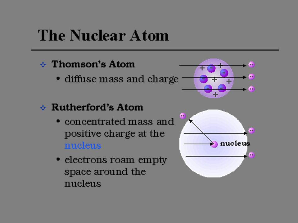
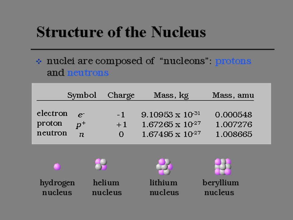
TEORÍA DE LA MATERIA -PARTÍCULA (~m) -MOLÉCULA (~A) -ÁTOMO (~A)
-PROTÓN, NEUTRÓN, ELECTRÓN (fm) -QUARK (am)
 -QUARK (am)")
7
MODELO ATÓMICO DE BOHR

8
LA MECÁNICA CUÁNTICA Y LA NUEVA TEORÍA DEL ÁTOMO
Werner Heisenberg Erwin Schrodinger

9
LAS MOLÉCULAS PRESENTAN NIVELES DE ENERGÍA MÁS COMPLEJOS

10
ENERGÍA INTERMOLECULAR DEPENDIENTE DE LA DISTANCIA

12
LA MATERIA TIENE UNA NATURALEZA ‘DUAL’… A VECES SE COMPORTA COMO PARTÍCULA, PERO OTRAS VECES SE COMPORTA COMO ONDA… LOUIS DE BROGLIE

13
HIPÓTESIS DE DE’BROGLIE
l=h/mv

14
EXPERIMENTO DE DOBLE RENDIJA CON ELECTRONES

15
QUE ES LA MATERIA, PARTÍCULA U ONDA. LAS DOS COSAS
QUE ES LA MATERIA, PARTÍCULA U ONDA? LAS DOS COSAS! LA MATERIA TIENE UN COMPORTAMIENTO DUAL. A VECES SE MANIFIESTA COMO PARTÍCULA, Y OTRAS VECES SE MANIFIESTA COMO ONDA!

16
LA RADIACIÓN ELECTROMAGNÉTICA
TEORÍA ONDULATORIA TEORÍA CORPUSCULAR

17
UNA ONDA ES LA PERTURBACIÓN DE UN CAMPO…

18
ONDAS LONGITUDINALES Y TRANSVERSALES

19
EN UNA ONDA NO HAY TRANSPORTE DE MATERIA, PERO SI SE TRANSPORTA ENERGÍA

20
A wave is a disturbance of a medium which transports energy through the medium without permanently transporting matter. In a wave, particles of the medium are temporily displaced and then return to their original position.

21
EL SONIDO ES UNA ONDA LONGITUDINAL

23
LA LUZ ES UNA ONDA ELECTROMAGNÉTICA
C = l n

24
REFLEXIÓN, REFRACCIÓN Y DIFRACCIÓN DE LA LUZ

25
POLARIZACIÓN DE LA LUZ

26
ESPECTRO DE LA RADIACIÓN ELECTROMAGNÉTICA

27
FUENTES DE LA RADIACIÓN E.M.

28
LA INTENSIDAD DE LA LUZ DEPENDE DEL NÚMERO DE FOTONES POR UNIDAD DE TIEMPO POR AREA

29
LAS ONDAS ELECTROMAGNÉTICAS (FOTONES), PUEDEN PROPAGARSE EN UN MEDIO MATERIAL ASÍ COMO EN EL VACÍO
, PUEDEN PROPAGARSE EN UN MEDIO MATERIAL ASÍ COMO EN EL VACÍO")
32
CON LA ABSORCIÓN DE LA LUZ OCURRE UNA EXITACIÓN ELECTRÓNICA

33
ABSORCIÓN DE LA LUZ

36
LA MATERIA PUEDE SER ‘TRANSPARENTE’ A LA RADIACIÓN

38
Cómo explica Usted este fenómeno ?

39
Purified Protein Solve the Phase Problem -MIR, MAD or MR (next class)
")
40
Proteins Can Form an Ordered Lattice

41
Celda unitaria tridimensional

42
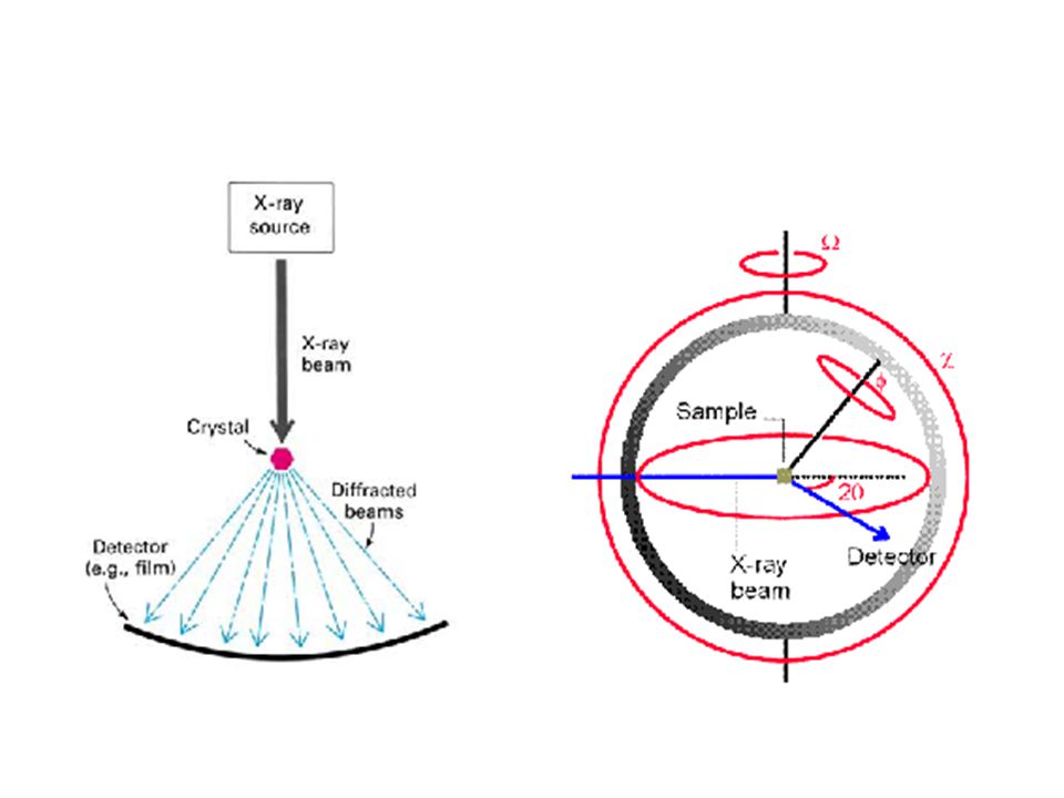
Overview of X-ray Experiment

43
h k l I Each reflection has an hkl index and a measured intensity 8 reflections selected from a 30,000 reflection data set shown to the right

45
X-Ray Crystallography
incoming x-ray detector diffracted protein crystal diffraction pattern goniometer controls crystal orientation

46
Crystal lattice “Real Space” Reflections “Reciprocal Space”
Fourier transform - FT Periodic r (x, y, z) Discrete, complex F (h, k, l) I (h, k, l) a (h, k, l) intensity phase
 Discrete, complex. F (h, k, l) I (h, k, l) a (h, k, l) intensity. phase.")
47
X-ray Equipment in Delaware
Area Detector RU-H3R X-ray generator

48
Close-up of Cryo Crystal

49
Patrón de difracción

50
Protein Crystal X-ray Diffraction

51
Zoom into a single reflection

52
The Diffraction Condition
Reflections are the result of constructive interference Geometry determines the constructive condition Bragg’s Law: n l = 2d sinQ sin Q = AB / d d sin Q = AB = 2d sin Q or n = 2d sin Q

53
Fase de la onda y la nterferencia constructiva/destructiva

54
Each Reflection Index Defines a Set of Parallel Planes that Slice Through the Crystal
Miller indices hkl Reciprocal Space Real h a k b l c Example: draw parallel planes that define the single reflection hkl 2 3 4 Reciprocal space has all reflections out to diffraction limit h 0 to 30 k 0 to just a hypothetical example l 0 to 43

55
Crystallography and Crystallization
To obtain X-ray diffraction data: First, need to grow a crystal Field of macromolecular crystallization Large parameter space (22 or so) Effects change over time Little theory Protein purification costly Tedious experimentation
 Effects change over time. Little theory. Protein purification costly. Tedious experimentation.")
56
Cristalización

57
Vapor Diffusion Method

58
Experimentation to grow protein crystals
Trial-and-error Experimentation Observables Partial Success Trial 1 Control Parameters Failure Trial 2 Success Trial 3

59
3. Crystals What defines a crystal?
Atoms, lattice points, symmetry, space groups Diffraction B-factors R-factors Resolution Refinement Modeling!

60
Crystals X-rays ‘see’ electrons (r) = (r+X)
What defines a crystal? 3D periodicity: anything (atom/molecule/void) present at some point in space, repeats at regular intervals, in three dimensions. X-rays ‘see’ electrons (r) = (r+X) (r): electron density at position r X: n1a + n2b + n3c n1, n2, n3: integers a, b, c: vectors
 present. at some point in space, repeats at regular intervals, in three dimensions. X-rays ‘see’ electrons (r) = (r+X) (r): electron density at position r. X: n1a + n2b + n3c. n1, n2, n3: integers. a, b, c: vectors.")
61
Crystals What defines a crystal? primary building block: lattice:
the unit cell lattice: set of points with identical environment crystal

62
Crystals organic versus inorganic
* lattice points need not coincide with atoms * symmetry can be ‘low’ * unit cell dimensions: 5-50Å, Å3 1 Å = m = 0.1 nm

63
Crystals: X-ray diffraction
diffraction: scattering of X-rays by periodic electron density diffraction ~ reflection against lattice planes, if: 2dhklsin = n X ~ Å Cu: 1.54Å dhkl Data set: list of intensities I and angles path: 2dhklsin

64
Crystals information contained in diffraction data
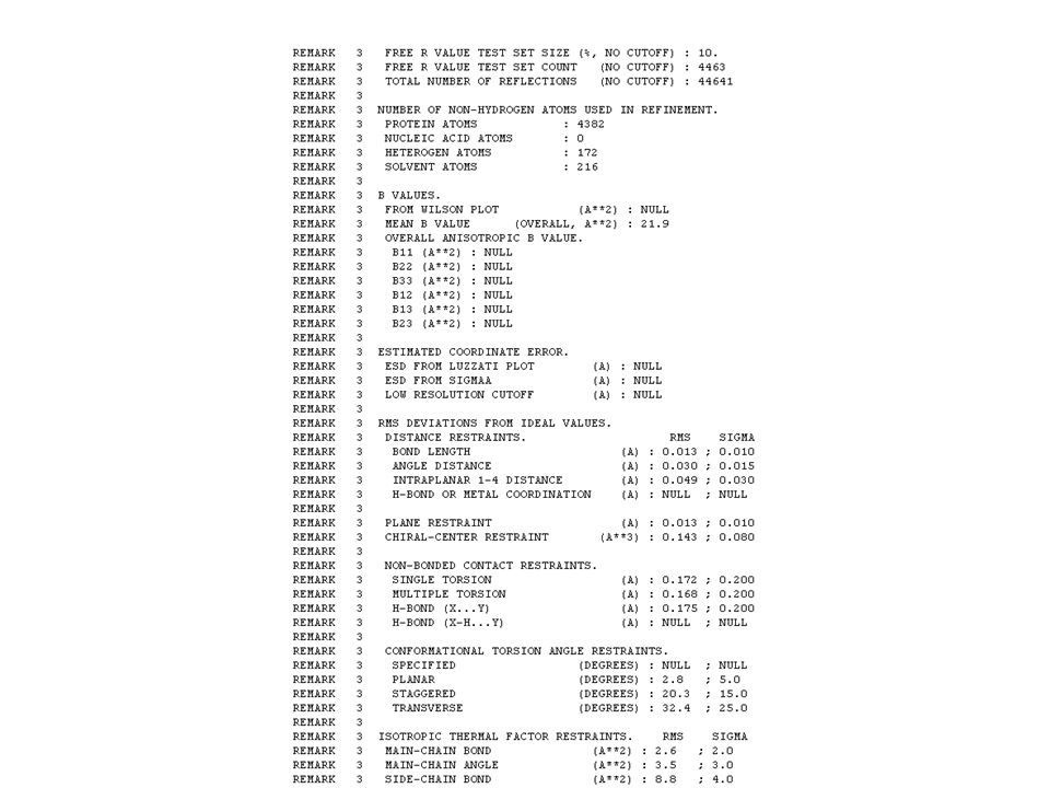
* How well does the proposed structure correspond to the experimental data? R-factor consider all (typically 5000) reflections, and compare calculated structure factors to observed ones. R = | Fhklobserved - Fhklcalculated | Fhkl = Ihkl Fhklobserved OK if 0.02 < R < 0.06 (small molecules) * Influence of movement due to temperature: atoms appear ‘smeared out’ compared to the static model ADP’s (‘B-factors’).
 reflections, and compare. calculated structure factors to observed ones. R = | Fhklobserved - Fhklcalculated | Fhkl = Ihkl. Fhklobserved. OK if 0.02 < R < 0.06 (small molecules) * Influence of movement due to temperature: atoms appear ‘smeared out’ compared to the static model ADP’s (‘B-factors’).")
65
The R-Factor: Measuring Convergence
To compare the generated electron density map and your model, you have to use the R-factor. The R-factor is a measure of convergence between the intensities given off by your model and the observed intensities. 0.6-VERY BAD 0.5 -BAD 0.4-Recoverable 0.2-Good for Protein 0.05-Good for small organic models 0-PERFECT FIT ||Fobs| - |Fcalc|| R= |Fobs| There is also an R-free, which is uses a test set of reflection points and corresponds well to the phase accuracy. R:

66
Cooling Protein Crystals
Crystals are damaged by x-ray radiation, resulting in loss of diffraction resolution due to formation of ion radicals or breaking of bonds within the protein. By cooling the crystal to liquid nitrogen temperatures, most of the radiation damage is eliminated. Cryocrystallography is thus an important method in determining protein structures. The common method for cooling is flash cooling. The crystal is held in a millimeter diameter loop (see picture) and quickly immersed in liquid nitrogen, liquid propane, or placed under a nitrogen boil-off cold stream. Flash cooling helps to prevent ice formation. Stresses during ice formation damage fragile protein crystals. Chemical cryoprotectants added to the solvent further suppress ice formation, keeping the surrounding solution glassy or vitreous. Finding the proper cryoprotectant is tedious because it is protein specific. Trial-and-error is the main method, and some crystals don’t have useable cryoprotectants. A general method for cooling without needing crystal specific cryoprotectants would be useful. Flash cooled crystal embedded in vitreous solvent.
 and quickly immersed in liquid nitrogen, liquid propane, or placed under a nitrogen boil-off cold stream. Flash cooling helps to prevent ice formation. Stresses during ice formation damage fragile protein crystals. Chemical cryoprotectants added to the solvent further suppress ice formation, keeping the surrounding solution glassy or vitreous. Finding the proper cryoprotectant is tedious because it is protein specific. Trial-and-error is the main method, and some crystals don’t have useable cryoprotectants. A general method for cooling without needing crystal specific cryoprotectants would be useful. Flash cooled crystal embedded in vitreous solvent.")
67
Pressure and Proteins Alpha helices in myoglobin rearrange when pressurized to 1500 atm. (yellow, 1 atm; green, 1500 atm) The lab uses pressure to elucidate the structural basis for pressure effects on proteins and to develop a novel method for cooling protein crystals. Pressures used in the lab are a few thousand atmospheres. EFFECTS ON PROTEIN STRUCTURE Fluctuations and the internal arrangements of atoms plays a crucial in protein function. Both can be probed with pressure. Pressure response of proteins is not compressive and is highly anisotropic. There are internal structural rearrangements. EFFECT ON CRYSTAL COOLING Pressure is known to slow kinetics of ice formation. Pressure also makes accessible other ice phases which contracts unlike normal hexagonal ice. Thus cooling under pressure might be a general method without needing chemical cryoprotectants. Phase diagram of water.

68
Various High Pressure Techniques in the Lab
beryllium pressure cell protein crystal pressurizing medium CELL FOR DIRECT X-RAY CRYSTALLOGRAPHY UNDER PRESSURE at room temperature CELL FOR COOLING CRYSTALS UNDER PRESSURE liquid pressurizing medium protein crystal Pressurised gas (blue arrow) is applied from a pump (not shown). protein crystal held on a loop using surface tension of solvent CELL FOR COOLING CRYSTALS UNDER PRESSURE gas pressurizing medium, cooling direclty onto loop pressure generator
 is applied from a pump (not shown). protein crystal held. on a loop using surface. tension of solvent. CELL FOR COOLING CRYSTALS. UNDER PRESSURE. gas pressurizing medium, cooling direclty onto loop. pressure generator.")
69
The Raw Data Every atom in a unit cell contributes to every reflection in the diffraction pattern. Two Pieces of Data The position of a reflection point on the reciprocal lattice, given by coordinates h,k,l. Determined by the direction reflected. The intensity of the reflection. Each spot is the sum of the contributions of all the scatterers in the unit cell. Reciprocal lattice is the coordinate system of the diffraction pattern, while the real lattice is the coordinate system within a unit cell. The real lattice spacing is inversely proportional to the spacing of the reflections. H, K, and L are expressed in reciprocal angstroms.

70
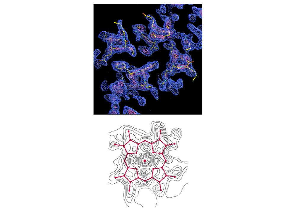
From diffraction to electron density map
Fourier Transform To get from the diffraction pattern to the electron density, you have to use a Fourier Transform. A transform is an eqn that transforms a function with some variables into a function with reciprocal variables. It is a way of going from the reciprocal lattice to the real lattice.

71
Once you have an electron density map, you can begin to fit models to it.

73
Resolution Resolution: another measure of how good your model is.
Resolution gives the size of the smallest molecule you can see or resolve. Dependent on the amount of data ultimately phased and used in structure determination.

74
Limitations and Difficulties, Besides the Phase Problem
Crystallizing Protein: Fragile Requires a crystal with shortest side 0.2 mm Flaws of Crystallization: Disorder in Unit Cell Vibrations of molecules Distortion in Crystallization Fragile-Protein Crystals held together by hydrogen bonds, not ionic bonds. Pictures of subtisilin and sperm whale myoglobin. Hanging Drop Technique Cryogenic Freezing--liquid nitrogen (-196 C), lowers vibration of molecules, increases the resolution Distortion in Crystallization--Testing to see whether the protein still has activity in the crystal form. An example is the shattering of deoxyhemoglobin crystals when oxygenated. Can’t see the H atoms…but can in what Trevor introduced, the Neutron Diffraction technique. Fix-Its: Cryogenic Cooling
, lowers vibration of molecules, increases the resolution. Distortion in Crystallization--Testing to see whether the protein still has activity in the crystal form. An example is the shattering of deoxyhemoglobin crystals when oxygenated. Can’t see the H atoms…but can in what Trevor introduced, the Neutron Diffraction technique. Fix-Its: Cryogenic Cooling.")
75
Steps of Protein X-ray Crystallography:
Conclusión Steps of Protein X-ray Crystallography: Crystallize your protein. Cryo-freeze your protein. Do an X-ray diffraction. Make a heavy atom derivative of protein. Take X-ray diffraction of the derivative. Do a Fourier Transform (or let a computer do it). Create models. Check R-Factor of models.
. Create models. Check R-Factor of models.")
76
Protein x-ray crystallography- practical point of view
A) cloning B) expression 6.5 14.4 21.5 31.0 45.0 66.2 kDa TBP Expression vector: a plasmid carrying the gene of interest Protein SDS PAGE gel: each band corresponds to one protein
 cloning. B) expression kDa. TBP. Expression vector: a plasmid carrying the gene of interest. Protein SDS PAGE gel: each band corresponds to one protein.")
77
Protein x-ray crystallography- practical point of view
C) purification D) crystallization E) solving the structure SDS PAGE showing a purified protein A protein crystal Ribbon representation of a protein structure (violet) bound to DNA
 purification. D) crystallization. E) solving the structure. SDS PAGE showing a purified protein. A protein crystal. Ribbon representation of a protein structure (violet) bound to DNA.")
78
Some important recent structures and what can we learn from them
The nucleosome core particle K. Luger et al, Nature 1997.

79
Some important recent structures and what can we learn from them
Aquaporin (water channel) H. Sui et al, Nature 2001.
 H. Sui et al, Nature")
80
Some important recent structures and what can we learn from them
The ribosome (large subunit) N. Ban et al, Science 2000.
 N. Ban et al, Science")
81
But it sometimes happens …
Rod Casey (Norwich): crystalloid in GM wheat with soya protein
: crystalloid in GM wheat with soya protein.")
82
Hydroxyapatite is the primary structural component of bone
Hydroxyapatite is the primary structural component of bone. As its formula suggests, it consists of Ca2+ ions surrounded by PO42– and OH– ions.

83
Estructura cristalina de la hidroxiapatita

86
PDB Protein Data Bank Currently contains about structures of macromolecules – proteins, nucleic acids, protein-DNA complexes and carbohydrates 11232 – X-Ray Diffraction & Other 2138 – NMR 302 – Theoretical Modeling

87
PDB: Growth

91
Unidad Asimétrica

Presentaciones similares


>")


 of verbs: those that end in –AR, those that end in –ER, and those that end in –IR. This.>")


