Descargar la presentación
La descarga está en progreso. Por favor, espere

1
Patología Definición: Estudio del sufrimiento
Estudio de los cambios estructurales y funcionales en células, tejidos y órganos que subyacen la enfermedad Proporciona bases para el cuidado clínico y la terapia Explica los por qué de los síntomas

2
Infarto al Miocardio A pale, whitish infarct is surrounded by a zone of hyperemia (vascular dilatation)
")
3
Estudio de la Patología
Patología general : Reacciones básicas de las células y tejidos a estímulos anormales Patología Especial o Sistémica: Respuesta específica de tejidos y órganos a estímulos definidos Pathology is literally the study (logos) of suffering (pathos). More specifically, it is a bridging discipline involving both basic science and clinical practice and is devoted to the study of the structural and functional changes in cells, tissues, and organs that underlie disease. By the use of molecular, microbiologic, immunologic, and morphologic techniques, pathology attempts to explain the whys and wherefores of the signs and symptoms manifested by patients while providing a sound foundation for rational clinical care and therapy. Traditionally, the study of pathology is divided into general pathology and special, or systemic, pathology. The former is concerned with the basic reactions of cells and tissues to abnormal stimuli that underlie all diseases. The latter examines the specific responses of specialized organs and tissues to more or less well-defined stimuli. In this book, we first cover the principles of general pathology and then proceed to specific disease processes as they affect particular organs or systems. The four aspects of a disease process that form the core of pathology are its cause (etiology), the mechanisms of its development (pathogenesis), the structural alterations induced in the cells and organs of the body (morphologic changes), and the functional consequences of the morphologic changes (clinical significance).
 of suffering (pathos). More specifically, it is a bridging discipline involving both basic science and clinical practice and is devoted to the study of the structural and functional changes in cells, tissues, and organs that underlie disease. By the use of molecular, microbiologic, immunologic, and morphologic techniques, pathology attempts to explain the whys and wherefores of the signs and symptoms manifested by patients while providing a sound foundation for rational clinical care and therapy. Traditionally, the study of pathology is divided into general pathology and special, or systemic, pathology. The former is concerned with the basic reactions of cells and tissues to abnormal stimuli that underlie all diseases. The latter examines the specific responses of specialized organs and tissues to more or less well-defined stimuli. In this book, we first cover the principles of general pathology and then proceed to specific disease processes as they affect particular organs or systems. The four aspects of a disease process that form the core of pathology are its cause (etiology), the mechanisms of its development (pathogenesis), the structural alterations induced in the cells and organs of the body (morphologic changes), and the functional consequences of the morphologic changes (clinical significance).")
4
Estudio de la Enfermedad
Etiología Patogénesis Cambios Morfológicos Significado Clínico The four aspects of a disease process that form the core of pathology are its cause (etiology), the mechanisms of its development (pathogenesis), the structural alterations induced in the cells and organs of the body (morphologic changes), and the functional consequences of the morphologic changes (clinical significance).
, the mechanisms of its development (pathogenesis), the structural alterations induced in the cells and organs of the body (morphologic changes), and the functional consequences of the morphologic changes (clinical significance).")
5
Etiología: “Causa” de la enfermedad
Factores Etiológicos: -Adquiridos ej. Infecciosa -Intrínsecos o Genéticos Patogenie: Secuencia de acontecimientos en la respuesta de las células o tejidos al agente etiológico.

6
Cambios Morfológicos: Alteraciones estructurales en células y tejidos que son característicos de la enfermedad o diagnósticas del proceso etiológico Significado Clínico: Los cambios morfológicos determinan manifestaciones clínicas (signos), la evolución y el pronóstico de la enfermedad
, la evolución y el pronóstico de la enfermedad.")
7
Necrosis, esteatosis, atrofia
The dark liver on the left is very flaccid on palpation, while the central liver is enlarged, and the right liver is dark, somewhat smaller than normal but still firm.Remarks:Acute starvation can also cause the central fatty appearance when the animal has fat to mobilize, but in chronic starvation without fat to mobilize the liver shrinks, gets darker, but is still firm.

8
Homeostasis Estado de tranquilidad celular derivada de la capacidad que tiene la célula de manejar estados fisiológicos normales

9
Célula Normal Definición Funcional y Estructural:
Programas Genéticos -Metabolismo Diferenciación -Especialización Relación con Células Vecinas Disponibilidad de Sustratos Metabólicos The normal cell is confined to a fairly narrow range of function and structure by its genetic programs of metabolism, differentiation, and specialization; by constraints of neighboring cells; and by the availability of metabolic substrates. It is nevertheless able to handle normal physiologic demands, maintaining a steady state called homeostasis.

10
Respuesta Celular Al Stress

11
Adaptaciones Celulares de Crecimiento y Diferenciación
Hiperplasia: Aumento número de células en órgano o tejido Hipertrofia: Aumento en el tamaño de las células Atrofia: Disminución en el tamaño celular por pérdida de sustancia celular Metaplasia: Cambio reversible en el cual una célula adulta de un tipo cambia a otra célula adulta de otro tipo

12
Hiperplasia Útero Grávido Útero Normal

13
Hipertrofia Corazón Normal Corazón Hipertrofiado

14
Atrofia Pérdida de masa Muscular

15
Atrofia Renal: Estenosis Ateroesclerótica

16
Metaplasia Células Metaplásicas Células Normales

17
Lesión Celular Momento en que la adaptación a los distintos estímulos es excedida Al alcanzar el “punto de no retorno” la célula sufre un daño irreversible y muere

18
Etapas de Alteración Progresiva
Homeostasis Adaptación Lesión Reversible Lesión Irreversible Muerte Celular Necrosis Apoptosis*

19
Respuesta a Estímulo Lesional I

20
Respuesta a Estímulo Lesional II
Reversible: R. Endoplásmico Tumefacción generalizada Dispersión Ribosomas Condensación Cromatina Vesículas M. Plasmática Irreversible: Autolisis R.Endoplásmico, Lisis Destrucción Membrana P. y Núcleo* Tumefacción Mitocondrial Figuras de mielina Figure 1-8 Schematic representation of a normal cell and the changes in reversible and irreversible cell injury. Depicted are morphologic changes, which are described in the following pages and shown in electron micrographs in Figure Reversible injury is characterized by generalized swelling of the cell and its organelles; blebbing of the plasma membrane; detachment of ribosomes from the endoplasmic reticulum; and clumping of nuclear chromatin. Transition to irreversible injury is characterized by increasing swelling of the cell; swelling and disruption of lysosomes; presence of large amorphous densities in swollen mitochondria; disruption of cellular membranes; and profound nuclear changes. The latter include nuclear codensation (pyknosis), followed by fragmentation (karyorrhexis) and dissolution of the nucleus (karyolysis). Laminated structures (myelin figures) derived from damaged membranes of organelles and the plasma membrane first appear during the reversible stage and become more pronounced in irreversibly damaged cells. The mechanisms underlying these changes are discussed in the text that follows. Reversible cell injury. Initially, injury is manifested as functional and morphologic changes that are reversible if the damaging stimulus is removed. The hallmarks of reversible injury are reduced oxidative phosphorylation, adenosine triphosphate (ATP) depletion, and cellular swelling caused by changes in ion concentrations and water influx. Irreversible injury and cell death. With continuing damage, the injury becomes irreversible, at which time the cell cannot recover. Is there a critical biochemical event (the "lethal hit") responsible for the point of no return? There are no clear answers to this question. However, as discussed later, in ischemic tissues such as the myocardium, certain structural changes (e.g., amorphous densities in mitochondria, indicative of severe mitochondrial damage) and functional changes (e.g., loss of membrane permeability) are indicative of cells that have suffered irreversible injury. Irreversibly injured cells invariably undergo morphologic changes that are recognized as cell death. There are two types of cell death, necrosis and apoptosis, which differ in their morphology, mechanisms, and roles in disease and physiology (Fig. 1-9 and Table 1-2). When damage to membranes is severe, lysosomal enzymes enter the cytoplasm and digest the cell, and cellular contents leak out, resulting in necrosis. Some noxious stimuli, especially those that damage DNA, induce another type of death, apoptosis, which is characterized by nuclear dissolution without complete loss of membrane integrity. Whereas necrosis is always a pathologic process, apoptosis serves many normal functions and is not necessarily associated with cell injury. Although we emphasize the distinctions between necrosis and apoptosis, there may be some overlaps and common mechanisms between these two pathways. In addition, at least some types of stimuli may induce either apoptosis or necrosis, depending on the intensity and duration of the stimulus, the rapidity of the death process, and the biochemical derangements induced in the injured cell. The mechanisms and significance of these two death pathways are discussed later in the chapter.
, followed by fragmentation (karyorrhexis) and dissolution of the nucleus (karyolysis). Laminated structures (myelin figures) derived from damaged membranes of organelles and the plasma membrane first appear during the reversible stage and become more pronounced in irreversibly damaged cells. The mechanisms underlying these changes are discussed in the text that follows. Reversible cell injury. Initially, injury is manifested as functional and morphologic changes that are reversible if the damaging stimulus is removed. The hallmarks of reversible injury are reduced oxidative phosphorylation, adenosine triphosphate (ATP) depletion, and cellular swelling caused by changes in ion concentrations and water influx. Irreversible injury and cell death. With continuing damage, the injury becomes irreversible, at which time the cell cannot recover. Is there a critical biochemical event (the lethal hit ) responsible for the point of no return There are no clear answers to this question. However, as discussed later, in ischemic tissues such as the myocardium, certain structural changes (e.g., amorphous densities in mitochondria, indicative of severe mitochondrial damage) and functional changes (e.g., loss of membrane permeability) are indicative of cells that have suffered irreversible injury. Irreversibly injured cells invariably undergo morphologic changes that are recognized as cell death. There are two types of cell death, necrosis and apoptosis, which differ in their morphology, mechanisms, and roles in disease and physiology (Fig. 1-9 and Table 1-2). When damage to membranes is severe, lysosomal enzymes enter the cytoplasm and digest the cell, and cellular contents leak out, resulting in necrosis. Some noxious stimuli, especially those that damage DNA, induce another type of death, apoptosis, which is characterized by nuclear dissolution without complete loss of membrane integrity. Whereas necrosis is always a pathologic process, apoptosis serves many normal functions and is not necessarily associated with cell injury. Although we emphasize the distinctions between necrosis and apoptosis, there may be some overlaps and common mechanisms between these two pathways. In addition, at least some types of stimuli may induce either apoptosis or necrosis, depending on the intensity and duration of the stimulus, the rapidity of the death process, and the biochemical derangements induced in the injured cell. The mechanisms and significance of these two death pathways are discussed later in the chapter.")
21
Tipos de Muerte Celular
Necrosis Tumefacción celular Núcleo experimenta daño* Autodigestión Lisosomal Origen Patológico Inflamación Asociada Apoptosis: Contracción tamaño celular Condensación cromatina Vesículas citoplasmáticas Cuerpos apoptóticos Fagocitosis de los c. apoptóticos Fisiológico, generalmente No-inflamación FeatureNecrosisApoptosisCell sizeEnlarged (swelling)Reduced (shrinkage)NucleusPyknosis → karyorrhexis → karyolysisFragmentation into nucleosome size fragmentsPlasma membraneDisruptedIntact; altered structure, especially orientation of lipidsCellular contentsEnzymatic digestion; may leak out of cellIntact; may be released in apoptotic bodiesAdjacent inflammationFrequentNoPhysiologic or pathologic roleInvariably pathologic (culmination of irreversible cell injury)Often physiologic, means of eliminating unwanted cells; may be pathologic after some forms of cell injury, especially DNA damage
Reduced (shrinkage)NucleusPyknosis → karyorrhexis → karyolysisFragmentation into nucleosome size fragmentsPlasma membraneDisruptedIntact; altered structure, especially orientation of lipidsCellular contentsEnzymatic digestion; may leak out of cellIntact; may be released in apoptotic bodiesAdjacent inflammationFrequentNoPhysiologic or pathologic roleInvariably pathologic (culmination of irreversible cell injury)Often physiologic, means of eliminating unwanted cells; may be pathologic after some forms of cell injury, especially DNA damage.")
22
Causas de Lesión Celular
Deprivación de Oxígeno (Hipoxia, Isquemia*) Agentes Físicos Agentes Químicos Agentes Infecciosos Reacciones Inmunológicas Desequilibrios Nutricionales Desórdenes Genéticos (Chemical Agents and Drugs. The list of chemicals that may produce cell injury defies compilation. Simple chemicals such as glucose or salt in hypertonic concentrations may cause cell injury directly or by deranging electrolyte homeostasis of cells. Even oxygen, in high concentrations, is severely toxic. Trace amounts of agents known as poisons, such as arsenic, cyanide, or mercuric salts, may destroy sufficient numbers of cells within minutes to hours to cause death. Other substances, however, are our daily companions: environmental and air pollutants, insecticides, and herbicides; industrial and occupational hazards, such as carbon monoxide and asbestos; social stimuli, such as alcohol and narcotic drugs; and the ever-increasing variety of therapeutic drugs. Infectious Agents. These agents range from the submicroscopic viruses to the large tapeworms. In between are the rickettsiae, bacteria, fungi, and higher forms of parasites. The ways by which this heterogeneous group of biologic agents cause injury are diverse and are discussed in Chapter 8. Immunologic Reactions. Although the immune system serves an essential function in defense against infectious pathogens, immune reactions may, in fact, cause cell injury. The anaphylactic reaction to a foreign protein or a drug is a prime example, and reactions to endogenous self-antigens are responsible for a number of autoimmune diseases (Chapter 6). Genetic Derangements. Genetic defects as causes of cell injury are of major interest to scientists and physicians today (Chapter 5). The genetic injury may result in a defect as severe as the congenital malformations associated with Down syndrome, caused by a chromosomal abnormality, or as subtle as the decreased life of red blood cells caused by a single amino acid substitution in hemoglobin S in sickle cell anemia. The many inborn errors of metabolism arising from enzymatic abnormalities, usually an enzyme lack, are excellent examples of cell damage due to subtle alterations at the level of DNA. Variations in the genetic makeup can also influence the susceptibility of cells to injury by chemicals and other environmental insults.
 Agentes Físicos. Agentes Químicos. Agentes Infecciosos. Reacciones Inmunológicas. Desequilibrios Nutricionales. Desórdenes Genéticos. (Chemical Agents and Drugs. The list of chemicals that may produce cell injury defies compilation. Simple chemicals such as glucose or salt in hypertonic concentrations may cause cell injury directly or by deranging electrolyte homeostasis of cells. Even oxygen, in high concentrations, is severely toxic. Trace amounts of agents known as poisons, such as arsenic, cyanide, or mercuric salts, may destroy sufficient numbers of cells within minutes to hours to cause death. Other substances, however, are our daily companions: environmental and air pollutants, insecticides, and herbicides; industrial and occupational hazards, such as carbon monoxide and asbestos; social stimuli, such as alcohol and narcotic drugs; and the ever-increasing variety of therapeutic drugs. Infectious Agents. These agents range from the submicroscopic viruses to the large tapeworms. In between are the rickettsiae, bacteria, fungi, and higher forms of parasites. The ways by which this heterogeneous group of biologic agents cause injury are diverse and are discussed in Chapter 8. Immunologic Reactions. Although the immune system serves an essential function in defense against infectious pathogens, immune reactions may, in fact, cause cell injury. The anaphylactic reaction to a foreign protein or a drug is a prime example, and reactions to endogenous self-antigens are responsible for a number of autoimmune diseases (Chapter 6). Genetic Derangements. Genetic defects as causes of cell injury are of major interest to scientists and physicians today (Chapter 5). The genetic injury may result in a defect as severe as the congenital malformations associated with Down syndrome, caused by a chromosomal abnormality, or as subtle as the decreased life of red blood cells caused by a single amino acid substitution in hemoglobin S in sickle cell anemia. The many inborn errors of metabolism arising from enzymatic abnormalities, usually an enzyme lack, are excellent examples of cell damage due to subtle alterations at the level of DNA. Variations in the genetic makeup can also influence the susceptibility of cells to injury by chemicals and other environmental insults.")
23
Lesión Celular Principios Comunes
La respuesta celular depende de: Naturaleza, Severidad, Duración del estímulo Las Consecuencias de la lesión dependen de: Tipo, Estado, Adaptabilidad de la célula El daño final depende de: Anormalidades bioquímicas y funcionales en uno o varios componentes celulares esenciales Mechanisms of Cell Injury The biochemical mechanisms responsible for cell injury are complex. There are, however, a number of principles that are relevant to most forms of cell injury: The cellular response to injurious stimuli depends on the type of injury, its duration, and its severity. Thus, small doses of a chemical toxin or brief periods of ischemia may induce reversible injury, whereas large doses of the same toxin or more prolonged ischemia might result either in instantaneous cell death or in slow, irreversible injury leading in time to cell death. The consequences of cell injury depend on the type, state, and adaptability of the injured cell. The cell's nutritional and hormonal status and its metabolic needs are important in its response to injury. How vulnerable is a cell, for example, to loss of blood supply and hypoxia? The striated muscle cell in the leg can be placed entirely at rest when it is deprived of its blood supply; not so the striated muscle of the heart. Exposure of two individuals to identical concentrations of a toxin, such as carbon tetrachloride, may produce no effect in one and cell death in the other. This may be due to genetic variations affecting the amount and activity of hepatic enzymes that convert carbon tetrachloride to toxic byproducts (Chapter 9). With the complete mapping of the human genome, there is great interest in identifying genetic polymorphisms that affect the cell's response to injurious agents. Cell injury results from functional and biochemical abnormalities in one or more of several essential cellular components (Fig. 1-10). The most important targets of injurious stimuli are: (1) aerobic respiration involving mitochondrial oxidative phosphorylation and production of ATP; (2) the integrity of cell membranes, on which the ionic and osmotic homeostasis of the cell and its organelles depends; (3) protein synthesis; (4) the cytoskeleton; and (5) the integrity of the genetic apparatus of the cell.
. With the complete mapping of the human genome, there is great interest in identifying genetic polymorphisms that affect the cell s response to injurious agents. Cell injury results from functional and biochemical abnormalities in one or more of several essential cellular components (Fig. 1-10). The most important targets of injurious stimuli are: (1) aerobic respiration involving mitochondrial oxidative phosphorylation and production of ATP; (2) the integrity of cell membranes, on which the ionic and osmotic homeostasis of the cell and its organelles depends; (3) protein synthesis; (4) the cytoskeleton; and (5) the integrity of the genetic apparatus of the cell.")
24
4.-Stress Oxidativo Daños más importantes
Peroxidación de lípidos de membrana Modificación oxidativa de proteínas Lesiones en ADN Lipid peroxidation of membranes. Free radicals in the presence of oxygen may cause peroxidation of lipids within plasma and organellar membranes. Oxidative damage is initiated when the double bonds in unsaturated fatty acids of membrane lipids are attacked by oxygen-derived free radicals, particularly by OH. The lipid-free radical interactions yield peroxides, which are themselves unstable and reactive, and an autocatalytic chain reaction ensues (called propagation), which can result in extensive membrane, organellar, and cellular damage. Other more favorable termination options take place when the free radical is captured by a scavenger, such as vitamin E , embedded in the cell membrane. Oxidative modification of proteins. Free radicals promote oxidation of amino acid residue side chains, formation of protein-protein cross-linkages (e.g., disulfide bonds), and oxidation of the protein backbone, resulting in protein fragmentation. Oxidative modification enhances degradation of critical proteins by the multicatalytic proteasome complex, raising havoc throughout the cell. Lesions in DNA. Reactions with thymine in nuclear and mitochondrial DNA produce single-stranded breaks in DNA. This DNA damage has been implicated in cell aging (discussed later in this chapter) and in malignant transformation of cells Figure 1-14 The role of reactive oxygen species in cell injury. O2 is converted to superoxide (O2-) by oxidative enzymes in the endoplasmic reticulum (ER), mitochondria, plasma membrane, peroxisomes, and cytosol. O2- is converted to H2O2 by dismutation and thence to OH by the Cu2+/Fe2+-catalyzed Fenton reaction. H2O2 is also derived directly from oxidases in peroxisomes. Not shown is another potentially injurious radical, singlet oxygen. Resultant free radical damage to lipid (peroxidation), proteins, and DNA leads to various forms of cell injury. Note that superoxide catalyzes the reduction of Fe3+ to Fe2+, thus enhancing OH generation by the Fenton reaction. The major antioxidant enzymes are superoxide dismutase (SOD), catalase, and glutathione peroxidase. GSH, reduced glutathione; GSSG, oxidized glutathione; NADPH, reduced form of nicotinamide adenine dinucleotide phosphate. ACCUMULATION OF OXYGEN-DERIVED FREE RADICALS (OXIDATIVE STRESS) Cells generate energy by reducing molecular oxygen to water. During this process, small amounts of partially reduced reactive oxygen forms are produced as an unavoidable byproduct of mitochondrial respiration. Some of these forms are free radicals that can damage lipids, proteins, and nucleic acids. They are referred to as reactive oxygen species. Cells have defense systems to prevent injury caused by these products. An imbalance between free radical-generating and radical-scavenging systems results in oxidative stress, a condition that has been associated with the cell injury seen in many pathologic conditions. Free radical-mediated damage contributes to such varied processes as chemical and radiation injury, ischemia-reperfusion injury (induced by restoration of blood flow in ischemic tissue), cellular aging, and microbial killing by phagocytes Free radicals are chemical species that have a single unpaired electron in an outer orbit. Energy created by this unstable configuration is released through reactions with adjacent molecules, such as inorganic or organic chemicals-proteins, lipids, carbohydrates-particularly with key molecules in membranes and nucleic acids. Moreover, free radicals initiate autocatalytic reactions, whereby molecules with which they react are themselves converted into free radicals to propagate the chain of damage. Free radicals may be initiated within cells in several ways (Fig. 1-14): Absorption of radiant energy (e.g., ultraviolet light, x-rays). For example, ionizing radiation can hydrolyze water into hydroxyl (OH) and hydrogen (H) free radicals. Enzymatic metabolism of exogenous chemicals or drugs (e.g., carbon tetrachloride [CCl4] can generate CCl3, described later in this chapter). The reduction-oxidation reactions that occur during normal metabolic processes. During normal respiration, molecular oxygen is sequentially reduced by the addition of four electrons to generate water. Such conversion occurs by oxidative enzymes in the endoplasmic reticulum, cytosol, mitochondria, peroxisomes, and lysosomes. In this process, small amounts of toxic intermediates are produced; these include superoxide anion radical (O2-), hydrogen peroxide (H2O2), and hydroxyl ions (OH). Rapid bursts of superoxide production occur in activated polymorphonuclear leukocytes during inflammation. This occurs by a precisely controlled reaction in a plasma membrane multiprotein complex that uses NADPH oxidase for the redox reaction (Chapter 2). In addition, some intracellular oxidases (such as xanthine oxidase) generate superoxide radicals as a consequence of their activity. Transition metals such as iron and copper donate or accept free electrons during intracellular reactions and catalyze free radical formation, as in the Fenton reaction (H2O2 + Fe2+ → Fe3+ + OH + OH-). Because most of the intracellular free iron is in the ferric (Fe3+) state, it must be first reduced to the ferrous (Fe2+) form to participate in the Fenton reaction. This reduction can be enhanced by superoxide, and thus sources of iron and superoxide are required for maximal oxidative cell damage. Nitric oxide (NO), an important chemical mediator generated by endothelial cells, macrophages, neurons, and other cell types (Chapter 2), can act as a free radical and can also be converted to highly reactive peroxynitrite anion (ONOO-) as well as NO2 and NO3-. page 16 page 17The effects of these reactive species are wide-ranging, but three reactions are particularly relevant to cell injury (see Fig. 1-14): Lesions in DNA. Reactions with thymine in nuclear and mitochondrial DNA produce single-stranded breaks in DNA. This DNA damage has been implicated in cell aging (discussed later in this chapter) and in malignant transformation of cells (Chapter 7).
, which can result in extensive membrane, organellar, and cellular damage. Other more favorable termination options take place when the free radical is captured by a scavenger, such as vitamin E , embedded in the cell membrane. Oxidative modification of proteins. Free radicals promote oxidation of amino acid residue side chains, formation of protein-protein cross-linkages (e.g., disulfide bonds), and oxidation of the protein backbone, resulting in protein fragmentation. Oxidative modification enhances degradation of critical proteins by the multicatalytic proteasome complex, raising havoc throughout the cell. Lesions in DNA. Reactions with thymine in nuclear and mitochondrial DNA produce single-stranded breaks in DNA. This DNA damage has been implicated in cell aging (discussed later in this chapter) and in malignant transformation of cells Figure 1-14 The role of reactive oxygen species in cell injury. O2 is converted to superoxide (O2-) by oxidative enzymes in the endoplasmic reticulum (ER), mitochondria, plasma membrane, peroxisomes, and cytosol. O2- is converted to H2O2 by dismutation and thence to OH by the Cu2+/Fe2+-catalyzed Fenton reaction. H2O2 is also derived directly from oxidases in peroxisomes. Not shown is another potentially injurious radical, singlet oxygen. Resultant free radical damage to lipid (peroxidation), proteins, and DNA leads to various forms of cell injury. Note that superoxide catalyzes the reduction of Fe3+ to Fe2+, thus enhancing OH generation by the Fenton reaction. The major antioxidant enzymes are superoxide dismutase (SOD), catalase, and glutathione peroxidase. GSH, reduced glutathione; GSSG, oxidized glutathione; NADPH, reduced form of nicotinamide adenine dinucleotide phosphate. ACCUMULATION OF OXYGEN-DERIVED FREE RADICALS (OXIDATIVE STRESS) Cells generate energy by reducing molecular oxygen to water. During this process, small amounts of partially reduced reactive oxygen forms are produced as an unavoidable byproduct of mitochondrial respiration. Some of these forms are free radicals that can damage lipids, proteins, and nucleic acids. They are referred to as reactive oxygen species. Cells have defense systems to prevent injury caused by these products. An imbalance between free radical-generating and radical-scavenging systems results in oxidative stress, a condition that has been associated with the cell injury seen in many pathologic conditions. Free radical-mediated damage contributes to such varied processes as chemical and radiation injury, ischemia-reperfusion injury (induced by restoration of blood flow in ischemic tissue), cellular aging, and microbial killing by phagocytes Free radicals are chemical species that have a single unpaired electron in an outer orbit. Energy created by this unstable configuration is released through reactions with adjacent molecules, such as inorganic or organic chemicals-proteins, lipids, carbohydrates-particularly with key molecules in membranes and nucleic acids. Moreover, free radicals initiate autocatalytic reactions, whereby molecules with which they react are themselves converted into free radicals to propagate the chain of damage. Free radicals may be initiated within cells in several ways (Fig. 1-14): Absorption of radiant energy (e.g., ultraviolet light, x-rays). For example, ionizing radiation can hydrolyze water into hydroxyl (OH) and hydrogen (H) free radicals. Enzymatic metabolism of exogenous chemicals or drugs (e.g., carbon tetrachloride [CCl4] can generate CCl3, described later in this chapter). The reduction-oxidation reactions that occur during normal metabolic processes. During normal respiration, molecular oxygen is sequentially reduced by the addition of four electrons to generate water. Such conversion occurs by oxidative enzymes in the endoplasmic reticulum, cytosol, mitochondria, peroxisomes, and lysosomes. In this process, small amounts of toxic intermediates are produced; these include superoxide anion radical (O2-), hydrogen peroxide (H2O2), and hydroxyl ions (OH). Rapid bursts of superoxide production occur in activated polymorphonuclear leukocytes during inflammation. This occurs by a precisely controlled reaction in a plasma membrane multiprotein complex that uses NADPH oxidase for the redox reaction (Chapter 2). In addition, some intracellular oxidases (such as xanthine oxidase) generate superoxide radicals as a consequence of their activity. Transition metals such as iron and copper donate or accept free electrons during intracellular reactions and catalyze free radical formation, as in the Fenton reaction (H2O2 + Fe2+ → Fe3+ + OH + OH-). Because most of the intracellular free iron is in the ferric (Fe3+) state, it must be first reduced to the ferrous (Fe2+) form to participate in the Fenton reaction. This reduction can be enhanced by superoxide, and thus sources of iron and superoxide are required for maximal oxidative cell damage. Nitric oxide (NO), an important chemical mediator generated by endothelial cells, macrophages, neurons, and other cell types (Chapter 2), can act as a free radical and can also be converted to highly reactive peroxynitrite anion (ONOO-) as well as NO2 and NO3-. page 16 page 17The effects of these reactive species are wide-ranging, but three reactions are particularly relevant to cell injury (see Fig. 1-14): Lesions in DNA. Reactions with thymine in nuclear and mitochondrial DNA produce single-stranded breaks in DNA. This DNA damage has been implicated in cell aging (discussed later in this chapter) and in malignant transformation of cells (Chapter 7).")
25
Inactivación de Radicales Libres
Antioxidantes : Vitamina E, A , C Unión a Proteínas : Almacenamiento y Transporte: Transferrina, lactoferrina etc. Enzimas: -Catalasas -Superóxido dismutasas -Glutatión peroxidadas Cells have developed multiple mechanisms to remove free radicals and thereby minimize injury. Free radicals are inherently unstable and generally decay spontaneously. Superoxide, for example, is unstable and decays (dismutates) spontaneously into oxygen and hydrogen peroxide in the presence of water. There are, however, several nonenzymatic and enzymatic systems that contribute to inactivation of free radical reactions (see Fig. 1-14). These include the following: Antioxidants either block the initiation of free radical formation or inactivate (e.g., scavenge) free radicals and terminate radical damage. Examples are the lipid-soluble vitamins E and A as well as ascorbic acid and glutathione in the cytosol. As we have seen, iron and copper can catalyze the formation of reactive oxygen species. The levels of these reactive forms are minimized by binding of the ions to storage and transport proteins (e.g., transferrin, ferritin, lactoferrin, and ceruloplasmin), thereby minimizing OH formation. A series of enzymes acts as free radical-scavenging systems and break down hydrogen peroxide and superoxide anion. These enzymes are located near the sites of generation of these oxidants and include the following: Catalase, present in peroxisomes, which decomposes H2O2 (2 H2O2 → O2 + 2 H2O). Superoxide dismutases are found in many cell types and convert superoxide to H2O2 (2 O H → H2O2 + O2).23 This group includes both manganese-superoxide dismutase, which is localized in mitochondria, and copper-zinc-superoxide dismutase, which is found in the cytosol. Glutathione peroxidase also protects against injury by catalyzing free radical breakdown (H2O2 + 2 GSH → GSSG [glutathione homodimer] + 2 H2O, or 2 OH + 2 GSH → GSSG + 2 H2O). The intracellular ratio of oxidized glutathione (GSSG) to reduced glutathione (GSH) is a reflection of the oxidative state of the cell and is an important aspect of the cell's ability to detoxify reactive oxygen species. In many pathologic processes, the final effects induced by free radicals depend on the net balance between free radical formation and termination. As stated earlier, free radicals are thought to be involved in many pathologic and physiologic processes, to be mentioned throughout this book.
 spontaneously into oxygen and hydrogen peroxide in the presence of water. There are, however, several nonenzymatic and enzymatic systems that contribute to inactivation of free radical reactions (see Fig. 1-14). These include the following: Antioxidants either block the initiation of free radical formation or inactivate (e.g., scavenge) free radicals and terminate radical damage. Examples are the lipid-soluble vitamins E and A as well as ascorbic acid and glutathione in the cytosol. As we have seen, iron and copper can catalyze the formation of reactive oxygen species. The levels of these reactive forms are minimized by binding of the ions to storage and transport proteins (e.g., transferrin, ferritin, lactoferrin, and ceruloplasmin), thereby minimizing OH formation. A series of enzymes acts as free radical-scavenging systems and break down hydrogen peroxide and superoxide anion. These enzymes are located near the sites of generation of these oxidants and include the following: Catalase, present in peroxisomes, which decomposes H2O2 (2 H2O2 → O2 + 2 H2O). Superoxide dismutases are found in many cell types and convert superoxide to H2O2 (2 O H → H2O2 + O2).23 This group includes both manganese-superoxide dismutase, which is localized in mitochondria, and copper-zinc-superoxide dismutase, which is found in the cytosol. Glutathione peroxidase also protects against injury by catalyzing free radical breakdown (H2O2 + 2 GSH → GSSG [glutathione homodimer] + 2 H2O, or 2 OH + 2 GSH → GSSG + 2 H2O). The intracellular ratio of oxidized glutathione (GSSG) to reduced glutathione (GSH) is a reflection of the oxidative state of the cell and is an important aspect of the cell s ability to detoxify reactive oxygen species. In many pathologic processes, the final effects induced by free radicals depend on the net balance between free radical formation and termination. As stated earlier, free radicals are thought to be involved in many pathologic and physiologic processes, to be mentioned throughout this book.")
26
LESIÓN CELULAR REVERSIBLE IRREVERSIBLE ? Varias formas de Lesión
Interdependencia de Organelos, enzimas y moléculas Punto de “no retorno” Indeterminado No existiría una causa común de muerte celular

27
Morfología de Injuria y Necrosis

28
Microscopía de Luz: Patrones de lesión Celular Reversible
Degeneración Hidrópica o Vacuolar pequeñas vacuolas claras en citoplasma Incapacidad para mantener equilibrio de fluidos órgano pálido y turgente Cambio graso, Esteatosis: injuria o lesión hipóxica lesiones tóxicometabólicas aparición de vacuolas lipídicas distinto tamaño

29
Necrosis Definición: Espectro de cambios morfológicos que siguen a la muerte celular en tejidos vivos, que resultan de la acción degradativa, progresiva, de enzimas sobre células letalmente lesionadas. *Necrosis no es sinónimo de Apoptosis

30
Infarto isquémico bazo Necrosis de Coagulación
Infarto Anémico - Bazo. Dos grandes infartos (áreas de necrosis isquémica, en este caso coagulativa) se observan en este bazo seccionado. Como la etiología de necrosis coagulativa es usualmente vascular con pérdida de aporte sanguíneo, el infarto ocurre en una zona de distribución vascular. Por esto los infartos son a menudo con forma de cuña con la base en la cápsula del órgano. Organo: Bazo Etiología: Se debe a la necrosis isquémica provocada por la falta de aporte sanguíneo adecuado al tejido. En general se deben a obstrucción del riego arterial (como en este caso) o en menor medida a interferencias en el retorno venoso (por congestión). La consistencia del bazo (que reduce la intensidad de la hemorragia) y la oclusión arterial del mismo provocan que el infarto sea anémico y no hemorrágico. Macroscópicamente primero hay una zona pálida, hinchada y mal delimitada, luego hay tejido amarillento y mejor delimitado. Por último hay sustitución con tejido fibroso. La necrosis presenta forma de cuña. Microscópicamente se observa necrosis coagulativa a caseosa en la zona afectada (no hay tejido linfoide, se preserva la arquitectura celular pero se pierden las pulpas blancas y rojas, los límites celulares están mal definidos e incluso se observa estructura fibrilar con algunos núcleos como único contenido). Alrededor de la zona de necrosis hay focos hemorrágicos (borde hiperémico que separa el tejido normal de la zona infartada) y más allá aparece una estructura basófila puntillada de núcleos redondos casi sin citoplasma (linfocitos). Desarrollo de respuesta inflamatoria aguda. Hay una posterior invasión de tejido de granulación y cicatriz colágena en la zona de necrosi
 se observan en este bazo seccionado. Como la etiología de necrosis coagulativa es usualmente vascular con pérdida de aporte sanguíneo, el infarto ocurre en una zona de distribución vascular. Por esto los infartos son a menudo con forma de cuña con la base en la cápsula del órgano. Organo: Bazo. Etiología: Se debe a la necrosis isquémica provocada por la falta de aporte sanguíneo adecuado al tejido. En general se deben a obstrucción del riego arterial (como en este caso) o en menor medida a interferencias en el retorno venoso (por congestión). La consistencia del bazo (que reduce la intensidad de la hemorragia) y la oclusión arterial del mismo provocan que el infarto sea anémico y no hemorrágico. Macroscópicamente primero hay una zona pálida, hinchada y mal delimitada, luego hay tejido amarillento y mejor delimitado. Por último hay sustitución con tejido fibroso. La necrosis presenta forma de cuña. Microscópicamente se observa necrosis coagulativa a caseosa en la zona afectada (no hay tejido linfoide, se preserva la arquitectura celular pero se pierden las pulpas blancas y rojas, los límites celulares están mal definidos e incluso se observa estructura fibrilar con algunos núcleos como único contenido). Alrededor de la zona de necrosis hay focos hemorrágicos (borde hiperémico que separa el tejido normal de la zona infartada) y más allá aparece una estructura basófila puntillada de núcleos redondos casi sin citoplasma (linfocitos). Desarrollo de respuesta inflamatoria aguda. Hay una posterior invasión de tejido de granulación y cicatriz colágena en la zona de necrosi.")
31
Necrosis isquémica del miocardio
Núcleo Linfocitos Miocardio Normal Necrosis de coagulación

32
Clasificación según patrón Morfológico de las necrosis
N. Coagulativa: Desnaturalización enzimática N.Licuefactiva: Proceso inflamatorio asociado a infección por bacterias y eventualmente hongos. No importando la causa, el cerebro siempre expresa Necrosis de Licuefacción. N.Caseosa: Forma específica de N. Coagulativa generalmente asociada a Tuberculosis. *Reacción granulomatosa. N. Grasa: Describe áreas focales de destrucción de grasas producto de liberación anormal de Lipasas pancreáticas

33
Necrosis caseosa Contenido similar a “quesillo”

34
Necrosis Caseosa Necrosis caseosa - Aspecto macroscópico. En la imagen de la izquierda se observa necrosis caseosa en un linfonódulo con tuberculosis. El nodulo tiene un aspecto blanco amarillento parecido a queso. Este tipo de necrosis es una combinación de necrosis coagulativa y licuefactiva que es característica en inflamaciones granulomatosas. En la imagen derecha la necrosis caseosa está más extendida, con granulomas amarronados y de color similar al queso, en la porción superior de este pulmón en un paciente con tuberculosis. La destrucción de tejido es tan extendida que hay áreas con cavidades siendo formadas mientras los restos necróticos (principalmente líquidos) son drenados por vía bronquial.
 son drenados por vía bronquial.")
35
Cirrosis hepática:

36
Necrosis grasa con saponificación
Jabones de calcio

37
Necrosis Urémica: Falla Renal

38
Necrosis en Intestino

39
Necrosis gastroduedodenal: AINE
Estómago Normal Estenosis Duodeno Necrosis y hemorragia The stomach and duodenum on the right are normal. The left shows marked loss of epithelium in a square pattern of the stratified epithelial portion of stomach and severe darkening with mucosal loss of the first part of the duodenum. The central specimen shows the pinker portion of stratified squamous epithelial loss along the margo plicata and much loss of duodenal mucosa with constriction of the duodenum.

40
1.-Isquemia e Hipoxia Definiciones:
Hipoxia: Cualquier estado de disponibilidad de oxígeno desminuida. Isquemia: Flujo sanguíneo reducido, usualmente como consecuencia de obstrucción del sistema arterial, caída de presión o pérdida de sangre. Bloqueo de vía glicolítica y acumulación de metabolitos Ej. bloqueo arteria coronaria

41
Infarto Renal: Necrosis de Coagulación
The infarcts are pale and wedge-shaped, reflecting the distribution of the blood supply. Histologically, infarction causes coagulative necrosis in most circumstances

42
Mecanismos de la Lesión Isquémica

43
Hígado Graso Hepatocito Vacuolas con grasa acumulada
Note the same changes as in the previous image. It is important to recognize that these are hepatocytes, not adipocytes.

44
Hígado graso The liver is enlarged, yellow, soft, and greasy, due to accumulation of fat in the hepatocytes, an example of reversible cell injury.

45
Apoptosis Definición: Es una vía de muerte celular que es inducida por un programa intracelular fuertemente regulado, en la cual las células destinadas a morir activan enzimas que degradan la propia célula, el ADN nuclear y las proteínas nucleares y citoplasmáticas. La membrana celular permanece intacta, pero alterada en su estructura, es muy atractiva para la fagocitosis No hay reacción inflamatoria asociada Afecta a células aisladas o pequeños grupos de células

46
Características Bioquímicas
Fragmentación Proteica: Activación de las Caspasas sobre la estructura nuclear y proteínas del citoesqueleto Enlaces cruzados en las proteínas: Tranglutaminasas, formación de enlaces covalentes fragmentables Fragmentación de ADN: Endonucleasas ( uso de técnicas citoquímicas Reconocimiento fagocitario: Macrófago reconoce la presencia de Fosfatidilserina y Trombospondina en capa externa de M. Plasmática.

47
Apoptosis Formación de vesículas y cuerpos apoptóticos
Fagocitosis de las células o cuerpos apoptóticos sin inflamación asociada

48
Electroforesis de ADN A Control B Apoptosis: Formación en “escalera”
C Necrosis: ADN difuso

49
Causas de Apoptosis El objetivo normal es eliminar células que ya no son necesarias, o potencialmente peligrosas o aquellas que han terminado su vida útil. También patológico en células no posibles de reparar, especialmente cuando existe daño al material genético

50
Apoptosis fisiológicas
Embriogénesis Involución Hormona-dependiente: endometrio Deleción celular en células altamente proliferativas: epitelio intestinal Término de uso útil: neutrófilos ( factores de crecimiento) Eliminación de Linfocitos autoreactivos Muerte celular inducida por Células T citotóxicas: Células infectadas con virus, células neoplásicas, rechazo injerto.
 Eliminación de Linfocitos autoreactivos. Muerte celular inducida por Células T citotóxicas: Células infectadas con virus, células neoplásicas, rechazo injerto.")
51
Apoptosis y Embriogénesis

52
Apoptosis Patológica En ciertas enfermedades virales: hepatitis
Obstrucción ductal órganos parenquimatosos En procesos tumorales: regresión y crecimiento Debido a diversos estímulos lesionales: Radiación, quimioterápicos, stress del retículo endoplásmico (acumulación de proteínas no-plegadas), calor, hipoxia (si estímulo es leve, sino necrosis secundaria).
, calor, hipoxia (si estímulo es leve, sino necrosis secundaria).")
53
Mecanismos

54
Mecanismos: Vía Intrínseca: Falta de factores crec. hormonas
Vía Extrínseca: Interacción Ligando-Receptor Linfocitos T-Citotóxicos: Perforinas GranzimaB Agentes Lesivos específicos: Radiación Toxinas Radicales libres

55
Defectos por Apoptosis Inhibida
Cáncer: Mutación p-53 Tumor Hormona dependiente: Mamario, Próstata Desorden Autoinmune: No-eliminación de Linfocitos autoreactivos

56
Defectos por Exceso de Apoptosis
Enfermedades neurodegenerativas Muerte de células infectadas por virus Lesión isquémica

57
Triglicéridos (Esteatosis)
Causas Alcoholismo Baja ingesta proteica Diabetes mellitus Obesidad Hepatotóxinas Ciertos fármacos

58
Calcificación Distrófica 1.-Paredes Vaso Aórtico
This image is looking down at the aortic valve. Nodular masses of deposited calcium are prominent here. Dystrophic calcification commonly develops in aging or damaged heart valves

59
2.-Válvula

60
3.-Masas Nodulares

61
Envejecimiento Celular

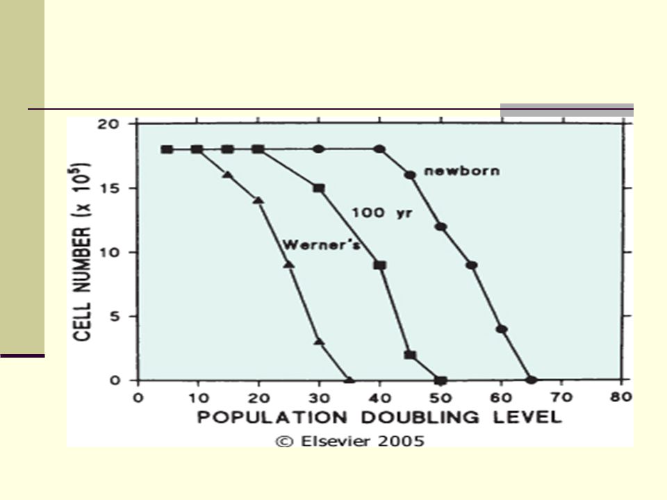
63
Telómero v/s Telomerasa

64
Telómero v/s Telomerasa

Presentaciones similares


 Variaciones del tamaño y forma de las células. 2) Aumento,>")


