Descargar la presentación
La descarga está en progreso. Por favor, espere

1
Constantes vitales Dra. Ma. Eugenia Icaza

2
SIGNOS VITALES Los toma la enfermera Verificarlos
Tomar la TA más de una vez si es anormal

3
Presión arterial Normal Circulation
Regulation of the Blood Pressure System The maintenance of blood pressure is dependent upon intrinsic, reflex, hormonal, renal, and microvascular control systems. The complexity in the control of blood pressure is dependent upon the interaction from each control system. Each system of control will be discussed separately. Where applicable, simple interactions of systems are discussed; however, the normal range of blood pressure from short term to long term is controlled by the precise integration of all control systems. According to Poisuille's law, [pressure is equal to flow times resistance], there are three ways to change blood pressure [P]: 1) altering flow [Q], 2) resistance[R] (i.e. total peripheral resistance [TPR]), or 3) both (73, 124). Thus, an increased blood flow and/or resistance to blood flow may alter blood pressure (35). This conclusion is based on the fact that mean arterial pressure [MAP] is the product of cardiac output; [Q] = [heart rate X stroke volume], and total peripheral resistance [TPR], thus, [MAP = CO X TPR](35). Blood pressure can increase or decrease only if the cardiac output and/or total peripheral resistance is altered. Regardless of the control systems used to adjust blood pressure, the resultant change in blood pressure is dependent on how these systems regulate [Q] or [TPR], or both. Intrinsic Regulation The integrity of the cardiovascular system is necessary to maintain life. Central determinants of blood pressure are stroke volume and heart rate; both of which influence cardiac output. Intrinsic mechanisms of the cardiovascular system stabilize cardiac output via stroke volume and heart rate. The intrinsic control of stroke volume is predominantly what regulates cardiac output (125). The volume of blood that fills the ventricles of the heart determines intrinsic control of stroke volume. Thus, peak wall tension is approximated by the peak pressure or volume [stroke volume] ejected during contraction (125). This relationship is known as the Frank Starling mechanism (108). The end?diastolic filling pressure [preload] is the most important determinant of stroke volume (125). Another intrinsic factor that determines stroke volume is the amount of resistance offered against the left ventricle during contraction. This is known as aortic pressure or afterload (125). A higher afterload will decrease stroke volume. Because the contraction velocity is decreased with a higher afterload, and because the time of contraction is regulated by the duration of depolarization, stroke volume will be lower at a higher afterload (125). Stroke volume is well regulated by the interaction of both preload and afterload. Both can have independent effects on stroke volume, but both rarely change separately. When cardiac output increases more volume is derived from the venous pool increasing preload. Thus, arterial pressure will then increase, increasing afterload. So, preload and afterload tend to oppose a rise in cardiac output [i.e., a balanced intrinsic regulation], (125). Heart rate is an intrinsic factor and central determinant of blood pressure. Heart rate properties that affect flow include: 1) an increase in heart rate which will increase the contractile strength and, 2) transient changes in heart rate where the contractility decreases with lower rates and increases with higher rates (125). Of the three central intrinsic control mechanisms, heart rate will have the least influence on cardiac output during rest. If heart rate is increased cardiac output will increase but this increase is not proportional to the increase in heart rate (125). This is because with an increase in heart rate: 1) there will be a decreased preload, and 2) an increased afterload. If peripheral resistance remains constant blood pressure will increase, like cardiac output, but proportionally less than heart rate (125). Intrinsic controls also exist in the periphery to regulate flow via changes in peripheral vascular resistance. In the arterial system, there is metabolic control of the smooth muscle cells of the arteries. An inadequate supply of oxygen or build up of other metabolites will promote vasodilation (125). Another important intrinsic control mechanism is autoregulation. Autoregulation of organs assures a constant blood flow under changing pressures and decreases peripheral resistance when arterial pressure or oxygen content of arterial blood falls (125). Reflex Regulation Intrinsic regulation is an effective control mechanism for maintaining blood pressure under resting conditions. A faster response system is needed under conditions of posture change, exercise, and moderate temperature changes. The reflex regulation system of baroreceptors [pressoreceptors] is able to control sudden changes in blood pressure with these conditions. The baroreflex system is composed of: 1) baroreceptors in the aortic arch and carotid sinuses; 2) sensory nerve fibers which relay signals both to and from the receptors to the medullary region of the brain; 3) and cardiac and smooth muscle cells on which the nerve fibers act to alter cardiac contractility, heart rate, and peripheral vascular resistance (125). The baroreceptors respond to changes in length, [stretch], of the tissue in which they lie (125). When there is an increase in blood pressure the receptors are stretched and the rate of firing or signaling to the medullary region of the brain is increased. When there is a decrease in pressure, the opposite occurs. These receptors have both a static and dynamic response: a) static responses are to maintained levels of pressure [ie. when rate of change of pressure is zero]; and, b) dynamic responses to phasic changes in pressure, [i.e. arterial pulse wave] (125). With the static response, firing rate will be minimal at a pressure of 20 to 50 mmHg, below which no firing will occur, and the frequency of the firing will increase as the pressure increases above this threshold (19, 40, 83, 133). As pressure increases, this firing rate will reach a plateau. In the dynamic response, the receptors fire with greatest frequency when the pressure is greatest during the arterial pulse wave and may cease to fire when pressure falls off. Thus, the receptor is sensitive to the mean pressure and rate of change in pressure. Because the receptor is sensitive to mean pressure and the rate of change in pressure, reflex responses are dependent upon mean arterial pressure, pulse pressure and heart rate (19, 40, 83, 133). Sympathetic and parasympathetic nerve fibers run from the baroreceptors to the medullary centers of the brain. Different regions of the medulla are responsible for stimulation of either the parasympathetic or sympathetic nerve fibers which, in turn, innervate the heart, arteries and adrenal gland (125). Sympathetic impulses to the medulla will decrease in firing frequency as arterial pressure and baroreceptor discharge increase (43, 74). The opposite is also true. As pressure increases, the parasympathetic activity increases (71, 133). The relationship between the two systems is linear and inverse (125). The different regions of the medulla will respond to the increase in sympathetic or parasympathetic activity. Increased sympathetic activity and decreased vagal activity to the medulla will result in a increased heart rate, and cardiac contractility, via norepinephrine release from adrenal gland on the cardiac pacemaker and myocardium. Increased sympathetic activity to the medulla will also cause a widespread vasoconstriction via norepinephrine on the peripheral arterioles (125). Increased parasympathetic activity to the medulla produces the opposite motor responses. Other Reflex Mechanisms Other reflex mechanisms involved in the regulation of blood pressure are the cardiopulmonary receptors and chemoreceptors. The cardiopulmonary receptors are located in the walls of the cardiac chambers and the pulmonary artery. The chemoreceptors are located close to the arterial baroreceptors and within the central nervous system (125). The firing pattern of the cardiopulmonary receptors parallels the pressure changes within the chambers or vessels where they are located. Impulses travel to the control centers in the brain through the vagus nerve (125). The most prominent cardiopulmonary receptor is located in the atria. Atrial receptors have a role in the regulation of the volume of body fluids (53, 128). The stronger reflex effects of the atrial baroreceptors mask the reflex effects of cardiopulmonary receptors of the atria. Experiments have been conducted on the canine model where the arterial baroreceptors have been denervated. Results from these experiments have shown cardiopulmonary receptors have effects on the peripheral resistance and heart rate in canine. Cardiopulmonary receptors of the atria can produce a global vasoconstriction (33), and stretching the atria may also produce tachycardia (7, 87). The prominent chemoreceptors include the arterial, ventricular, and medullary chemoreceptors. Collectively, these receptors are sensitive to changes in pH, blood gasses, or changes in plasma composition. The arterial chemoreceptors are composed of the aortic and carotid bodies. The carotid body is more sensitive. The carotid body is a small mass of tissue but receives a high volume of blood flow and is located close to the carotid baroreceptors (125). It is sensitive to changes in the partial pressure of oxygen and pH. The bodies in the aortic arch respond in a similar fashion as the carotid, but their response is attenuated under comparison. Both chemoreceptors have marked effects on respiration and under conditions of extreme depravation of oxygen will cause severe vasoconstriction and bradycardia (125). The chemoreceptors of the ventricles will respond to a series of pharmacological agents including: Veratrum alkaloids, nicotine, and serotonin. Exposure to these agents will cause a marked bradycardia and vasodilation, known as the Bezold?Jarisch reflex (125). Its significance in circulatory regulation remains unknown. The chemoreceptors of the medulla are sensitive to a decrease in pH or an increase in plasma carbon dioxide concentration. Inadequate profusion of the medullary region, increased plasma carbon dioxide, or hydrogen ion concentration, can cause a marked vasoconstriction (125). Hormonal Regulation Hormones also operate in maintaining the blood pressure. Two hormones of concern are the hormones included in the renin?angiotensin system, and vasopressin. Both are slower regulatory mechanisms of blood pressure. Renin is an enzyme produced in the juxtaglomerular cells of the kidney. A decrease in the pressure at the renal artery, an activation of the sympathetic nerve fibers to the kidney, or a decrease in the amount of sodium passing through the distal tubule of the kidney, will prompt the release of renin. The release will convert renin to angiotensin I and angiotensin I is converted to angiotensin II in the lung and other organs. Angiotensin II is a potent vasoconstrictor and will also stimulate the release of aldosterone from the adrenal gland. Aldosterone will increase sodium absorption at the kidney, thus allowing for fluid retention and increasing plasma volume (125). Vasopressin, better known as antidiuretic hormone ,[ADH], is housed in the pituitary gland. Two major stimuli promoting the release of ADH are increase in the plasma osmolarity, and decreases in plasma volume by an indirect route from pressoreceptors in the carotid sinus and aortic arch. These pressoreceptors communicate with the hypothalamus, and a decrease in the firing rate from the receptors will somehow initiate the release of ADH (125). ADH is both a potent vasoconstrictor and plasma volume regulator (107). ADH will regulate plasma volume by resorption of water at the distal tubule of the nephron (125). Renal Regulation The kidneys control blood pressure through the retention and excretion of extracellular fluid. The glomerular filtration rate is dependent upon the mean arterial pressure; however, a large increase in arterial pressure will only slightly increase glomerular filtration (103, 155). Filtration is not greatly increased because renal arterial pressure is autoregulated before reaching the glomerular capillary. The loss of extracellular fluid is more pronounced by a lack of resorption. As previously discussed, resorption in the kidney is controlled by aldosterone and ADH. Extracellular volume is determined by the osmolarity of the plasma. Sodium and coanions constitute 95% of the osmotically active solute of the extracellular fluid and osmolarity is regulated by water excretion. Thus, extracellular volume is controlled by the amount of sodium retained by the body (136). Aldosterone is the most important determinant of sodium retention in the kidney. The kidneys, under hormonal regulation, will control the extracellular volume. An increase in renal output will decrease the extracellular volume, decreasing venous return and subsequently cardiac output and arterial pressure. An increase in extracellular volume without compensation from the kidney has been shown to maintain elevated arterial pressure (136). Atrial natriuretic factor [ANF] has also been shown to increase glomerular filtration rate and the filtered load of sodium and sodium excretion in mammals (32, 104). ANF may play an important indirect role in regulating intravascular volume and decreasing blood pressure. An increased intravascular volume (ie. extracellular fluid) will stretch the atria and stimulate the release of ANF. ANF will inhibit the release of vasopressin. Other circulating substances with natriuretic activity have also been reported (21). Agents which control the synthesis and release of ANF have not been determined (104). Thus, renal regulation of blood pressure is by control of extracellular volume. This is accomplished through the hormonal regulation of resorption, and to a lesser extent, glomerular filtration rate. Hormonal control of resorption is through the action of ADH and aldosterone, whereas glomerular filtration rate is effected to some degree by filtration pressure, ANF, and possibly other circulating natriuretic factors. Microvascular Circulation Vascular fluid shifts occur with the microvascular circulation (ie. at the capillaries). The rate of fluid movement across exchange vessel walls is proportional to the difference between the hydrostatic and colloid osmotic pressures (100). In other words, high arterial pressure tends to decrease plasma volume by driving fluid into the interstitium, while high plasma proteins tend to increase plasma volume by osmotically drawing fluid from the interstitium (136). This mechanism would allow a decrease in intravascular volume from the higher hydrostatic pressure and a concomitant decrease in cardiac output and arterial pressure (125). The opposite is also true. All things being equal, a low plasma volume would increase the concentration of positive colloid pressure drawing fluid from the interstitium to the capillary, increasing intravascular volume. Integration The mechanisms which regulate the blood pressure do not respond simultaneously. Some mechanisms require only a few seconds to respond whereas others may require a period of days. The mechanisms for the control of blood pressure may work somewhat independently, such as the baroreflexive control of blood pressure in posture changes. However, in the maintenance of normal pressure from changes in vascular volume, integration of the mechanisms becomes crucial. Short term mechanisms may be activated within seconds. These include all of the reflex mechanisms and chemoreceptor control. Intermediate mechanisms are usually activated within hours. These include fluid shifts in the microvascular circulation and the renin?angiotensin system. Long term mechanisms are the hormones which direct the renal control of extracellular volume. Under normal conditions, these control mechanisms should function to keep the blood pressure within a normal range indefinitely.
 altering flow [Q], 2) resistance[R] (i.e. total peripheral resistance [TPR]), or 3) both (73, 124). Thus, an increased blood flow and/or resistance to blood flow may alter blood pressure (35). This conclusion is based on the fact that mean arterial pressure [MAP] is the product of cardiac output; [Q] = [heart rate X stroke volume], and total peripheral resistance [TPR], thus, [MAP = CO X TPR](35). Blood pressure can increase or decrease only if the cardiac output and/or total peripheral resistance is altered. Regardless of the control systems used to adjust blood pressure, the resultant change in blood pressure is dependent on how these systems regulate [Q] or [TPR], or both. Intrinsic Regulation. The integrity of the cardiovascular system is necessary to maintain life. Central determinants of blood pressure are stroke volume and heart rate; both of which influence cardiac output. Intrinsic mechanisms of the cardiovascular system stabilize cardiac output via stroke volume and heart rate. The intrinsic control of stroke volume is predominantly what regulates cardiac output (125). The volume of blood that fills the ventricles of the heart determines intrinsic control of stroke volume. Thus, peak wall tension is approximated by the peak pressure or volume [stroke volume] ejected during contraction (125). This relationship is known as the Frank Starling mechanism (108). The end diastolic filling pressure [preload] is the most important determinant of stroke volume (125). Another intrinsic factor that determines stroke volume is the amount of resistance offered against the left ventricle during contraction. This is known as aortic pressure or afterload (125). A higher afterload will decrease stroke volume. Because the contraction velocity is decreased with a higher afterload, and because the time of contraction is regulated by the duration of depolarization, stroke volume will be lower at a higher afterload (125). Stroke volume is well regulated by the interaction of both preload and afterload. Both can have independent effects on stroke volume, but both rarely change separately. When cardiac output increases more volume is derived from the venous pool increasing preload. Thus, arterial pressure will then increase, increasing afterload. So, preload and afterload tend to oppose a rise in cardiac output [i.e., a balanced intrinsic regulation], (125). Heart rate is an intrinsic factor and central determinant of blood pressure. Heart rate properties that affect flow include: 1) an increase in heart rate which will increase the contractile strength and, 2) transient changes in heart rate where the contractility decreases with lower rates and increases with higher rates (125). Of the three central intrinsic control mechanisms, heart rate will have the least influence on cardiac output during rest. If heart rate is increased cardiac output will increase but this increase is not proportional to the increase in heart rate (125). This is because with an increase in heart rate: 1) there will be a decreased preload, and 2) an increased afterload. If peripheral resistance remains constant blood pressure will increase, like cardiac output, but proportionally less than heart rate (125). Intrinsic controls also exist in the periphery to regulate flow via changes in peripheral vascular resistance. In the arterial system, there is metabolic control of the smooth muscle cells of the arteries. An inadequate supply of oxygen or build up of other metabolites will promote vasodilation (125). Another important intrinsic control mechanism is autoregulation. Autoregulation of organs assures a constant blood flow under changing pressures and decreases peripheral resistance when arterial pressure or oxygen content of arterial blood falls (125). Reflex Regulation. Intrinsic regulation is an effective control mechanism for maintaining blood pressure under resting conditions. A faster response system is needed under conditions of posture change, exercise, and moderate temperature changes. The reflex regulation system of baroreceptors [pressoreceptors] is able to control sudden changes in blood pressure with these conditions. The baroreflex system is composed of: 1) baroreceptors in the aortic arch and carotid sinuses; 2) sensory nerve fibers which relay signals both to and from the receptors to the medullary region of the brain; 3) and cardiac and smooth muscle cells on which the nerve fibers act to alter cardiac contractility, heart rate, and peripheral vascular resistance (125). The baroreceptors respond to changes in length, [stretch], of the tissue in which they lie (125). When there is an increase in blood pressure the receptors are stretched and the rate of firing or signaling to the medullary region of the brain is increased. When there is a decrease in pressure, the opposite occurs. These receptors have both a static and dynamic response: a) static responses are to maintained levels of pressure [ie. when rate of change of pressure is zero]; and, b) dynamic responses to phasic changes in pressure, [i.e. arterial pulse wave] (125). With the static response, firing rate will be minimal at a pressure of 20 to 50 mmHg, below which no firing will occur, and the frequency of the firing will increase as the pressure increases above this threshold (19, 40, 83, 133). As pressure increases, this firing rate will reach a plateau. In the dynamic response, the receptors fire with greatest frequency when the pressure is greatest during the arterial pulse wave and may cease to fire when pressure falls off. Thus, the receptor is sensitive to the mean pressure and rate of change in pressure. Because the receptor is sensitive to mean pressure and the rate of change in pressure, reflex responses are dependent upon mean arterial pressure, pulse pressure and heart rate (19, 40, 83, 133). Sympathetic and parasympathetic nerve fibers run from the baroreceptors to the medullary centers of the brain. Different regions of the medulla are responsible for stimulation of either the parasympathetic or sympathetic nerve fibers which, in turn, innervate the heart, arteries and adrenal gland (125). Sympathetic impulses to the medulla will decrease in firing frequency as arterial pressure and baroreceptor discharge increase (43, 74). The opposite is also true. As pressure increases, the parasympathetic activity increases (71, 133). The relationship between the two systems is linear and inverse (125). The different regions of the medulla will respond to the increase in sympathetic or parasympathetic activity. Increased sympathetic activity and decreased vagal activity to the medulla will result in a increased heart rate, and cardiac contractility, via norepinephrine release from adrenal gland on the cardiac pacemaker and myocardium. Increased sympathetic activity to the medulla will also cause a widespread vasoconstriction via norepinephrine on the peripheral arterioles (125). Increased parasympathetic activity to the medulla produces the opposite motor responses. Other Reflex Mechanisms. Other reflex mechanisms involved in the regulation of blood pressure are the cardiopulmonary receptors and chemoreceptors. The cardiopulmonary receptors are located in the walls of the cardiac chambers and the pulmonary artery. The chemoreceptors are located close to the arterial baroreceptors and within the central nervous system (125). The firing pattern of the cardiopulmonary receptors parallels the pressure changes within the chambers or vessels where they are located. Impulses travel to the control centers in the brain through the vagus nerve (125). The most prominent cardiopulmonary receptor is located in the atria. Atrial receptors have a role in the regulation of the volume of body fluids (53, 128). The stronger reflex effects of the atrial baroreceptors mask the reflex effects of cardiopulmonary receptors of the atria. Experiments have been conducted on the canine model where the arterial baroreceptors have been denervated. Results from these experiments have shown cardiopulmonary receptors have effects on the peripheral resistance and heart rate in canine. Cardiopulmonary receptors of the atria can produce a global vasoconstriction (33), and stretching the atria may also produce tachycardia (7, 87). The prominent chemoreceptors include the arterial, ventricular, and medullary chemoreceptors. Collectively, these receptors are sensitive to changes in pH, blood gasses, or changes in plasma composition. The arterial chemoreceptors are composed of the aortic and carotid bodies. The carotid body is more sensitive. The carotid body is a small mass of tissue but receives a high volume of blood flow and is located close to the carotid baroreceptors (125). It is sensitive to changes in the partial pressure of oxygen and pH. The bodies in the aortic arch respond in a similar fashion as the carotid, but their response is attenuated under comparison. Both chemoreceptors have marked effects on respiration and under conditions of extreme depravation of oxygen will cause severe vasoconstriction and bradycardia (125). The chemoreceptors of the ventricles will respond to a series of pharmacological agents including: Veratrum alkaloids, nicotine, and serotonin. Exposure to these agents will cause a marked bradycardia and vasodilation, known as the Bezold Jarisch reflex (125). Its significance in circulatory regulation remains unknown. The chemoreceptors of the medulla are sensitive to a decrease in pH or an increase in plasma carbon dioxide concentration. Inadequate profusion of the medullary region, increased plasma carbon dioxide, or hydrogen ion concentration, can cause a marked vasoconstriction (125). Hormonal Regulation. Hormones also operate in maintaining the blood pressure. Two hormones of concern are the hormones included in the renin angiotensin system, and vasopressin. Both are slower regulatory mechanisms of blood pressure. Renin is an enzyme produced in the juxtaglomerular cells of the kidney. A decrease in the pressure at the renal artery, an activation of the sympathetic nerve fibers to the kidney, or a decrease in the amount of sodium passing through the distal tubule of the kidney, will prompt the release of renin. The release will convert renin to angiotensin I and angiotensin I is converted to angiotensin II in the lung and other organs. Angiotensin II is a potent vasoconstrictor and will also stimulate the release of aldosterone from the adrenal gland. Aldosterone will increase sodium absorption at the kidney, thus allowing for fluid retention and increasing plasma volume (125). Vasopressin, better known as antidiuretic hormone ,[ADH], is housed in the pituitary gland. Two major stimuli promoting the release of ADH are increase in the plasma osmolarity, and decreases in plasma volume by an indirect route from pressoreceptors in the carotid sinus and aortic arch. These pressoreceptors communicate with the hypothalamus, and a decrease in the firing rate from the receptors will somehow initiate the release of ADH (125). ADH is both a potent vasoconstrictor and plasma volume regulator (107). ADH will regulate plasma volume by resorption of water at the distal tubule of the nephron (125). Renal Regulation. The kidneys control blood pressure through the retention and excretion of extracellular fluid. The glomerular filtration rate is dependent upon the mean arterial pressure; however, a large increase in arterial pressure will only slightly increase glomerular filtration (103, 155). Filtration is not greatly increased because renal arterial pressure is autoregulated before reaching the glomerular capillary. The loss of extracellular fluid is more pronounced by a lack of resorption. As previously discussed, resorption in the kidney is controlled by aldosterone and ADH. Extracellular volume is determined by the osmolarity of the plasma. Sodium and coanions constitute 95% of the osmotically active solute of the extracellular fluid and osmolarity is regulated by water excretion. Thus, extracellular volume is controlled by the amount of sodium retained by the body (136). Aldosterone is the most important determinant of sodium retention in the kidney. The kidneys, under hormonal regulation, will control the extracellular volume. An increase in renal output will decrease the extracellular volume, decreasing venous return and subsequently cardiac output and arterial pressure. An increase in extracellular volume without compensation from the kidney has been shown to maintain elevated arterial pressure (136). Atrial natriuretic factor [ANF] has also been shown to increase glomerular filtration rate and the filtered load of sodium and sodium excretion in mammals (32, 104). ANF may play an important indirect role in regulating intravascular volume and decreasing blood pressure. An increased intravascular volume (ie. extracellular fluid) will stretch the atria and stimulate the release of ANF. ANF will inhibit the release of vasopressin. Other circulating substances with natriuretic activity have also been reported (21). Agents which control the synthesis and release of ANF have not been determined (104). Thus, renal regulation of blood pressure is by control of extracellular volume. This is accomplished through the hormonal regulation of resorption, and to a lesser extent, glomerular filtration rate. Hormonal control of resorption is through the action of ADH and aldosterone, whereas glomerular filtration rate is effected to some degree by filtration pressure, ANF, and possibly other circulating natriuretic factors. Microvascular Circulation. Vascular fluid shifts occur with the microvascular circulation (ie. at the capillaries). The rate of fluid movement across exchange vessel walls is proportional to the difference between the hydrostatic and colloid osmotic pressures (100). In other words, high arterial pressure tends to decrease plasma volume by driving fluid into the interstitium, while high plasma proteins tend to increase plasma volume by osmotically drawing fluid from the interstitium (136). This mechanism would allow a decrease in intravascular volume from the higher hydrostatic pressure and a concomitant decrease in cardiac output and arterial pressure (125). The opposite is also true. All things being equal, a low plasma volume would increase the concentration of positive colloid pressure drawing fluid from the interstitium to the capillary, increasing intravascular volume. Integration. The mechanisms which regulate the blood pressure do not respond simultaneously. Some mechanisms require only a few seconds to respond whereas others may require a period of days. The mechanisms for the control of blood pressure may work somewhat independently, such as the baroreflexive control of blood pressure in posture changes. However, in the maintenance of normal pressure from changes in vascular volume, integration of the mechanisms becomes crucial. Short term mechanisms may be activated within seconds. These include all of the reflex mechanisms and chemoreceptor control. Intermediate mechanisms are usually activated within hours. These include fluid shifts in the microvascular circulation and the renin angiotensin system. Long term mechanisms are the hormones which direct the renal control of extracellular volume. Under normal conditions, these control mechanisms should function to keep the blood pressure within a normal range indefinitely.")
4
Importancia de la presión arterial
Debe mantenerse estrechamente regulada La PA baja produce hipoperfusión (hipotensión arterial) La PA alta hace que el flujo sanguíneo sea excesivo en relación a la demanda y puede dañar a los órganos (hipertensión arterial)
 La PA alta hace que el flujo sanguíneo sea excesivo en relación a la demanda y puede dañar a los órganos (hipertensión arterial)")
5
¿Qué es la presión arterial
Es el producto del gasto cardiaco por la resistencia periférica

6
Presión arterial P = Q X R (Ley de Poisuille)
presión = flujo X resistencia Un incremento del flujo o de la resistencia eleva la presión arterial Flujo = gasto cardiaco = volumen latido X frecuencia cardiaca Resistencia periférica: la ejercida por las arteriolas According to Poisuille's law, [pressure is equal to flow times resistance], there are three ways to change blood pressure [P]: 1) altering flow [Q], 2) resistance[R] (i.e. total peripheral resistance [TPR]), or 3) both (73, 124). Thus, an increased blood flow and/or resistance to blood flow may alter blood pressure (35). This conclusion is based on the fact that mean arterial pressure [MAP] is the product of cardiac output; [Q] = [heart rate X stroke volume], and total peripheral resistance [TPR], thus, [MAP = CO X TPR](35). Blood pressure can increase or decrease only if the cardiac output and/or total peripheral resistance is altered. Regardless of the control systems used to adjust blood pressure, the resultant change in blood pressure is dependent on how these systems regulate [Q] or [TPR], or both. Intrinsic Regulation The integrity of the cardiovascular system is necessary to maintain life. Central determinants of blood pressure are stroke volume and heart rate; both of which influence cardiac output. Intrinsic mechanisms of the cardiovascular system stabilize cardiac output via stroke volume and heart rate. The intrinsic control of stroke volume is predominantly what regulates cardiac output (125). The volume of blood that fills the ventricles of the heart determines intrinsic control of stroke volume. Thus, peak wall tension is approximated by the peak pressure or volume [stroke volume] ejected during contraction (125). This relationship is known as the Frank Starling mechanism (108). The end?diastolic filling pressure [preload] is the most important determinant of stroke volume (125). Another intrinsic factor that determines stroke volume is the amount of resistance offered against the left ventricle during contraction. This is known as aortic pressure or afterload (125). A higher afterload will decrease stroke volume. Because the contraction velocity is decreased with a higher afterload, and because the time of contraction is regulated by the duration of depolarization, stroke volume will be lower at a higher afterload (125). Stroke volume is well regulated by the interaction of both preload and afterload. Both can have independent effects on stroke volume, but both rarely change separately. When cardiac output increases more volume is derived from the venous pool increasing preload. Thus, arterial pressure will then increase, increasing afterload. So, preload and afterload tend to oppose a rise in cardiac output [i.e., a balanced intrinsic regulation], (125). Heart rate is an intrinsic factor and central determinant of blood pressure. Heart rate properties that affect flow include: 1) an increase in heart rate which will increase the contractile strength and, 2) transient changes in heart rate where the contractility decreases with lower rates and increases with higher rates (125). Of the three central intrinsic control mechanisms, heart rate will have the least influence on cardiac output during rest. If heart rate is increased cardiac output will increase but this increase is not proportional to the increase in heart rate (125). This is because with an increase in heart rate: 1) there will be a decreased preload, and 2) an increased afterload. If peripheral resistance remains constant blood pressure will increase, like cardiac output, but proportionally less than heart rate (125). Intrinsic controls also exist in the periphery to regulate flow via changes in peripheral vascular resistance. In the arterial system, there is metabolic control of the smooth muscle cells of the arteries. An inadequate supply of oxygen or build up of other metabolites will promote vasodilation (125). Another important intrinsic control mechanism is autoregulation. Autoregulation of organs assures a constant blood flow under changing pressures and decreases peripheral resistance when arterial pressure or oxygen content of arterial blood falls (125). Reflex Regulation Intrinsic regulation is an effective control mechanism for maintaining blood pressure under resting conditions. A faster response system is needed under conditions of posture change, exercise, and moderate temperature changes. The reflex regulation system of baroreceptors [pressoreceptors] is able to control sudden changes in blood pressure with these conditions. The baroreflex system is composed of: 1) baroreceptors in the aortic arch and carotid sinuses; 2) sensory nerve fibers which relay signals both to and from the receptors to the medullary region of the brain; 3) and cardiac and smooth muscle cells on which the nerve fibers act to alter cardiac contractility, heart rate, and peripheral vascular resistance (125). The baroreceptors respond to changes in length, [stretch], of the tissue in which they lie (125). When there is an increase in blood pressure the receptors are stretched and the rate of firing or signaling to the medullary region of the brain is increased. When there is a decrease in pressure, the opposite occurs. These receptors have both a static and dynamic response: a) static responses are to maintained levels of pressure [ie. when rate of change of pressure is zero]; and, b) dynamic responses to phasic changes in pressure, [i.e. arterial pulse wave] (125). With the static response, firing rate will be minimal at a pressure of 20 to 50 mmHg, below which no firing will occur, and the frequency of the firing will increase as the pressure increases above this threshold (19, 40, 83, 133). As pressure increases, this firing rate will reach a plateau. In the dynamic response, the receptors fire with greatest frequency when the pressure is greatest during the arterial pulse wave and may cease to fire when pressure falls off. Thus, the receptor is sensitive to the mean pressure and rate of change in pressure. Because the receptor is sensitive to mean pressure and the rate of change in pressure, reflex responses are dependent upon mean arterial pressure, pulse pressure and heart rate (19, 40, 83, 133). Sympathetic and parasympathetic nerve fibers run from the baroreceptors to the medullary centers of the brain. Different regions of the medulla are responsible for stimulation of either the parasympathetic or sympathetic nerve fibers which, in turn, innervate the heart, arteries and adrenal gland (125). Sympathetic impulses to the medulla will decrease in firing frequency as arterial pressure and baroreceptor discharge increase (43, 74). The opposite is also true. As pressure increases, the parasympathetic activity increases (71, 133). The relationship between the two systems is linear and inverse (125). The different regions of the medulla will respond to the increase in sympathetic or parasympathetic activity. Increased sympathetic activity and decreased vagal activity to the medulla will result in a increased heart rate, and cardiac contractility, via norepinephrine release from adrenal gland on the cardiac pacemaker and myocardium. Increased sympathetic activity to the medulla will also cause a widespread vasoconstriction via norepinephrine on the peripheral arterioles (125). Increased parasympathetic activity to the medulla produces the opposite motor responses. Other Reflex Mechanisms Other reflex mechanisms involved in the regulation of blood pressure are the cardiopulmonary receptors and chemoreceptors. The cardiopulmonary receptors are located in the walls of the cardiac chambers and the pulmonary artery. The chemoreceptors are located close to the arterial baroreceptors and within the central nervous system (125). The firing pattern of the cardiopulmonary receptors parallels the pressure changes within the chambers or vessels where they are located. Impulses travel to the control centers in the brain through the vagus nerve (125). The most prominent cardiopulmonary receptor is located in the atria. Atrial receptors have a role in the regulation of the volume of body fluids (53, 128). The stronger reflex effects of the atrial baroreceptors mask the reflex effects of cardiopulmonary receptors of the atria. Experiments have been conducted on the canine model where the arterial baroreceptors have been denervated. Results from these experiments have shown cardiopulmonary receptors have effects on the peripheral resistance and heart rate in canine. Cardiopulmonary receptors of the atria can produce a global vasoconstriction (33), and stretching the atria may also produce tachycardia (7, 87). The prominent chemoreceptors include the arterial, ventricular, and medullary chemoreceptors. Collectively, these receptors are sensitive to changes in pH, blood gasses, or changes in plasma composition. The arterial chemoreceptors are composed of the aortic and carotid bodies. The carotid body is more sensitive. The carotid body is a small mass of tissue but receives a high volume of blood flow and is located close to the carotid baroreceptors (125). It is sensitive to changes in the partial pressure of oxygen and pH. The bodies in the aortic arch respond in a similar fashion as the carotid, but their response is attenuated under comparison. Both chemoreceptors have marked effects on respiration and under conditions of extreme depravation of oxygen will cause severe vasoconstriction and bradycardia (125). The chemoreceptors of the ventricles will respond to a series of pharmacological agents including: Veratrum alkaloids, nicotine, and serotonin. Exposure to these agents will cause a marked bradycardia and vasodilation, known as the Bezold?Jarisch reflex (125). Its significance in circulatory regulation remains unknown. The chemoreceptors of the medulla are sensitive to a decrease in pH or an increase in plasma carbon dioxide concentration. Inadequate profusion of the medullary region, increased plasma carbon dioxide, or hydrogen ion concentration, can cause a marked vasoconstriction (125). Hormonal Regulation Hormones also operate in maintaining the blood pressure. Two hormones of concern are the hormones included in the renin?angiotensin system, and vasopressin. Both are slower regulatory mechanisms of blood pressure. Renin is an enzyme produced in the juxtaglomerular cells of the kidney. A decrease in the pressure at the renal artery, an activation of the sympathetic nerve fibers to the kidney, or a decrease in the amount of sodium passing through the distal tubule of the kidney, will prompt the release of renin. The release will convert renin to angiotensin I and angiotensin I is converted to angiotensin II in the lung and other organs. Angiotensin II is a potent vasoconstrictor and will also stimulate the release of aldosterone from the adrenal gland. Aldosterone will increase sodium absorption at the kidney, thus allowing for fluid retention and increasing plasma volume (125). Vasopressin, better known as antidiuretic hormone ,[ADH], is housed in the pituitary gland. Two major stimuli promoting the release of ADH are increase in the plasma osmolarity, and decreases in plasma volume by an indirect route from pressoreceptors in the carotid sinus and aortic arch. These pressoreceptors communicate with the hypothalamus, and a decrease in the firing rate from the receptors will somehow initiate the release of ADH (125). ADH is both a potent vasoconstrictor and plasma volume regulator (107). ADH will regulate plasma volume by resorption of water at the distal tubule of the nephron (125). Renal Regulation The kidneys control blood pressure through the retention and excretion of extracellular fluid. The glomerular filtration rate is dependent upon the mean arterial pressure; however, a large increase in arterial pressure will only slightly increase glomerular filtration (103, 155). Filtration is not greatly increased because renal arterial pressure is autoregulated before reaching the glomerular capillary. The loss of extracellular fluid is more pronounced by a lack of resorption. As previously discussed, resorption in the kidney is controlled by aldosterone and ADH. Extracellular volume is determined by the osmolarity of the plasma. Sodium and coanions constitute 95% of the osmotically active solute of the extracellular fluid and osmolarity is regulated by water excretion. Thus, extracellular volume is controlled by the amount of sodium retained by the body (136). Aldosterone is the most important determinant of sodium retention in the kidney. The kidneys, under hormonal regulation, will control the extracellular volume. An increase in renal output will decrease the extracellular volume, decreasing venous return and subsequently cardiac output and arterial pressure. An increase in extracellular volume without compensation from the kidney has been shown to maintain elevated arterial pressure (136). Atrial natriuretic factor [ANF] has also been shown to increase glomerular filtration rate and the filtered load of sodium and sodium excretion in mammals (32, 104). ANF may play an important indirect role in regulating intravascular volume and decreasing blood pressure. An increased intravascular volume (ie. extracellular fluid) will stretch the atria and stimulate the release of ANF. ANF will inhibit the release of vasopressin. Other circulating substances with natriuretic activity have also been reported (21). Agents which control the synthesis and release of ANF have not been determined (104). Thus, renal regulation of blood pressure is by control of extracellular volume. This is accomplished through the hormonal regulation of resorption, and to a lesser extent, glomerular filtration rate. Hormonal control of resorption is through the action of ADH and aldosterone, whereas glomerular filtration rate is effected to some degree by filtration pressure, ANF, and possibly other circulating natriuretic factors. Microvascular Circulation Vascular fluid shifts occur with the microvascular circulation (ie. at the capillaries). The rate of fluid movement across exchange vessel walls is proportional to the difference between the hydrostatic and colloid osmotic pressures (100). In other words, high arterial pressure tends to decrease plasma volume by driving fluid into the interstitium, while high plasma proteins tend to increase plasma volume by osmotically drawing fluid from the interstitium (136). This mechanism would allow a decrease in intravascular volume from the higher hydrostatic pressure and a concomitant decrease in cardiac output and arterial pressure (125). The opposite is also true. All things being equal, a low plasma volume would increase the concentration of positive colloid pressure drawing fluid from the interstitium to the capillary, increasing intravascular volume. Integration The mechanisms which regulate the blood pressure do not respond simultaneously. Some mechanisms require only a few seconds to respond whereas others may require a period of days. The mechanisms for the control of blood pressure may work somewhat independently, such as the baroreflexive control of blood pressure in posture changes. However, in the maintenance of normal pressure from changes in vascular volume, integration of the mechanisms becomes crucial. Short term mechanisms may be activated within seconds. These include all of the reflex mechanisms and chemoreceptor control. Intermediate mechanisms are usually activated within hours. These include fluid shifts in the microvascular circulation and the renin?angiotensin system. Long term mechanisms are the hormones which direct the renal control of extracellular volume. Under normal conditions, these control mechanisms should function to keep the blood pressure within a normal range indefinitely.
 altering flow [Q], 2) resistance[R] (i.e. total peripheral resistance [TPR]), or 3) both (73, 124). Thus, an increased blood flow and/or resistance to blood flow may alter blood pressure (35). This conclusion is based on the fact that mean arterial pressure [MAP] is the product of cardiac output; [Q] = [heart rate X stroke volume], and total peripheral resistance [TPR], thus, [MAP = CO X TPR](35). Blood pressure can increase or decrease only if the cardiac output and/or total peripheral resistance is altered. Regardless of the control systems used to adjust blood pressure, the resultant change in blood pressure is dependent on how these systems regulate [Q] or [TPR], or both. Intrinsic Regulation. The integrity of the cardiovascular system is necessary to maintain life. Central determinants of blood pressure are stroke volume and heart rate; both of which influence cardiac output. Intrinsic mechanisms of the cardiovascular system stabilize cardiac output via stroke volume and heart rate. The intrinsic control of stroke volume is predominantly what regulates cardiac output (125). The volume of blood that fills the ventricles of the heart determines intrinsic control of stroke volume. Thus, peak wall tension is approximated by the peak pressure or volume [stroke volume] ejected during contraction (125). This relationship is known as the Frank Starling mechanism (108). The end diastolic filling pressure [preload] is the most important determinant of stroke volume (125). Another intrinsic factor that determines stroke volume is the amount of resistance offered against the left ventricle during contraction. This is known as aortic pressure or afterload (125). A higher afterload will decrease stroke volume. Because the contraction velocity is decreased with a higher afterload, and because the time of contraction is regulated by the duration of depolarization, stroke volume will be lower at a higher afterload (125). Stroke volume is well regulated by the interaction of both preload and afterload. Both can have independent effects on stroke volume, but both rarely change separately. When cardiac output increases more volume is derived from the venous pool increasing preload. Thus, arterial pressure will then increase, increasing afterload. So, preload and afterload tend to oppose a rise in cardiac output [i.e., a balanced intrinsic regulation], (125). Heart rate is an intrinsic factor and central determinant of blood pressure. Heart rate properties that affect flow include: 1) an increase in heart rate which will increase the contractile strength and, 2) transient changes in heart rate where the contractility decreases with lower rates and increases with higher rates (125). Of the three central intrinsic control mechanisms, heart rate will have the least influence on cardiac output during rest. If heart rate is increased cardiac output will increase but this increase is not proportional to the increase in heart rate (125). This is because with an increase in heart rate: 1) there will be a decreased preload, and 2) an increased afterload. If peripheral resistance remains constant blood pressure will increase, like cardiac output, but proportionally less than heart rate (125). Intrinsic controls also exist in the periphery to regulate flow via changes in peripheral vascular resistance. In the arterial system, there is metabolic control of the smooth muscle cells of the arteries. An inadequate supply of oxygen or build up of other metabolites will promote vasodilation (125). Another important intrinsic control mechanism is autoregulation. Autoregulation of organs assures a constant blood flow under changing pressures and decreases peripheral resistance when arterial pressure or oxygen content of arterial blood falls (125). Reflex Regulation. Intrinsic regulation is an effective control mechanism for maintaining blood pressure under resting conditions. A faster response system is needed under conditions of posture change, exercise, and moderate temperature changes. The reflex regulation system of baroreceptors [pressoreceptors] is able to control sudden changes in blood pressure with these conditions. The baroreflex system is composed of: 1) baroreceptors in the aortic arch and carotid sinuses; 2) sensory nerve fibers which relay signals both to and from the receptors to the medullary region of the brain; 3) and cardiac and smooth muscle cells on which the nerve fibers act to alter cardiac contractility, heart rate, and peripheral vascular resistance (125). The baroreceptors respond to changes in length, [stretch], of the tissue in which they lie (125). When there is an increase in blood pressure the receptors are stretched and the rate of firing or signaling to the medullary region of the brain is increased. When there is a decrease in pressure, the opposite occurs. These receptors have both a static and dynamic response: a) static responses are to maintained levels of pressure [ie. when rate of change of pressure is zero]; and, b) dynamic responses to phasic changes in pressure, [i.e. arterial pulse wave] (125). With the static response, firing rate will be minimal at a pressure of 20 to 50 mmHg, below which no firing will occur, and the frequency of the firing will increase as the pressure increases above this threshold (19, 40, 83, 133). As pressure increases, this firing rate will reach a plateau. In the dynamic response, the receptors fire with greatest frequency when the pressure is greatest during the arterial pulse wave and may cease to fire when pressure falls off. Thus, the receptor is sensitive to the mean pressure and rate of change in pressure. Because the receptor is sensitive to mean pressure and the rate of change in pressure, reflex responses are dependent upon mean arterial pressure, pulse pressure and heart rate (19, 40, 83, 133). Sympathetic and parasympathetic nerve fibers run from the baroreceptors to the medullary centers of the brain. Different regions of the medulla are responsible for stimulation of either the parasympathetic or sympathetic nerve fibers which, in turn, innervate the heart, arteries and adrenal gland (125). Sympathetic impulses to the medulla will decrease in firing frequency as arterial pressure and baroreceptor discharge increase (43, 74). The opposite is also true. As pressure increases, the parasympathetic activity increases (71, 133). The relationship between the two systems is linear and inverse (125). The different regions of the medulla will respond to the increase in sympathetic or parasympathetic activity. Increased sympathetic activity and decreased vagal activity to the medulla will result in a increased heart rate, and cardiac contractility, via norepinephrine release from adrenal gland on the cardiac pacemaker and myocardium. Increased sympathetic activity to the medulla will also cause a widespread vasoconstriction via norepinephrine on the peripheral arterioles (125). Increased parasympathetic activity to the medulla produces the opposite motor responses. Other Reflex Mechanisms. Other reflex mechanisms involved in the regulation of blood pressure are the cardiopulmonary receptors and chemoreceptors. The cardiopulmonary receptors are located in the walls of the cardiac chambers and the pulmonary artery. The chemoreceptors are located close to the arterial baroreceptors and within the central nervous system (125). The firing pattern of the cardiopulmonary receptors parallels the pressure changes within the chambers or vessels where they are located. Impulses travel to the control centers in the brain through the vagus nerve (125). The most prominent cardiopulmonary receptor is located in the atria. Atrial receptors have a role in the regulation of the volume of body fluids (53, 128). The stronger reflex effects of the atrial baroreceptors mask the reflex effects of cardiopulmonary receptors of the atria. Experiments have been conducted on the canine model where the arterial baroreceptors have been denervated. Results from these experiments have shown cardiopulmonary receptors have effects on the peripheral resistance and heart rate in canine. Cardiopulmonary receptors of the atria can produce a global vasoconstriction (33), and stretching the atria may also produce tachycardia (7, 87). The prominent chemoreceptors include the arterial, ventricular, and medullary chemoreceptors. Collectively, these receptors are sensitive to changes in pH, blood gasses, or changes in plasma composition. The arterial chemoreceptors are composed of the aortic and carotid bodies. The carotid body is more sensitive. The carotid body is a small mass of tissue but receives a high volume of blood flow and is located close to the carotid baroreceptors (125). It is sensitive to changes in the partial pressure of oxygen and pH. The bodies in the aortic arch respond in a similar fashion as the carotid, but their response is attenuated under comparison. Both chemoreceptors have marked effects on respiration and under conditions of extreme depravation of oxygen will cause severe vasoconstriction and bradycardia (125). The chemoreceptors of the ventricles will respond to a series of pharmacological agents including: Veratrum alkaloids, nicotine, and serotonin. Exposure to these agents will cause a marked bradycardia and vasodilation, known as the Bezold Jarisch reflex (125). Its significance in circulatory regulation remains unknown. The chemoreceptors of the medulla are sensitive to a decrease in pH or an increase in plasma carbon dioxide concentration. Inadequate profusion of the medullary region, increased plasma carbon dioxide, or hydrogen ion concentration, can cause a marked vasoconstriction (125). Hormonal Regulation. Hormones also operate in maintaining the blood pressure. Two hormones of concern are the hormones included in the renin angiotensin system, and vasopressin. Both are slower regulatory mechanisms of blood pressure. Renin is an enzyme produced in the juxtaglomerular cells of the kidney. A decrease in the pressure at the renal artery, an activation of the sympathetic nerve fibers to the kidney, or a decrease in the amount of sodium passing through the distal tubule of the kidney, will prompt the release of renin. The release will convert renin to angiotensin I and angiotensin I is converted to angiotensin II in the lung and other organs. Angiotensin II is a potent vasoconstrictor and will also stimulate the release of aldosterone from the adrenal gland. Aldosterone will increase sodium absorption at the kidney, thus allowing for fluid retention and increasing plasma volume (125). Vasopressin, better known as antidiuretic hormone ,[ADH], is housed in the pituitary gland. Two major stimuli promoting the release of ADH are increase in the plasma osmolarity, and decreases in plasma volume by an indirect route from pressoreceptors in the carotid sinus and aortic arch. These pressoreceptors communicate with the hypothalamus, and a decrease in the firing rate from the receptors will somehow initiate the release of ADH (125). ADH is both a potent vasoconstrictor and plasma volume regulator (107). ADH will regulate plasma volume by resorption of water at the distal tubule of the nephron (125). Renal Regulation. The kidneys control blood pressure through the retention and excretion of extracellular fluid. The glomerular filtration rate is dependent upon the mean arterial pressure; however, a large increase in arterial pressure will only slightly increase glomerular filtration (103, 155). Filtration is not greatly increased because renal arterial pressure is autoregulated before reaching the glomerular capillary. The loss of extracellular fluid is more pronounced by a lack of resorption. As previously discussed, resorption in the kidney is controlled by aldosterone and ADH. Extracellular volume is determined by the osmolarity of the plasma. Sodium and coanions constitute 95% of the osmotically active solute of the extracellular fluid and osmolarity is regulated by water excretion. Thus, extracellular volume is controlled by the amount of sodium retained by the body (136). Aldosterone is the most important determinant of sodium retention in the kidney. The kidneys, under hormonal regulation, will control the extracellular volume. An increase in renal output will decrease the extracellular volume, decreasing venous return and subsequently cardiac output and arterial pressure. An increase in extracellular volume without compensation from the kidney has been shown to maintain elevated arterial pressure (136). Atrial natriuretic factor [ANF] has also been shown to increase glomerular filtration rate and the filtered load of sodium and sodium excretion in mammals (32, 104). ANF may play an important indirect role in regulating intravascular volume and decreasing blood pressure. An increased intravascular volume (ie. extracellular fluid) will stretch the atria and stimulate the release of ANF. ANF will inhibit the release of vasopressin. Other circulating substances with natriuretic activity have also been reported (21). Agents which control the synthesis and release of ANF have not been determined (104). Thus, renal regulation of blood pressure is by control of extracellular volume. This is accomplished through the hormonal regulation of resorption, and to a lesser extent, glomerular filtration rate. Hormonal control of resorption is through the action of ADH and aldosterone, whereas glomerular filtration rate is effected to some degree by filtration pressure, ANF, and possibly other circulating natriuretic factors. Microvascular Circulation. Vascular fluid shifts occur with the microvascular circulation (ie. at the capillaries). The rate of fluid movement across exchange vessel walls is proportional to the difference between the hydrostatic and colloid osmotic pressures (100). In other words, high arterial pressure tends to decrease plasma volume by driving fluid into the interstitium, while high plasma proteins tend to increase plasma volume by osmotically drawing fluid from the interstitium (136). This mechanism would allow a decrease in intravascular volume from the higher hydrostatic pressure and a concomitant decrease in cardiac output and arterial pressure (125). The opposite is also true. All things being equal, a low plasma volume would increase the concentration of positive colloid pressure drawing fluid from the interstitium to the capillary, increasing intravascular volume. Integration. The mechanisms which regulate the blood pressure do not respond simultaneously. Some mechanisms require only a few seconds to respond whereas others may require a period of days. The mechanisms for the control of blood pressure may work somewhat independently, such as the baroreflexive control of blood pressure in posture changes. However, in the maintenance of normal pressure from changes in vascular volume, integration of the mechanisms becomes crucial. Short term mechanisms may be activated within seconds. These include all of the reflex mechanisms and chemoreceptor control. Intermediate mechanisms are usually activated within hours. These include fluid shifts in the microvascular circulation and the renin angiotensin system. Long term mechanisms are the hormones which direct the renal control of extracellular volume. Under normal conditions, these control mechanisms should function to keep the blood pressure within a normal range indefinitely.")
7
Presión arterial Aorta y arterias: 120/80 mmHg Capilares: 40 a 20 mmHg
Venas 20 mmHg La presión arterial refleja qué tanto las arterias elásticas cercanas al corazón pueden distenderse (distensibilidad) que tanto volumen llega a las arterias/tiempo La sangre que llega a la aorta la distiende. Esto es la presión sistólica. La sangre fluye a las demás arterias por la diferencia de presión entre la aorta y esas arterias. Durante la diástole, las paredes de la aorta regresan a su tamaño normal, y mantienen la presión a pesar de reducir su volumen debido a esa distensibilidad. En este momento la presión cae a su mínimo y se llama presión diastólica. Cuando las arterias pierden su elasticidad, la presión sistólica se eleva.
 que tanto volumen llega a las arterias/tiempo. La sangre que llega a la aorta la distiende. Esto es la presión sistólica. La sangre fluye a las demás arterias por la diferencia de presión entre la aorta y esas arterias. Durante la diástole, las paredes de la aorta regresan a su tamaño normal, y mantienen la presión a pesar de reducir su volumen debido a esa distensibilidad. En este momento la presión cae a su mínimo y se llama presión diastólica. Cuando las arterias pierden su elasticidad, la presión sistólica se eleva.")
8
Control de la TA Los factores principales que determinan las variaciones en la TA incluyen: Edad Género IMC Ingestión de sodio

9
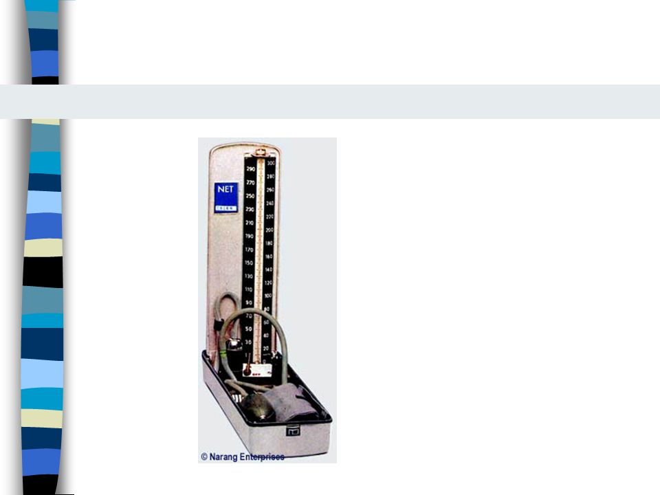
Tipos de baumanómetros
De mercurio Aneroides (requieren verificar periódicamente la calibración)
")
12
Partes del baumanómetro
Cámara de aire Manguito Columna de mercurio o carátula Bomba para insuflar Estetoscopio incluido o por separado

13
Técnica de medición Evitar fumar e ingerir cafeína 30 minutos antes y un descanso de 5 minutos Brazo sin ropa Sin alteraciones vasculares Palpar la arteria braquial La arteria braquial en el pliegue antecubital debe estar a nivel del corazón en el cuarto espacio intercostal en su unión con el esternón Sentado: elevar brazo en una mesa, de pié: apoyar brazo en nuestro brazo

14
Técnica de medición Centrar la cámara insuflable sobre la arteria humeral El borde inferior del manguito 2.5 cm por arriba del pliegue antecubital Ajustar el manguito Leve flexión del codo del paciente Estimar la presión sistólica por palpación a nivel radial y agregar 30 mmHg (se evita la presión excesiva innecesaria y el error de la brecha auscultatoria)
")
15
Presión sistólica 200 160 120 Presión diastólica 80 40

16
Presión arterial 1 200 160 2 120 3 80 40 mmHg Sonidos

17
Técnica de medición Colocar campana del estetoscopio sobre la arteria braquial sin presionarla demasiado Insuflara manguito Desinsuflara lentamente (2-3 mmHg/segundo) Registrar la presión en que se escuchen los latidos (sistólica)
 Registrar la presión en que se escuchen los latidos (sistólica)")
18
Técnica de medición Continuar descenso de presión hasta que desaparezcan los ruidos (presión diastólica) Escuchar 10 a 20 mmHg más abajo Desinsuflar el manguito Repetir 2 minutos después

19
Técnica de medición Esfigmomanómetro de mercurio: manómetro vertical
Ojos a nivel del menisco Carátula a nivel de los ojos Evitar insuflaciones lentas o repetidas (la congestión venosa origina registros falsos)
")
20
Técnica de medición Tomar en ambos brazos por lo menos una vez
Diferencia normal de 5 mmHg Hipertensos, historia de desmayo, mareo postural o hipovolemia: medir la presión en posición supina, sedente y de pié Al ponerse de pié la sistólica cae un poco o no cambia y la diastólica aumenta un poco

21
Elección del manguito La anchura de la bolsa insuflable del manguito debe ser del 40% del perímetro del brazo (12 a 14 cm en el adulto promedio) La longitud de la bolsa debe ser del 80% del perímetro del brazo (casi lo debe rodear)
")
22
Tamaño incorrecto Los manguitos demasiado cortos o angostos pueden generar registros elevados falsos El uso de un manguito normal en un brazo obeso puede conducir al falso diagnóstico de hipertensión Pacientes obesos (15 cm), si el perímetro es mayor de 41 cm usar manguito para muslo (18 cm)
, si el perímetro es mayor de 41 cm usar manguito para muslo (18 cm)")
23
Frecuencia cardiaca Auscultación del corazón
Pulso radial: pulpejos de los dedos índice y medio Comprimir la arteria radial Si el ritmo es regular: 15 segundos Si el ritmo es irregular, rápido o lento: 60 segundos de preferencia auscultación cardiaca Normal: 60 a 100 por minuto

24
Frecuencia cardiaca en niños
Al nacer 140 0-6 meses 130 6-12 meses 115 1-2 años 110 2-6 años 103 6-10 años 95 10-14 años 85

25
Frecuencia respiratoria
Frecuencia, ritmo, profundidad y esfuerzo respiratorio Número de respiraciones por minuto Por inspección o durante la exploración del corazón Normal 14 a 20 respiraciones por minuto

26
TEMPERATURA

27
Regulación de la temperatura
Poiquilotermia: la temperatura corporal es = a la temperatura ambiente Homeotermia: la tempreratura corporal es constante a pesar de marcadas diferencias en la temperatura ambiente

28
¿Porqué es dañina la fiebre?
La temperatura mayor a 41oC: Desnaturaliza a las enzimas Altera la función mitocondrial Desestabiliza las membranas celulares When temperatures are above 41° C, enzymes are denatured, mitochondrial function is disturbed, cell membranes are destabilized, and oxygen-dependent metabolic pathways are disrupted. Multisystem failure regularly occurs concomitantly with heat injury syndromes, along with significant associated morbidity and mortality. Patients with these syndromes usually require admission to the intensive care unit.

29
GLOSARIO Temperatura central: la temperatura que tienen las regiones profundas del cuerpo y las porciones proximales de las extremidades. Temperatura periférica: temperatura exterior (piel y tejido subcutaneo). La temperatura de estas zonas cambia cuando se exponen a cambios de temperatura exterior.
. La temperatura de estas zonas cambia cuando se exponen a cambios de temperatura exterior.")
30
GLOSARIO Conducción: Transferencia directa de energía por el contacto entre dos cuerpos de diferente temperatura. Convección: Pérdida de temperatura por el contacto entre la piel y un medio que se mueve: aire o agua. Evaporación: pérdida de calor por medio de la evaporación de la superficie corporal o los pulmones Radiación: Transferencia de energía calorífica entre dos objetos separados por medio de ondas electromagnéticas. No requiere un medio.

31
GLOSARIO Fiebre: Incremento de la temperatura central por arriba de los niveles normales. El cuerpo se comporta como si hiciera frío. La temperatura aumenta por la contracción muscular y vasoconstricción.

32
Temperatura del cuerpo
El cuerpo tiene múltiples temperaturas El cuerpo tiene una región central y una región periférica El contenido de calor de un cuerpo humano se refleja en su temperatura El termómetro mide la temperatura del termómetro, por lo tanto su localización es importante La temperatura central promedio es de 37oC en adultos en reposo 1. The temperatures of the body The human body consists of a peripheral shell and a central core (Fig. 21-1). The heat content (H or enthalpy) of the human body is reflected by its temperature. By definition a thermometer only measures the temperature of the thermometer, so its location is essential. The mean core temperature is 37 oC in healthy adults at rest, but small children have larger diurnal variations. The skin is the main heat exchanger of the body. The skin temperature is determined by the core temperature and by the environment (temperature, humidity, air velocity). Thus the shell temperature is governed by the needs of the body to exchange heat energy.
. The heat content (H or enthalpy) of the human body is reflected by its temperature. By definition a thermometer only measures the temperature of the thermometer, so its location is essential. The mean core temperature is 37 oC in healthy adults at rest, but small children have larger diurnal variations. The skin is the main heat exchanger of the body. The skin temperature is determined by the core temperature and by the environment (temperature, humidity, air velocity). Thus the shell temperature is governed by the needs of the body to exchange heat energy.")
33
Temperatura corporal The shell temperature is measured on the skin surface and at the hands and feet to approach the room temperature of 19oC in a person standing in a cold room for hours (Fig. 21-1, left). The shell temperature is several degrees lower than the temperature in the central core. The limbs have both a longitudinal and a radial temperature gradient. The shell temperature and the size of the shell vary with the environmental temperature and the termal state of the person. A naked person, standing on a cold floor in 19oC air has a small core and a thick shell compared to the same person in a warm environment (Fig. 21-1). The shell temperature of the skin and distal extremities is difficult to evaluate. The best estimate is measurement of the infrared heat radiation flux with a radiometer. The core temperature is the rather constant temperature in the deeper parts of the body and in the proximal extremity portions (see the red stippled lines of Fig. 21-1). However, the core temperature may vary several Centigrades between different regions depending on the cellular activity. The brain has a radial temperature gradient between its deep and superficial parts. In a sense, the temperature of the mixed venous blood represents an essential core temperature.
. The shell temperature is several degrees lower than the temperature in the central core. The limbs have both a longitudinal and a radial temperature gradient. The shell temperature and the size of the shell vary with the environmental temperature and the termal state of the person. A naked person, standing on a cold floor in 19oC air has a small core and a thick shell compared to the same person in a warm environment (Fig. 21-1). The shell temperature of the skin and distal extremities is difficult to evaluate. The best estimate is measurement of the infrared heat radiation flux with a radiometer. The core temperature is the rather constant temperature in the deeper parts of the body and in the proximal extremity portions (see the red stippled lines of Fig. 21-1). However, the core temperature may vary several Centigrades between different regions depending on the cellular activity. The brain has a radial temperature gradient between its deep and superficial parts. In a sense, the temperature of the mixed venous blood represents an essential core temperature.")
34
Temperatura del cuerpo
La piel es un intercambiador de calor Su temperatura depende de las necesidades de intercambio de calor del cuerpo La temperatura periférica depende de la temperatura central y del ambiente Temperatura Humedad Velocidad del aire The skin is the main heat exchanger of the body. The skin temperature is determined by the core temperature and by the environment (temperature, humidity, air velocity). Thus the shell temperature is governed by the needs of the body to exchange heat energy.
. Thus the shell temperature is governed by the needs of the body to exchange heat energy.")
35
Temperatura central Mejor representada por la temperatura de la sangre venosa mixta – aurícula derecha Es estrechamente regulada Tiene ciclos circadianos En promedio es de 37oC Las variaciones no exceden 0.6oC

36
Temperatura normal 1992: análisis de 700 tomas de temperatura oral en 148 hombres y mujeres sanos Intervalo de 35.6oC a 38.2oC Promedio global oC Mediana 36.8oC Nadir a las 6 a.m. y cenit a las 6 p.m. A 1992 descriptive analysis of 700 baseline oral temperature observations from 148 healthy men and women found a range of 35.6°C (96.0°F) to 38.2°C (100.8°F), an overall mean of 36.8 ± 0.4°C (98.2 ± 0.7°F), a median of 36.8°C (98.2°F), and a mode of 36.7°C (98.0°F); 37°C (98.6°F) accounted for only 56 of the 700 (8%) oral temperature observations recorded (Fig. 43-2) (Figure Not Available) . [8] The mean temperature varied diurnally with a 6 AM nadir and a 4 to 6 PM apogee (Fig. 43-3) (Figure Not Available) . The maximal temperature (as reflected by the 99th percentile) varied from a low of 37.2°C (98.9°F) at 6 AM to a high of 37.7°C (99.9°F) at 4 PM.
 to 38.2°C (100.8°F), an overall mean of 36.8 ± 0.4°C (98.2 ± 0.7°F), a median of 36.8°C (98.2°F), and a mode of 36.7°C (98.0°F); 37°C (98.6°F) accounted for only 56 of the 700 (8%) oral temperature observations recorded (Fig. 43-2) (Figure Not Available) . [8] The mean temperature varied diurnally with a 6 AM nadir and a 4 to 6 PM apogee (Fig. 43-3) (Figure Not Available) . The maximal temperature (as reflected by the 99th percentile) varied from a low of 37.2°C (98.9°F) at 6 AM to a high of 37.7°C (99.9°F) at 4 PM.")
38
Homeotermia Región pre-óptica del hipotálamo anterior: mantiene el punto térmico

39
Hipotálamo

40
Núcleo preóptico Preóptico

41
Hipotálamo Mecanismo local que percibe la temperatura de la sangre
La temperatura se eleva por una descarga simpática que provoca vasoconstricción y contracciones musculares La temperatura se disminuye por medio de la sudoración y la vasodilatación por vía parasimpática The neural pathways responsible for thermoregulation originate in the hypothalamus. A local sensing mechanism exists wherein the temperature of blood is coupled to the development of autonomic discharge. Elevation of body temperature depends primarily on sympathetic outflow and leads to shivering thermogenesis and dermal vasoconstriction, whereas cooling mechanisms (sweating and dermal vasodilation) involve a mixture of sympathetic and parasympathetic pathways. Certain neurotropic drugs can disrupt the hypothalamic thermosensory mechanism—or blunt the hypothalamic response—and thus may interfere with the development of fever. Among these drugs, phenothiazines are the best known for their "poikilothermic" effect. These agents are not specifically active in febrile states; rather, they act to disable thermoregulatory mechanisms.
 involve a mixture of sympathetic and parasympathetic pathways. Certain neurotropic drugs can disrupt the hypothalamic thermosensory mechanism—or blunt the hypothalamic response—and thus may interfere with the development of fever. Among these drugs, phenothiazines are the best known for their poikilothermic effect. These agents are not specifically active in febrile states; rather, they act to disable thermoregulatory mechanisms.")
42
Control de la temperatura
Escalofríos Piloerección Sudoración Vasoconstricción Vasodilatación Mecanismo de contracorriente Elevación del metabolismo basal

43
Medición de la temperatura
Los termómetros pueden fallar Pocos médicos verifican la calibración de los termómetros Abbey y cols encontraron que ¼ de los termómetros de mercurio no medían correctamente la temperatura después de estar guardados o ser usados por 8 meses.

44
Tiempo que debe permanecer el termómetro
Con los termómetros de mercurio se requiere entre 1 a 9 minutos en lograr el equilibrio térmico. En un estudio realizado por Kucha y Nichols, solo 13% de las lecturas del termómetro alcanzaron el máximo después de 3 minutos y 90% después de 8 minutos.

45
Sitios donde se toma la temperatura
¿Hay alguno mejor que otro? ¿Comodidad? ¿Certeza? ¿Seguridad?

46
Rectal Se ha considerado tradicionalmente como un lugar bueno para tomar la temperatura La temperatura rectal es más alta que la obtenida en otros sitios Si el termómetro se introduce en la materia fecal del recto, pueden mitigar los cambios de temperatura rectal Durante el choque la perfusión rectal puede estar muy alterada Muy bajo riesgo de perforación Requiere higiene

47
Temperatura oral La boca es un sitio cómodo, accesible
Responde rápidamente a cambios en la temperatura corporal La región sublingual está irrigada por una rama de la arteria carótida externa. Requiere la cooperación del paciente (intubados, niños ancianos) Termómetro de chupón: muy malo pues solo identificó correctamente al 10% de niños con fiebre
 Termómetro de chupón: muy malo pues solo identificó correctamente al 10% de niños con fiebre.")
48
Temperatura oral Masticar y fumar eleva la temperatura oral
Ingerir líquidos fríos la disminuye

50
Axilar y cutánea Axilar: Toma mucho tiempo en alcanzar la temperatura máxima (hasta 12 minutos) Algunos opinan que no debe usarse este sitio, excepto en neonatos Cutánea: no sirve

51
Palpación Insensible: las enfermeras detectaron fiebre en solo 42% de 138 niños con más de 38oC De 1000 niños afebriles, solo se sospechó fiebre en 1.8% por las enfermeras Las mamás tienen mejor sensibilidad (con sus propios hijos): 74%
: 74%")
52
Comparación entre sitios
En promedio, la temperatura rectal es 0.4oC mayor a la oral y 0.8oC más alta que la timpánica. Hay gran variabilidad y no es fácil establecer un factor de conversión

Presentaciones similares