Descargar la presentación
La descarga está en progreso. Por favor, espere

1
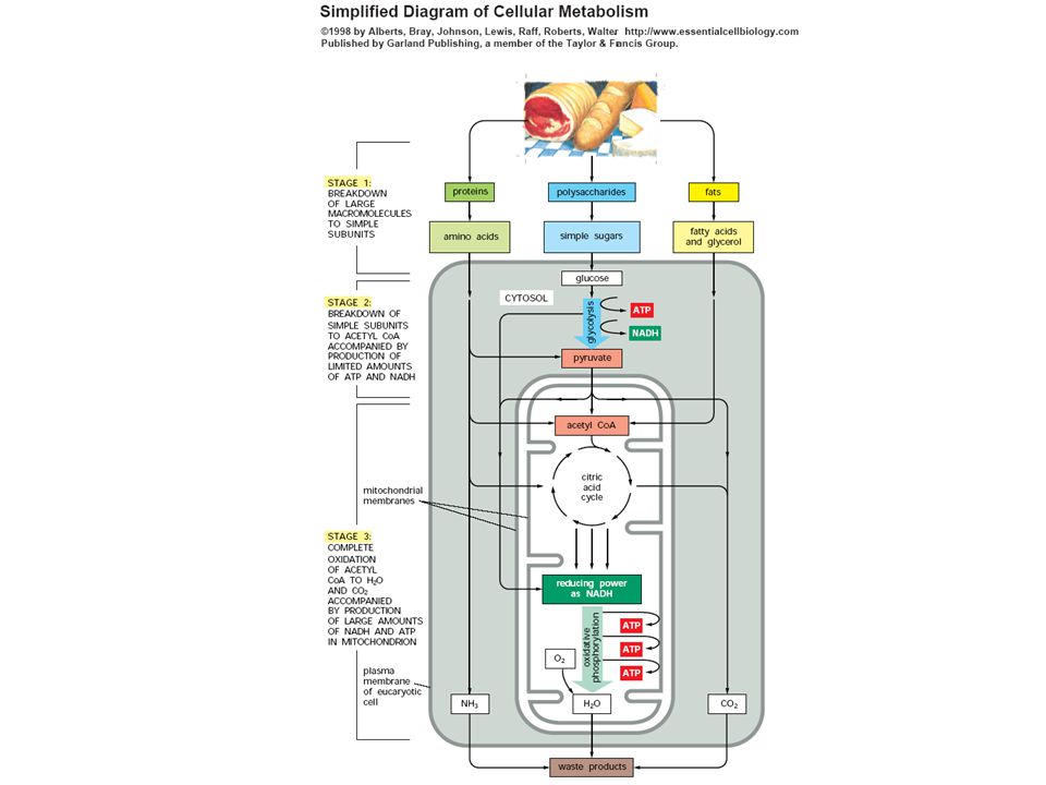
La teoría quimiosmótica y la síntesis de ATP en la mitocondria
José G. Sampedro Instituto de Física, UASLP

4
Typical prokaryotic (left) and eukaryotic (right) cells.
W. Ford Doolittle Nature 392, 15-16, 1998

5
The endosymbiont hypothesis for the origin of mitochondria (1970)
W. Ford Doolittle Nature 392, 15-16, 1998

6
Endosymbiosis theory (Lynn Margulis, 1970’s)
Model for origin of eukaryotes (A) endomembrane system of eukaryotes may have evolved from specialized infoldings of plasma membrane of ancestral prokaryotes (B) chloroplasts are descendants of photosynthetic prokaryotes, probably cyanobacteria - proposed ancestors of mitochondria were endosymbiotic bacteria that were aerobic heterotrophs a. may have first gained entry into larger cells as undigested prey or internal parasites Be a good skeptic! Where’s the evidence?
 endomembrane system of eukaryotes may have evolved from specialized infoldings of plasma membrane of ancestral prokaryotes. (B) chloroplasts are descendants of photosynthetic prokaryotes, probably cyanobacteria. - proposed ancestors of mitochondria were endosymbiotic bacteria that were aerobic heterotrophs. a. may have first gained entry into larger cells as undigested prey or internal parasites. Be a good skeptic! Where’s the evidence")
7
Lynn Margulis

8
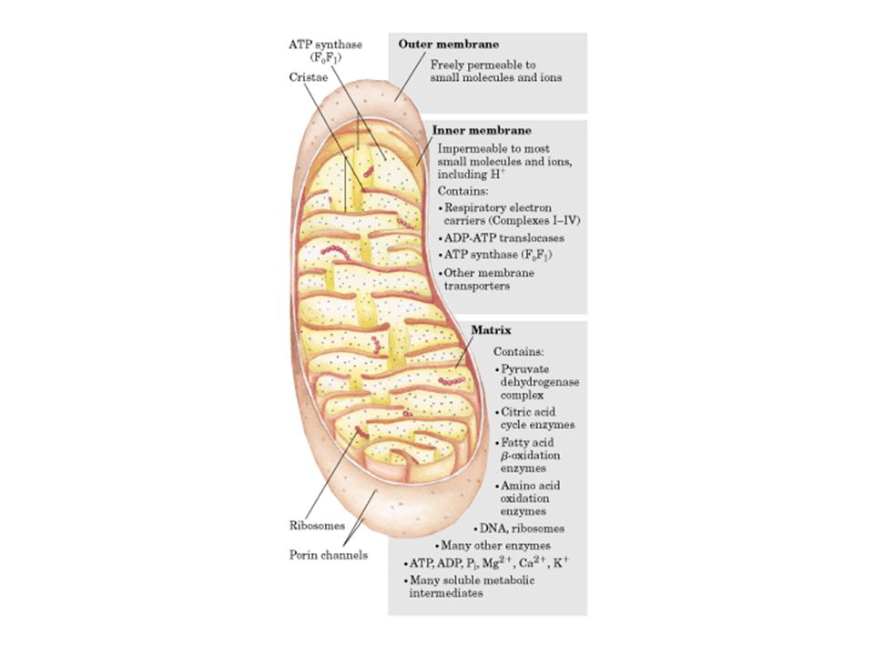
Topology of mitochondrial inner membranes
Carmen Manella, 2008

9
Dr. Carmen Manella Topología de la membrana interna mitocondrial

10
García JJ, 2006 Facultad de Química, UNAM

11
Mitochondrial volume homeostasis mediated by K+ transport and the PTP
Mitochondrial volume homeostasis mediated by K+ transport and the PTP. The scheme summarizes the role of K+ fluxes and PTP opening in mitochondrial volume homeostasis. Kc, potassium channels; ETC, electron transfer chain; Mkh1, essential mitochondrial KHE factor; Δψ, mitochondrial membrane potential difference; PTP, permeability transition pore

13
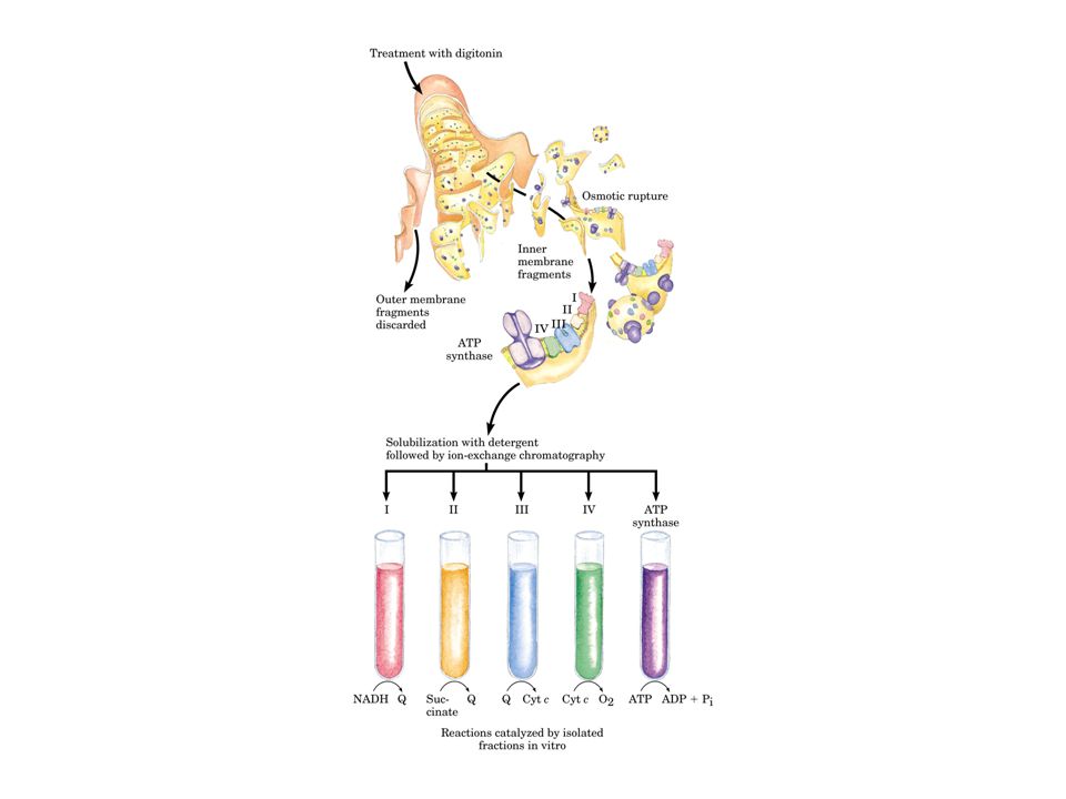
Cadena transportadora de electrones

14
Cadena transportadora de electrones

16
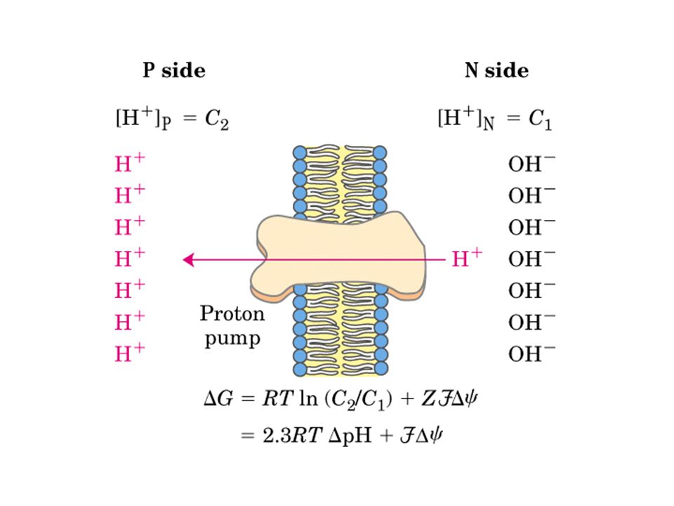
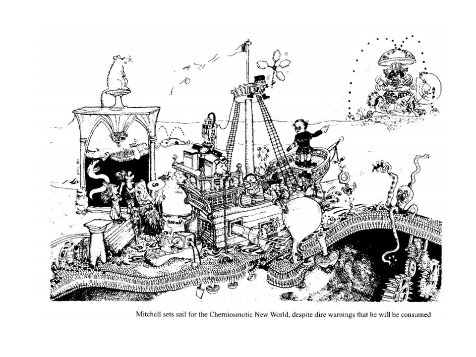
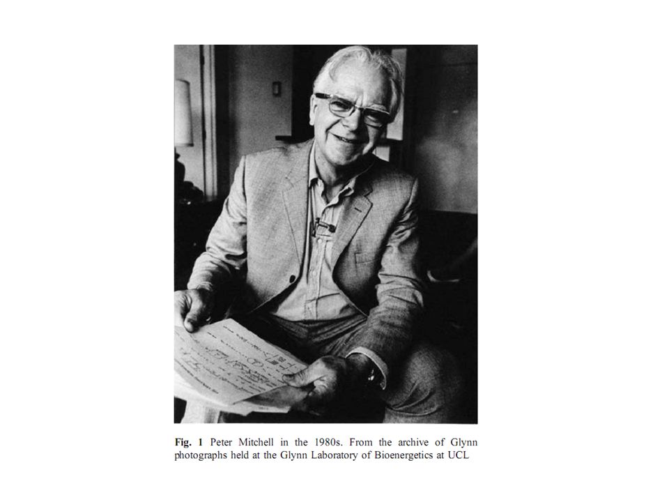
La teoría quimiosmótica (1961)
Peter Mitchel

17
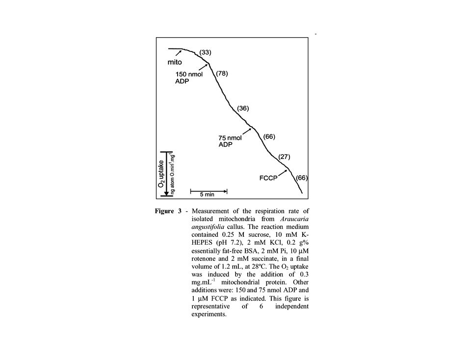
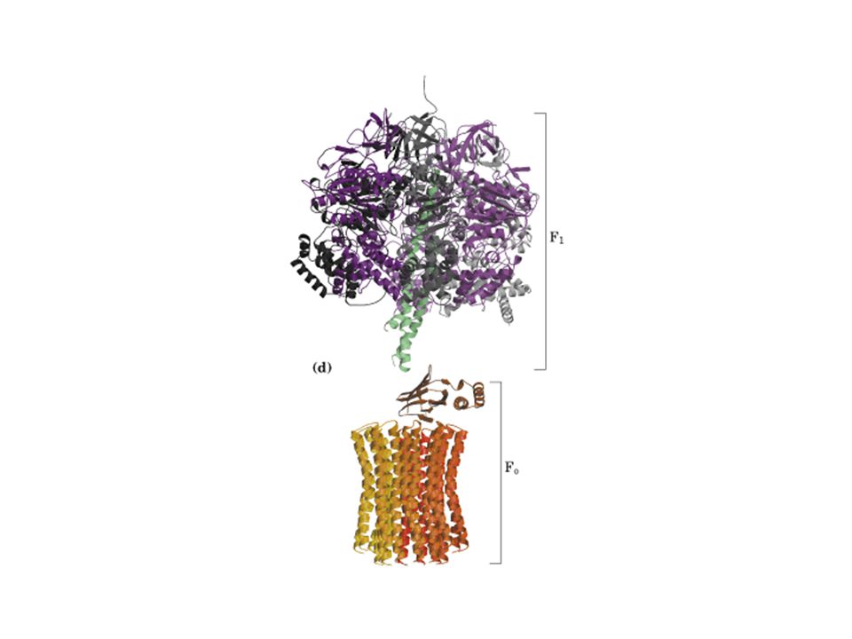
Acoplamiento de la respiración con la síntesis de ATP

18
desacoplamiento de la respiración de la síntesis de ATP!

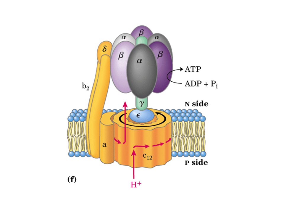
25
hidrólisis ATP → ADP + Pi

26
ATP → ADP + Pi Rotation of the actin filament attached to the ε subunit in the presence of 2 mm ATP. Rotation of the actin filament attached to the ε subunit in the presence of 2 mm ATP. a, an example of sequential images of a rotating actin filament attached to the ε subunit in α3β3γεSA. Length of the filament from rotational axis to the tip was 1.6 μm. Rotational rate was 1.0 rps. Time interval between images was 100 ms. Thescale bar denotes 5 μm. b, examples of time course of the rotation of the actin filament. Length of the filaments presented here is 0.5–1.4 μm. Only the filaments that rotated around one end are shown. Each line represents one filament. Solid lines indicate the rotation of the ε subunit in α3β3γεSAwhile dotted lines and broken lines indicate the rotation of the γ subunit in α3β3γSA and α3β3γSAε, respectively.c, same as b except that the length of the filaments is more than 1.5 μm. Details of the experiments are described under “Experimental Procedures.” Kato-Yamada Y et al. J. Biol. Chem. 1998;273: ©1998 by American Society for Biochemistry and Molecular Biology

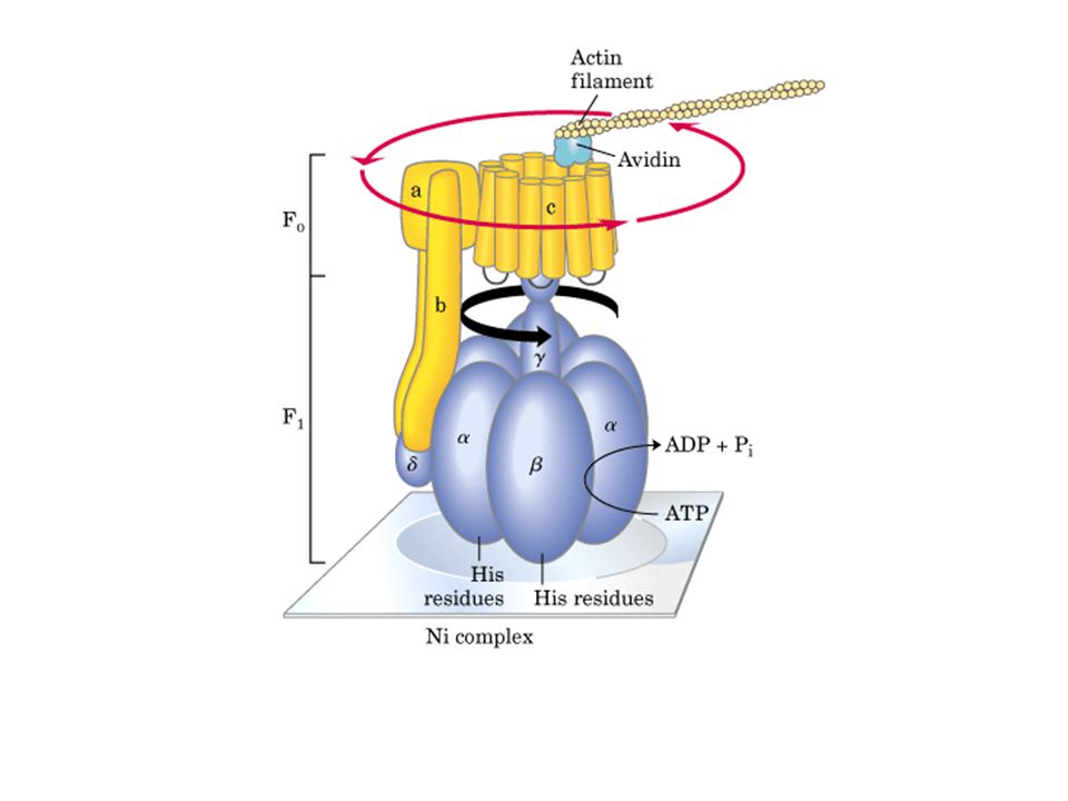
27
síntesis ADP + Pi → ATP Hiroyasu et al, 2004

28
Hiroyasu et al, 2004

29
Luciernaga

30
Hiroyasu et al, 2004

31
Esto ocurre en la grasa parda!

32
Prefiero jugar! sip!

33
y la cobija amá? Quiero mi chambrita!

34
La generación de calor en la grasa parda

35
Bacteria con flagelos

36
La fotofosforilación oxidativa

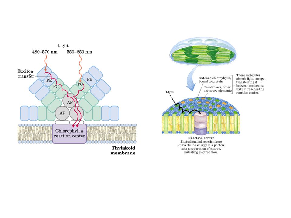
37
Espectro de absorbencia de la luz

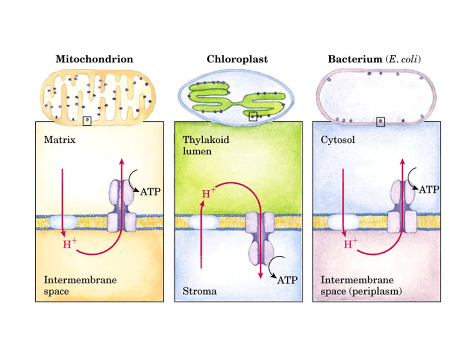
39
El acoplamiento del transporte de electrones a la foto-fosforilación oxidativa

Presentaciones similares


.>")


